. 2018 May 30:7:10.
doi: 10.1038/s41377-018-0003-3. eCollection 2018.

High-resolution multimodal flexible coherent Raman endoscope

Affiliations

High-resolution multimodal flexible coherent Raman endoscope

Alberto Lombardini et al. Light Sci Appl. .

Abstract

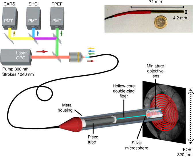
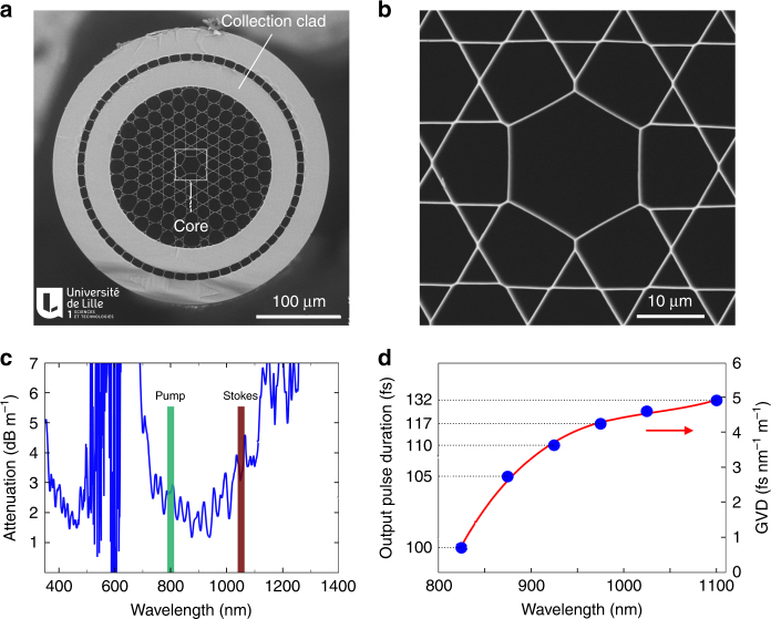
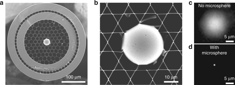
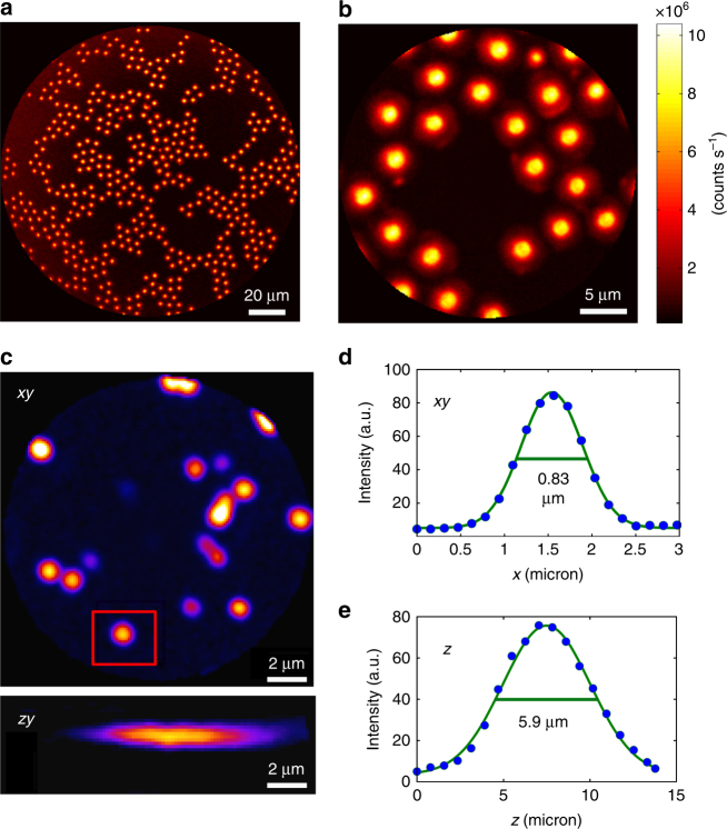
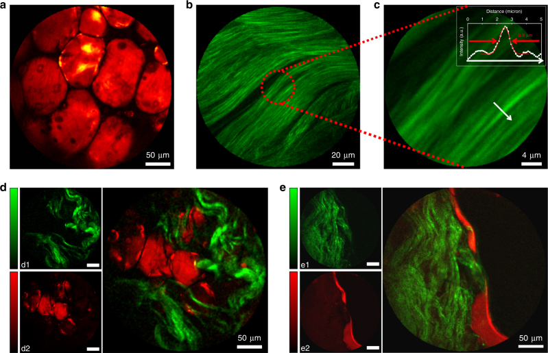
Coherent Raman scattering microscopy is a fast, label-free, and chemically specific imaging technique that shows high potential for future in vivo optical histology. However, the imaging depth in tissues is limited to the sub-millimeter range because of absorption and scattering. Realization of coherent Raman imaging using a fiber endoscope system is a crucial step towards imaging deep inside living tissues and providing information that is inaccessible with current microscopy tools. Until now, the development of coherent Raman endoscopy has been hampered by several issues, mainly related to the fiber delivery of the excitation pulses and signal collection. Here, we present a flexible, compact, coherent Raman, and multimodal nonlinear endoscope (4.2 mm outer diameter, 71 mm rigid length) based on a resonantly scanned hollow-core Kagomé-lattice double-clad fiber. The fiber design enables distortion-less, background-free delivery of femtosecond excitation pulses and back-collection of nonlinear signals through the same fiber. Sub-micrometer spatial resolution over a large field of view is obtained by combination of a miniature objective lens with a silica microsphere lens inserted into the fiber core. We demonstrate high-resolution, high-contrast coherent anti-Stokes Raman scattering, and second harmonic generation endoscopic imaging of biological tissues over a field of view of 320 µm at a rate of 0.8 frames per second. These results pave the way for intraoperative label-free imaging applied to real-time histopathology diagnosis and surgery guidance.


Conflict of interest statement

The authors declare that they have no conflict of interest.

Figures

Fig. 1
Fig. 1. Coherent Raman endoscope overview.
A tunable femtosecond laser provides two synchronized pulse trains that are injected into the hollow-core fiber probe to perform CARS, SHG, and TPEF endoscopy. The light emitted by the sample is collected by and back-propagates through the same fiber (1 m long) before detection by large area photomultiplier detectors. The inset picture shows the endoscope probe head inserted into its stainless steel housing
Fig. 2
Fig. 2. Hollow-core photonic crystal fiber for multimodal nonlinear endoscopy.
a Electron microscope image of the hollow-core (HC) fiber featuring a Kagomé lattice together with a double cladding (DC) separated by air holes. The excitation pulses propagate through the HC, whereas the nonlinear signal is collected and transmitted through the DC. b Close-up view of the fiber core area. c Attenuation of the hollow fiber core; the wavelengths used for the pump and Stokes beams are highlighted. d Expected pulse duration after propagation over 1 m of fiber, for initially transform-limited 100 fs pulses. The output pulse duration is computed using the  experimental GVD values (blue dots—right scale), which are  obtained from the measurement of the group delay versus wavelength. The expected duration for a 150 fs input pulse centered at 1040 nm (the Stokes pulse) is 157 fs after 1 m of fiber. The red line is a third-order polynomial fit to the GVD data
Fig. 3
Fig. 3. Microsphere lens inserted into the hollow fiber core provides a submicron focus spot required for imaging.
a Scanning electron microscope image of the Kagomé HC fiber with a 30 µm silica microsphere inserted and sealed into the fiber core. b Close-up view of the microsphere inserted into the hollow core. c In the absence of the microsphere, the HC mode diameter is 15 µm, which is inappropriate for high-resolution nonlinear imaging. d The microsphere acts as a ball lens and focuses the light exiting the fiber core into a ~1 µm diameter spot
Fig. 4
Fig. 4. High-contrast submicron image resolution.
a, b CARS images of 5-µm diameter polystyrene beads deposited onto a glass coverslip with two different FoVs: 155 µm (a) and 25 µm (b) corresponding to 7.5 V and 1.5 V pk-pk driving voltages, respectively. Note the vanishing background between the beads; powers: 10 mW (pump) and 5 mW (Stokes); excitation wavelengths: 800 nm (pump) and 1040 nm (Stokes). c Lateral (x,y) and axial (x,z) TPEF images (forward detected) of 200-nm-diameter fluorescent nanoparticles (excitation: 800 nm, 10 mW; detection: 500–600 nm). The nanoparticle highlighted in the red rectangle is used to obtain the image cross-cuts (d, e), which are used to deduce the lateral and axial two-photon PSFs. The values shown on the graph indicate the full width at half maximum (FWHM)
Fig. 5
Fig. 5. Multimodal nonlinear endoscopy in biological tissues.
a CARS image of fresh fatty tissues obtained from human colon. b SHG image of rat tail tendons featuring collagen fibers. c Close-up view of b confirming the 0.8 µm lateral resolution for nonlinear imaging. d, e Multimodal SHG (green, d1, e1) and CARS (red, d2, e2) images of fresh human colon tissues taken 50 µm below the sample surface. The large images are overlaps of the SHG and CARS images. SHG images required higher powers and were acquired immediately after taking the CARS images. Laser powers incident on the sample: CARS pump 20 mW, Stokes 10 mW; SHG 60 mW (45 mW for images b and c). Excitation wavelengths: 800 nm (pump CARS, SHG) and 1040 nm (Stokes). All images were averaged over five acquisitions for a total acquisition time of 6.5 s, except b and c that were averaged 10 times (13 s). The color scale in (counts per second) is provided on Fig. S14

References

    1. Cheng JX, Xie XS. Vibrational spectroscopic imaging of living systems: an emerging platform for biology and medicine. Science. 2015;350:aaa8870. doi: 10.1126/science.aaa8870. - DOI - PubMed
    1. Camp CH, Jr., Cicerone MT. Chemically sensitive bioimaging with coherent Raman scattering. Nat. Photonics. 2015;9:295–305. doi: 10.1038/nphoton.2015.60. - DOI
    1. Gao LS, et al. On-the-spot lung cancer differential diagnosis by label-free, molecular vibrational imaging and knowledge-based classification. J. Biomed. Opt. 2011;16:096004. doi: 10.1117/1.3619294. - DOI - PubMed
    1. Ji MB, et al. Detection of human brain tumor infiltration with quantitative stimulated Raman scattering microscopy. Sci. Transl. Med. 2015;7:309ra163. doi: 10.1126/scitranslmed.aab0195. - DOI - PMC - PubMed
    1. Orringer DA, et al. Rapid intraoperative histology of unprocessed surgical specimens via fibre-laser-based stimulated Raman scattering microscopy. Nat. Biomed. Eng. 2017;1:0027. doi: 10.1038/s41551-016-0027. - DOI - PMC - PubMed