

Monoamine Oxidase B Total Distribution Volume in the Prefrontal Cortex of Major Depressive Disorder: An [11C]SL25.1188 Positron Emission Tomography Study
- PMID: 30840042
- PMCID: PMC6551845
- DOI: 10.1001/jamapsychiatry.2019.0044
Monoamine Oxidase B Total Distribution Volume in the Prefrontal Cortex of Major Depressive Disorder: An [11C]SL25.1188 Positron Emission Tomography Study
Erratum in
-
Error in Scale of Y-Axis in Figure 2.JAMA Psychiatry. 2019 Jun 1;76(6):659. doi: 10.1001/jamapsychiatry.2019.0688. JAMA Psychiatry. 2019. PMID: 30969324 Free PMC article. No abstract available.
Abstract
Importance: Monoamine oxidase B (MAO-B) is an important, high-density enzyme in the brain that generates oxidative stress by hydrogen peroxide production, alters mitochondrial function, and metabolizes nonserotonergic monoamines. Recent advances in positron emission tomography radioligand development for MAO-B in humans enable highly quantitative measurement of MAO-B distribution volume (MAO-B VT), an index of MAO-B density. To date, this is the first investigation of MAO-B in the brain of major depressive disorder that evaluates regions beyond the raphe and amygdala.
Objective: To investigate whether MAO-B VT is elevated in the prefrontal cortex in major depressive episodes (MDEs) of major depressive disorder.
Design, setting, and participants: This case-control study was performed at a tertiary care psychiatric hospital from April 1, 2014, to August 30, 2018. Twenty patients with MDEs without current psychiatric comorbidities and 20 age-matched controls underwent carbon 11-labeled [11C]SL25.1188 positron emission tomography scanning to measure MAO-B VT. All participants were drug and medication free, nonsmoking, and otherwise healthy.
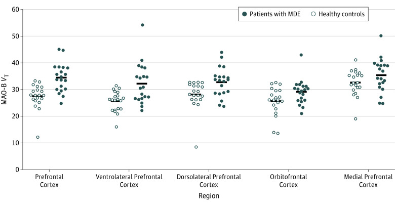
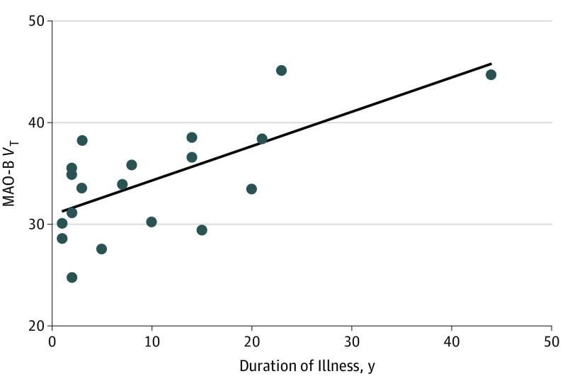
Main outcomes and measures: The MAO-B VT in the prefrontal cortex (PFC). The second main outcome was to evaluate the association between MAO-B VT in the PFC and duration of major depressive disorder illness.
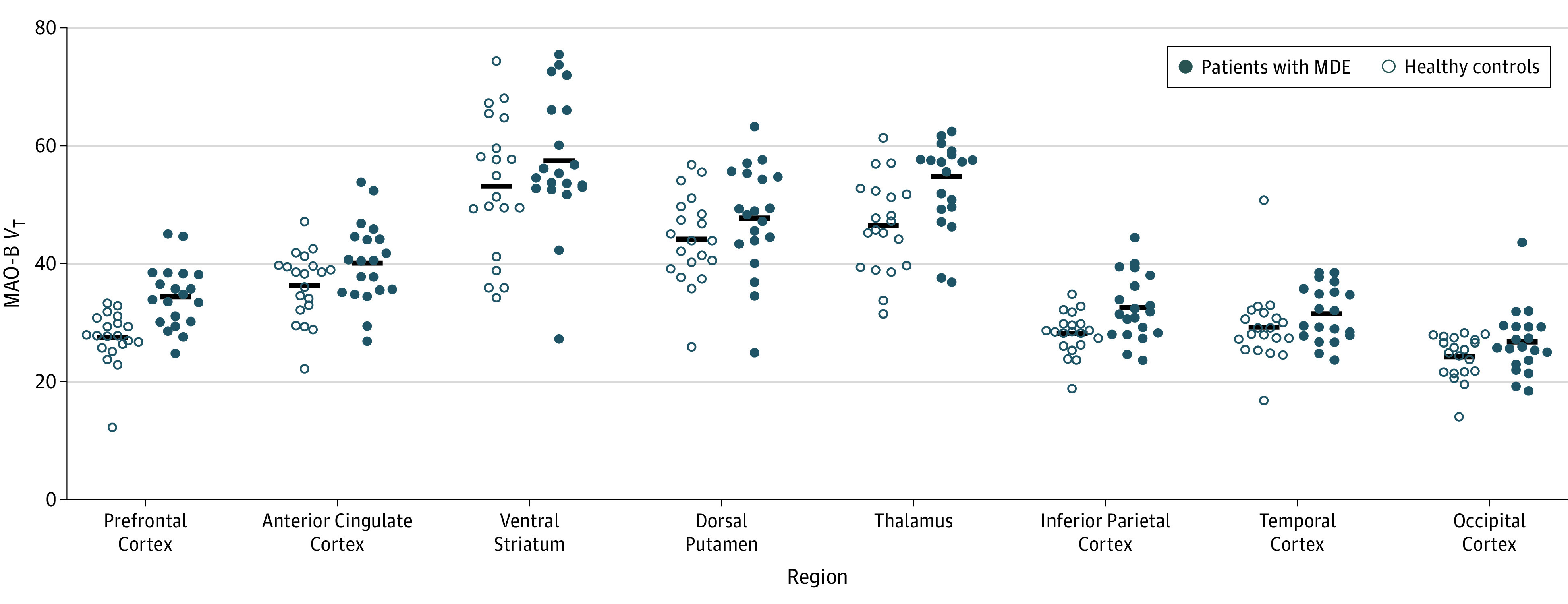
Results: Twenty patients with MDEs (mean [SD] age, 34.2 [13.2] years; 11 women) and 20 healthy controls (mean [SD] age, 33.7 [13.1] years; 10 women) were recruited. Patients with MDEs had significantly greater MAO-B VT in the PFC (mean, 26%; analysis of variance, F1,38 = 19.6, P < .001). In individuals with MDEs, duration of illness covaried positively with MAO-B VT in the PFC (analysis of covariance, F1,18 = 15.2, P = .001), as well as most other cortex regions and the thalamus.
Conclusions and relevance: Fifty percent (10 of 20) of patients with MDEs had MAO-B VT values in the PFC exceeding those of healthy controls. Greater MAO-B VT is an index of MAO-B overexpression, which may contribute to pathologies of mitochondrial dysfunction, elevated synthesis of neurotoxic products, and increased metabolism of nonserotonergic monoamines. Hence, this study identifies a common pathological marker associated with downstream consequences poorly targeted by the common selective serotonin reuptake inhibitor treatments. It is also recommended that the highly selective MAO-B inhibitor medications that are compatible for use with other antidepressants and have low risk for hypertensive crisis should be developed or repurposed as adjunctive treatment for MDEs.
Conflict of interest statement
Figures
References
-
- Mathers C, Fat DM, Boerma JT. The Global Burden of Disease: 2004 Update. Geneva, Switzerland: World Health Organization; 2008. doi: 10.1016/B978-012373960-5.00335-X - DOI
Publication types
MeSH terms
Substances
Grants and funding
LinkOut - more resources
Full Text Sources
Miscellaneous
