. 2019 Jul 1;125(13):2222-2232.
doi: 10.1002/cncr.32033. Epub 2019 Mar 6.

Targeting Burkitt lymphoma with a tumor cell-specific heptamethine carbocyanine-cisplatin conjugate

Affiliations

Targeting Burkitt lymphoma with a tumor cell-specific heptamethine carbocyanine-cisplatin conjugate

Stefan Mrdenovic et al. Cancer. .

Abstract

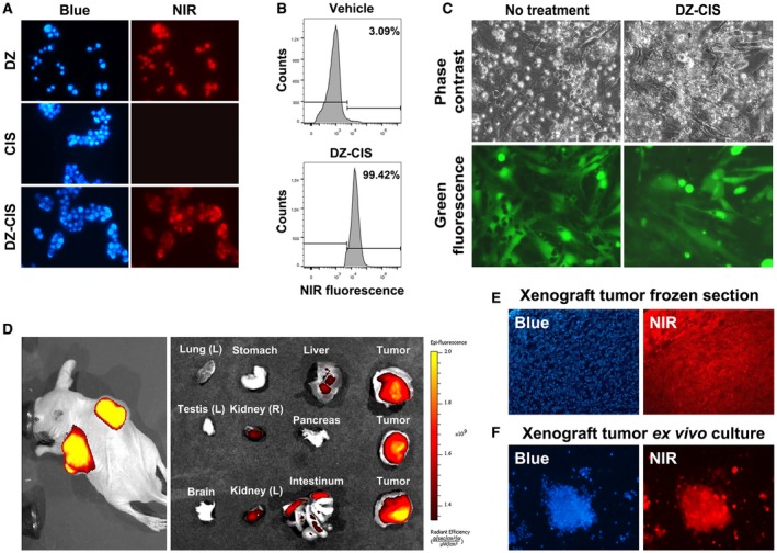
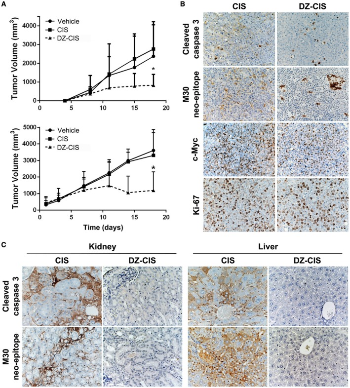
Background: Burkitt lymphoma is a fast-growing mature B cell malignancy, whose genetic hallmark is translocation and activation of the c-myc gene. Prompt multiagent immunochemotherapy regimens can have favorable outcomes, but prognosis is poor in refractory or relapsed disease. We previously identified a novel family of near-infrared heptamethine carbocyanine fluorescent dyes (HMCD or DZ) with tumor-homing properties via organic anion-transporting peptides. These membrane carriers have uptake in tumor cells but not normal cells in cell culture, mouse and dog tumor models, patient-derived xenografts, and perfused kidney cancers in human patients.

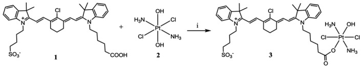
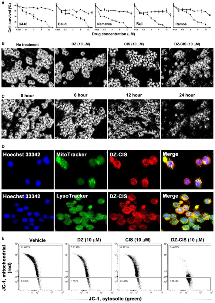
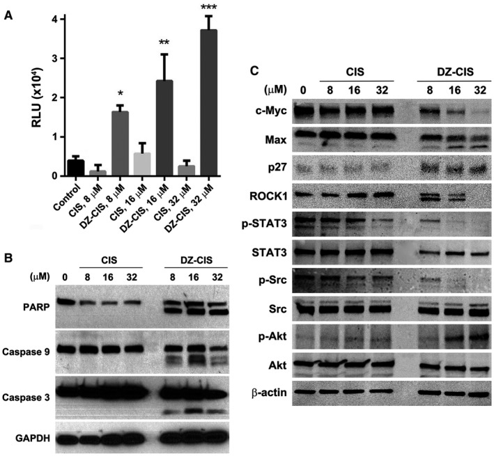
Methods: Here we report the cytotoxic effects of a synthesized conjugate of DZ with cisplatin (CIS) on B cell lymphoma CA46, Daudi, Namalwa, Raji, and Ramos cell lines in cell culture and in xenograft tumor formation. Impaired mitochondrial membrane permeability was examined as the mechanism of DZ-CIS-induced lymphoma cell death.

Results: The new conjugate, DZ-CIS, is cytotoxic against Burkitt lymphoma cell lines and tumor models. DZ-CIS retains tumor-homing properties to mitochondrial and lysosomal compartments, does not accumulate in normal cells and tissues, and has no nephrotoxicity in mice. DZ-CIS accumulated in Burkitt lymphoma cells and tumors induces apoptosis and retards tumor cell growth in culture and xenograft tumor growth in mice.

Conclusion: DZ-CIS downregulated c-myc and overcame CIS resistance in myc-driven TP53-mutated aggressive B cell Burkitt lymphoma. We propose that DZ-CIS could be used to treat relapsed/refractory aggressive Burkitt lymphomas.

Keywords: Burkitt lymphoma; cell death; cisplatin; conjugate; heptamethine carbocyanine.


Conflict of interest statement

DZ‐CIS is licensed to DaZen Theranostics, Inc., where Leland W. K. Chung is currently serving as the Chairman of the Board and Chief Scientist for the development of one of the licensed lead compounds from laboratory to the clinic and for which Stefan Mrdenovic, Yi Zhang, Ruoxiang Wang, and Gina Chia‐Yi Chu are shareholders. Leland W. K. Chung also has a patent pending (US 2016/0310604 A1).

Figures

Figure 1
Figure 1
Synthesis of the heptamethine carbocyanine fluorescent dye–cisplatin conjugate (DZ‐CIS). DZ‐CIS conjugate was synthesized through a succinic ester linker. DZ‐1 cis‐ and trans‐diaminedichloro‐dihydroxyplatinum (IV) (oxoplatin) 2 were mixed and reacted as described in Materials and Methods by adding N’‐ethylcarbodiimide hydrochloride and dimethylaminopyridine in dimethyl sulfoxide solution.
Figure 2
Figure 2
Heptamethine carbocyanine fluorescent dye–cisplatin conjugate (DZ‐CIS) cytotoxicity. (A) Dose‐effect curve. All Burkitt lymphoma cell lines were insensitive to CIS (circles on solid line) but dose‐dependently sensitive to DZ‐ CIS (squares on dotted line). (B) DZ‐CIS‐induced death of Namalwa cells 24 hours after treatment. Black and white images of trypan blue–stained cells (magnification ×100). (C) DZ‐CIS (10 μM) caused marked lymphoma cell death with morphological changes starting 6 hours into the treatment. Cell death became conspicuous around 12 hours (trypan blue stain, magnification ×100). (D) DZ‐CIS preferentially accumulates to mitochondria and lysosomes, demonstrated by colocalization (magnification ×600) of the NIR signal with MitoTracker (upper row) and LysoTracker (lower row). (E) Flow cytometry indicates that 6‐hour treatment with DZ‐CIS disrupts mitochondrial membrane integrity. JC‐1 in the cytosol lost its red fluorescence as monomeric JC‐1 (which emits green fluorescence).
Figure 3
Figure 3
The heptamethine carbocyanine fluorescent dye–cisplatin conjugate (DZ‐CIS) acts specifically on Burkitt lymphoma cells. (A) Near‐infrared (NIR) microscopy of Namalwa cells after treatment with 4 μM DZ‐CIS for 15 minutes. DZ‐CIS accumulated in all lymphoma cells. Blue fluorescence of the nuclei was from Hoechst 33342 co‐stain (200×). (B) Namalwa cells treated with 4 μM DZ‐CIS for 15 minutes were assayed for rapid uptake of the fluorescent dye–drug conjugate in live cells using flow cytometry. (C) Images of lymphoma and stromal cell coculture show that DZ‐CIS preferentially kills lymphoma but not stromal cells. In coculture with GFP‐tagged CCD16Lu normal human lung mesenchymal stromal cells, 24‐hour DZ‐CIS treatment preferentially killed Burkitt lymphoma cells (Namalwa), while stromal cells survived and displayed healthy morphology (magnification ×200). DZ‐CIS–induced death of lymphoma cells in coculture was confirmed with trypan blue stain. (D) Tumor‐specific uptake and retention of DZ‐CIS in vivo were evaluated with NCrnu/nu mice bearing Namalwa xenograft tumors. After treatment with DZ‐CIS for 2 weeks, mice were subjected to whole body NIR imaging (left). Necropsy tumor samples together with host organs were subjected to ex vivo NIR imaging (right). (E) Frozen sections of xenograft tumors were stained with 4’,6‐diamidino‐2‐phenyindole dihydrochloride (DAPI) and examined for retention of DZ‐CIS by NIR microscopy (magnification ×100). (F) Xenograft tumors diced in ex vivo culture were stained with DAPI (magnification ×100). Most tumor cells still carried DZ‐CIS signals after 3 days in the culture.
Figure 4
Figure 4
Heptamethine carbocyanine fluorescent dye–cisplatin conjugate (DZ‐CIS)–induced caspase cascade activation. (A) Namalwa cells treated with DZ‐CIS were assayed for the involvement of caspases with a CaspaseGlo 3/7 luminescent assay 24 hours after treatment. Results are presented with the mean ± standard deviation of quadruple measurements (* P < .05, ** P < .01, *** P < .0001). (B) Whole cell lysates of the treated cells were subjected to western blotting to detect activation of the caspase cascade. (C) DZ‐CIS–treated Namalwa cells were examined for protein level and protein modification changes by western blotting. DZ‐CIS treatment showed decreased expression of c‐myc and induced Max protein cleavage and increased p27 protein expression, suggesting growth arrest of treated cells. DZ‐CIS treatment caused cleavage of the ROCK1 protein. c‐Jun, STAT3, and Src protein phosphorylation was suppressed. Paradoxically, DZ‐CIS caused increased phosphorylation of AKT, suggesting its activation.
Figure 5
Figure 5
The heptamethine carbocyanine fluorescent dye–cisplatin conjugate (DZ‐CIS) inhibited xenograft tumor growth. (A) NCrnu/nu mice (top, n = 5 nude mice bearing 8‐10 Namalwa tumors per treatment group) and NOD Scid mice (bottom, n = 5 Scid mice bearing 16 Namalwa tumors per treatment group) were treated intraperitoneally with either 5 (NOD Scid) or 10 (NCr nude) mg/kg DZ‐CIS twice a week, from day 4 and day 1, respectively, resulting in significantly decreased tumor volume compared with CIS or vehicle treatment groups (P < .05). (B) CIS and DZ‐CIS–treated xenograft tumors were analyzed by immunohistochemistry for cell proliferation, survival, and death biomarker protein expression. (C) Mice were evaluated for renal and hepatic injuries with immunohistochemistry for cell death markers.

Similar articles

Cited by

References

    1. Molyneux EM, Rochford R, Griffin B, et al. Burkitt’s lymphoma. Lancet. 2012;379:1234‐1244. - PubMed
    1. Swerdlow SH, Campo E, Harris NL, et al. WHO classification of tumors of haematopoietic and lymphoid tissues, Revised Fourth Edition. Geneva, Switzerland: World Health Organization; 2017.
    1. Brown JR, Freedman AS, Aster JC. Pathobiology of Burkitt lymphoma. UpToDate. https://www.uptodate.com/contents/pathobiology-of-burkitt-lymphoma. Accessed July 1, 2018.
    1. ar‐Rushdi A, Nishikura K, Erikson J, Watt R, Rovera G, Croce CM. Differential expression of the translocated and the untranslocated c‐myc oncogene in Burkitt lymphoma. Science. 1983;222:390‐393. - PubMed
    1. Yang F, Du J, Zhang H, et al. Serum metabolomics of Burkitt lymphoma mouse models. PLoS One. 2017;12:e0170896. - PMC - PubMed

Publication types