. 2019 Mar 3;53(1):49-56.
doi: 10.2478/raon-2019-0012.

MRI prognostic factors of tongue cancer: potential predictors of cervical lymph nodes metastases

Affiliations

MRI prognostic factors of tongue cancer: potential predictors of cervical lymph nodes metastases

Mohamed A F Mourad et al. Radiol Oncol. .

Abstract

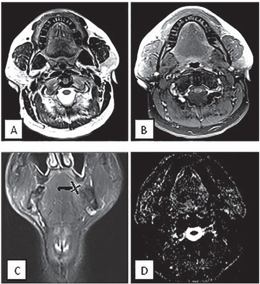
Background This study aimed to evaluate the efficacy of three MR imaging parameters, which are tumour thickness, para-lingual distance and apparent diffusion coefficient (ADC) value for prediction of cervical lymph nodes metastasis in cancer tongue patients. Patients and methods Fifty patients with proved cancer tongue by histopathological examination underwent MRI examination. T1 and T2- weighted MRI, diffusion-weighted images and post-contrast T1 fat suppression sequences were used. Results The patients were classified according to lymph nodes involvement as seen by MRI into two groups. Significant differences between positive and negative nodes groups were observed regarding tumour thickness and para-lingual distance (p-values = 0.008 and 0.003 respectively). ROC curve analyses revealed cut-off values >13.8 mm and ≤ 3.3 mm for tumour thickness and para-lingual distance respectively for prediction of nodes involvement. No significant differences between patients with and without cervical lymph nodes metastasis were found regarding corresponding ADC value of the tumour (p-value = 0.518). Conclusions Para-lingual distance and tumour thickness are factors that could influence pre-operative judgment and prognosis of tongue cancer patients. ADC value of the tumour itself seem not to be a reliable index of cancer progression to regional lymph nodes.

Keywords: apparent diffusion coefficient; cervical lymph nodes metastases; para-lingual distance; tongue cancer; tumour thickness.


Figures

Figure 1
Figure 1
Scatter plot showing strong negative correlation between MR tumour thickness and para-lingual distance (p-value < 0.001 and r = 0.84).
Figure 2
Figure 2
Comparison graphs illustrating the significant differences between tumour thickness and para-lingual distance among nodes positive (N1) and negative (N0) patients (p-values 0.008 and 0.003 respectively).
Figure 3
Figure 3
Receiver Operator Characteristic (ROC) curve analyses for tumour thickness and para-lingual distance predicting nodes spread (p-values < 0.001 and AUC 0.864 and 0.848 respectively).
Figure 4
Figure 4
MRI of a male patient 65-years-old with small lesion at left hemi-tongue (T1N0) disease. (A) Axial T2 (B) Axial T1 fat suppression post contrast (C) T2 coronal (D) Axial DWI. MRI and elective dissected neck revealed no positive cervical lymph nodes spread. The vertical black line was drawn as a reference line connecting maximum tumour-mucosa junctions. Two horizontal lines were drawn perpendicular to the reference line. Tumour thickness is the sum of both of these horizontal lines and was determined as 5.5 mm. The thick black line between the tumour and the para-lingual space represented the para-lingual distance = 10.5 mm.
Figure 5
Figure 5
MRI of a 75-years-old female with sizable tongue mass crossing the midline (T4N1 disease). (A) Axial T1 post contrast fat suppression (B) Axial DWI (C) Coronal T1 post contrast fat suppression (D) Coronal T1 post contrast shows metastatic cervical lymph nodes. Tumour thickness is the sum of the two horizontal black lines drawn perpendicular to the vertical black line connecting maximum tumour junction distance and was determined as 30 mm. The thick black line representing para-lingual distance between the tumour and the para-lingual space was determined as – 10 as the tumour margin extends beyond the midline by 10 mm

References

    1. Funk GF, Karnell LH, Robinson RA, Zhen WK, Trask DK, Hoffman HT. Presentation, treatment, and outcome of oral cavity cancer: a National Cancer Data Base report. Head Neck. 2002;24:165–80. - PubMed
    1. Lubek JE, Clayman L. An update on squamous carcinoma of the oral cavity, oropharynx, and maxillary sinus. Oral Maxillofac Surg Clin North Am. 2012;24:307–16. doi: 10.1016/j.coms.2012.01.003. - DOI - PubMed
    1. Syrjanen S. Human papillomavirus infections and oral tumors. Med Microbiol Immunol. 2003;192:123–8. doi: 10.1007/s00430-002-0173-7. - DOI - PubMed
    1. Tshering Vogel DW, Zbaeren P. Thoeny HC. Cancer of the oral cavity and oropharynx. Cancer Imaging. 2010;10:62–72. doi: 10.1102/14707330.2010.0008. - DOI - PMC - PubMed
    1. Stone M, Davis EP, Douglas AS, Aiver MN, Gullapalli R, Levine WS. Modeling tongue surface contours from Cine-MRI images. J Speech Lang Hear Res. 2001;44:1026–40. - PubMed

MeSH terms