. 2019 Jun;33(6):7061-7071.
doi: 10.1096/fj.201802535R. Epub 2019 Mar 6.

Examination of the role of sphingosine kinase 2 in a murine model of systemic lupus erythematosus

Affiliations

Examination of the role of sphingosine kinase 2 in a murine model of systemic lupus erythematosus

Sabira Mohammed et al. FASEB J. 2019 Jun.

Abstract

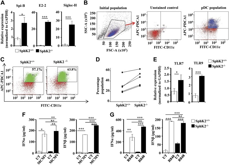
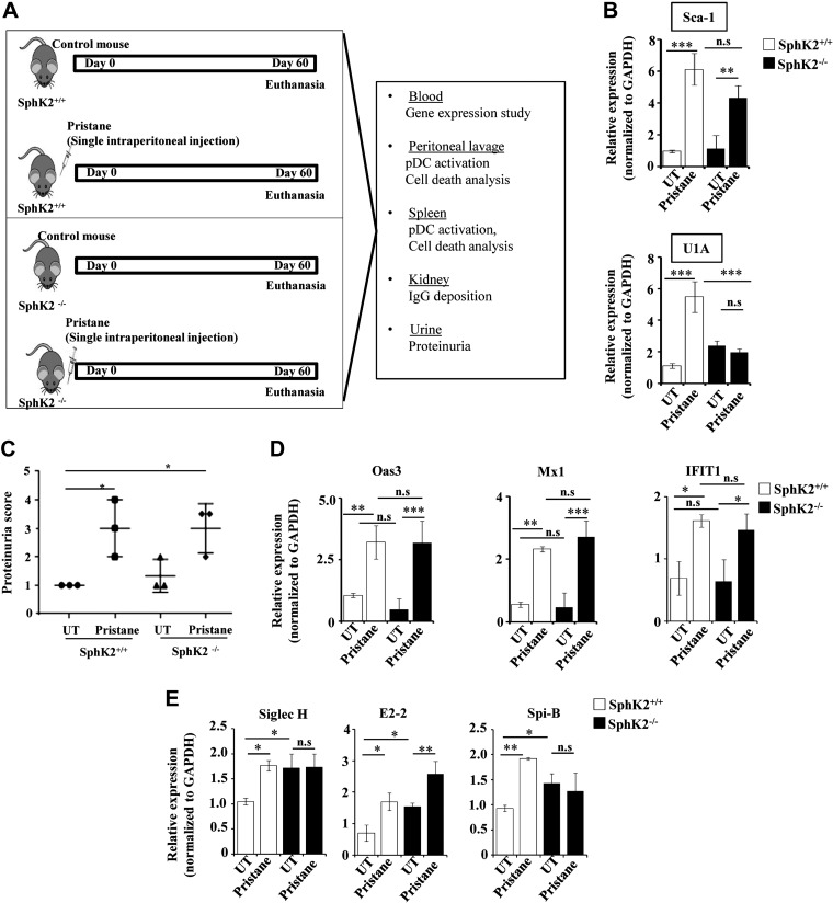
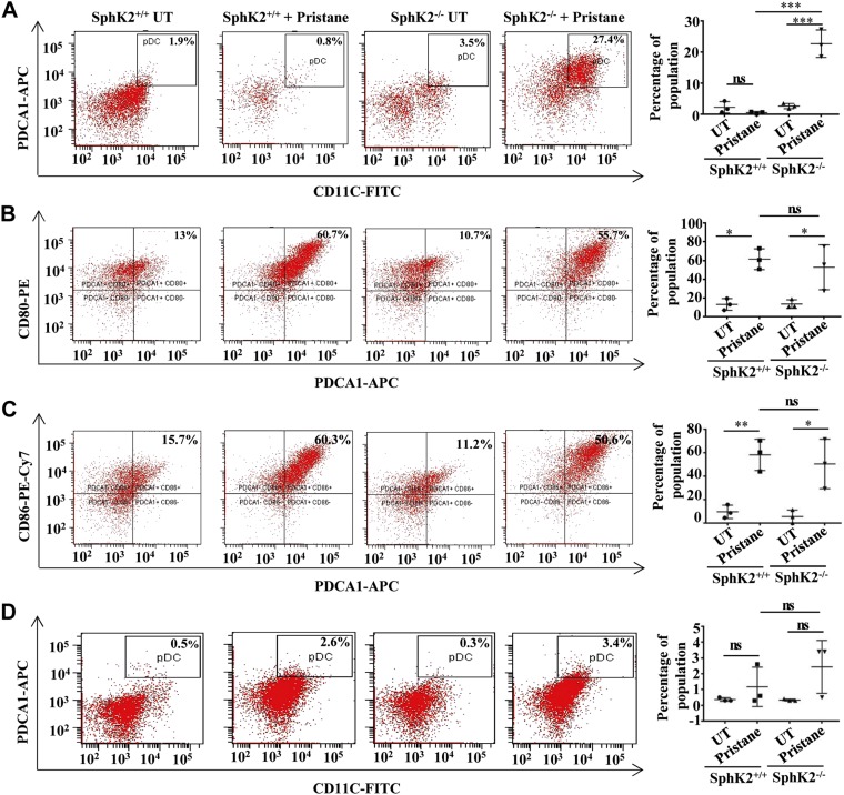
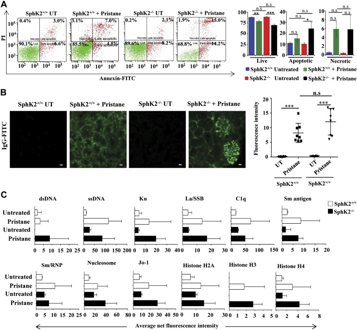
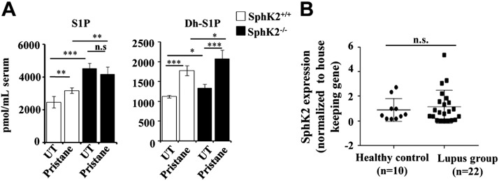
Systemic lupus erythematosus is an autoimmune disease characterized by overproduction of type 1 IFN that causes multiple organ dysfunctions. Plasmacytoid dendritic cells (pDCs) that secrete large amounts of IFN have recently been implicated in the initiation of the disease in preclinical mouse models. Sphingosine-1-phosphate, a bioactive sphingolipid metabolite, is produced by 2 highly conserved isoenzymes, sphingosine kinase (SphK) 1 and SphK2, and regulates diverse processes important for immune responses and autoimmunity. However, not much is known about the role of SphK2 in autoimmune disorders. In this work, we examined the role of SphK2 in pDC development and activation and in the pristane-induced lupus model in mice that mimics the hallmarks of the human disease. Increases in pDC-specific markers were observed in peripheral blood of SphK2 knockout mice. In agreement, the absence of SphK2 increased the differentiation of FMS-like tyrosine kinase 3 ligand dendritic cells as well as expression of endosomal TLRs, TLR7 and TLR9, that modulate production of IFN. Surprisingly, however, SphK2 deficiency did not affect the initiation or progression of pristane-induced lupus. Moreover, although absence of SphK2 increased pDC frequency in pristane-induced lupus, there were no major changes in their activation status. Additionally, SphK2 expression was unaltered in lupus patients. Taken together, our results suggest that SphK2 may play a role in dendritic cell development. Yet, because its deletion had no effect on the clinical lupus parameters in this preclinical model, inhibitors of SphK2 might not be useful for treatment of this devastating disease.-Mohammed, S., Vineetha, N. S., James, S., Aparna, J. S., Lankadasari, M. B., Allegood, J. C., Li, Q.-Z., Spiegel, S., Harikumar, K. B. Examination of the role of sphingosine kinase 2 in a murine model of systemic lupus erythematosus.

Keywords: IFN; S1P; autoimmunity; pDCs; pristane.


Conflict of interest statement

The authors are thankful to the personnel of the Flow Cytometry Core Facility and Bioimaging Facility for excellent technical assistance, and the Animal Research Facility for mouse colony maintenance. S.M. and M.B.L. acknowledge the senior research fellowship from the Indian Council of Medical Research and the University Grant Commission, respectively. S.M. acknowledges the Doctoral Advisory Committee for valuable suggestions. The work was supported by the U.S. National Institutes of Health, National Institute of General Medical Sciences Grant R01GM043880 (to S.S.), a fast-track grant from the Indian Government’s Department of Science and Technology (SR/FT/LS-159/2012), a grand-in-aid scheme of the Council for Scientific and Industrial Research [37 (1720)/18/EMR-II], and, in part, by a faculty start-up grant and Department of Biotechnology (DBT) Ramalingaswami Re-Entry Fellowship (BT/RLF/Re-entry/38/2011) to K.B.H. The authors declare no conflicts of interest.

Figures

Figure 1
Figure 1
Effect of SphK2 on pDC development and function. A) pDC-specific markers SpiB, E2-2, and Siglec H were examined in the peripheral blood of SphK2+/+ and SphK2−/− mice by quantitative PCR. The fold change was calculated after normalizing with GAPDH. Data are expressed as means ± sd of 3 independent experiments (n = 7 mice/group). BD) FACS analysis of pDCs. Flt3L-DCs from SphK2+/+ and SphK2−/− mice were analyzed by FACS by staining for both CD11c and PDCA-1. B) The gating strategy of FACS is shown. The cells were first gated using FSC and SSC plots. Unstained samples were used to determine the gating for both FITC and APC. See Materials and Methods section for more details. C) The dot plots are representative of 5 different experiments showing Flt3L-pDCs generated from SphK2+/+ and SphK2−/− mice. D) Percent populations of Flt3L-pDCs positive for both CD11c and PDCA-1, sorted out by FACS (D). Data from 5 independent experiments are shown. E) Expression of TLR7 and TLR9 in Flt3L-DCs generated from SphK2+/+ and SphK2−/− mice was determined by quantitative PCR and expressed as fold change after normalizing with GAPDH. Similar results were obtained in 3 independent experiments (n = 5 mice/group). F) Flt3L-DCs (1 × 10⁵ cells) isolated by cell sorting from SphK2+/+ to SphK2−/− mice were treated without or with MCMV (multiplicity of infection = 1). After 18 h, cell supernatants were collected and analyzed by ELISA for IFN-α and β. Similar results were obtained in 3 independent experiments (n = 5 mice/group). G) SphK2+/+ and SphK2−/− mice were tail-vein injected with 2 µg of R848 per mouse, and blood was collected after 2 h. Serum was separated and analyzed by ELISA for type I IFN secretion. Statistical significance was assessed by a Student's unpaired t test (A, E) or by ANOVA (F, G) followed by a Tukey’s post hoc test; n.s., not significant. *P < 0.05, **P < 0.005, ***P < 0.001.
Figure 2
Figure 2
Lupus disease markers are similar in pristane-treated SphK2+/+ and SphK2−/− mice. A) Schematic representation of the pristane-induced lupus model, depicting the experimental conditions and the endpoint analyses. B) Expression of disease markers Sca-1 and U1A was determined in peripheral blood of mice by quantitative PCR and expressed as fold change after normalizing with GAPDH (n = 7/group). C) Renal function was evaluated by measurement of urinary protein levels using Uristix colorimetric strips. Proteinuria scores were based on color development that signifies the amount of protein in urine (n = 3/group). D) Expression of IFN-stimulated genes 2'-5′-oligoadenylate synthase 3 (Oas3), myxovirus resistance 1 (Mx1), and IFN-induced protein with tetratricopeptide repeats (IFIT1) in peripheral blood from SphK2+/+ to SphK2−/− mice (n = 7/group) was determined by quantitative PCR. Data were expressed as relative expressions normalized to GAPDH. E) Expression of the pDC markers SpiB, E2-2, and Siglec H in peripheral blood was determined by quantitative PCR and expressed as relative levels after normalizing to GAPDH. Data are expressed as means ± sd from 3 independent experiments. Statistical significance was determined by ANOVA with a post hoc Tukey’s test; n.s., not significant. *P < 0.05, **P < 0.005, ***P < 0.001.
Figure 3
Figure 3
SphK2 deficiency increases the pDC population in a diseased state but not their activation status. A) The percentage of pDC population identified as CD11c+ and PDCA-1+ in the peritoneal lavage of the experimental mice was assessed by FACS. B, C) The activation status of peritoneal pDCs indicated by the expression of CD80 (B) and CD86 (C) was assessed by FACS analysis. Representative dot plots of 1 of 3 independent experiments are shown in the left panels. Data shown are averages of 3 independent experiments (A–C). D) Percentage of pDC population double-positive for CD11c and PDCA-1 in splenocytes isolated from SphK2+/+ to SphK2−/− mice. Representative dot plots of 1 of 3 independent experiments are shown in the left panels, and averaged data from 3 independent experiments are shown in the right panel. Statistical significance was determined by ANOVA with a post hoc Tukey’s test; n.s., not significant. *P < 0.05, **P < 0.005, ***P < 0.001.
Figure 4
Figure 4
Absence of SphK2 enhances pristane-induced apoptosis of peritoneal cells. A) Peritoneal-lavage cells from SphK2+/+ to SphK2−/− mice untreated or injected with pristane were stained with PI and annexin V–FITC and analyzed by FACS. Representative FACS analyses are shown in the left panels. The right panel shows average populations of live cells (PI and annexin negative), necrotic cells (PI positive and annexin negative), and early and late apoptotic cells (annexin positive). Data are expressed as means ± sem (n = 7 mice each in SphK2+/+ groups, and n = 6 mice each in SphK2−/− groups). Statistical significance was determined by ANOVA with a post hoc Tukey’s test. B) Glomerulonephritis in renal sections were determined by deposition of IgG-FITC immunofluorescence after staining with goat anti-mouse IgG antibody. Scale bars, 10 µm. Data from 8 fields were quantified by ImageJ software. Statistical significance was determined by ANOVA with a post hoc Tukey’s test. C) IgG autoantibodies in serum from SphK2+/+ to SphK2−/− mice untreated or injected with pristane were measured fluorometrically with an autoantigen microarray as described in Materials and Methods. Data are presented as average net fluorescence intensities; n.s., not significant. *P < 0.05, **P < 0.005, ***P < 0.001.
Figure 5
Figure 5
Levels of the sphingolipid metabolites S1P and dihydro-S1P in pristane-induced lupus in mice. A) S1P (left panel) and dihydro-S1P (right panel) levels in the serum from untreated and pristane-treated SphK2+/+ and SphK2−/− mice were measured by LC-ESI-MS/MS. Data are expressed as picomoles per milliliter and are means ± sd (n = 4 mice/group). B) Expression of SphK2 in clinical lupus patients. Expression of SphK2 in PBMCs isolated from healthy volunteers (n = 10) and lupus patients (n = 22). mRNA levels of SphK2 were determined by quantitative PCR and normalized to β-actin. Statistical significance was determined by ANOVA with a post hoc Tukey’s test; n.s., not significant. *P < 0.05, **P < 0.005, ***P < 0.001.

References

    1. Swiecki M., Colonna M. (2015) The multifaceted biology of plasmacytoid dendritic cells. Nat. Rev. Immunol. 15, 471–485 - PMC - PubMed
    1. Siegal F. P., Kadowaki N., Shodell M., Fitzgerald-Bocarsly P. A., Shah K., Ho S., Antonenko S., Liu Y. J. (1999) The nature of the principal type 1 interferon-producing cells in human blood. Science 284, 1835–1837 - PubMed
    1. Fitzgerald-Bocarsly P., Feng D. (2007) The role of type I interferon production by dendritic cells in host defense. Biochimie 89, 843–855 - PMC - PubMed
    1. Rönnblom L., Alm G. V. (2003) Systemic lupus erythematosus and the type I interferon system. Arthritis Res. Ther. 5, 68–75 - PMC - PubMed
    1. Rönnblom L., Eloranta M. L., Alm G. V. (2006) The type I interferon system in systemic lupus erythematosus. Arthritis Rheum. 54, 408–420 - PubMed

Publication types