. 2019;53(4):467-474.
doi: 10.1159/000496983. Epub 2019 Mar 6.

Cup-Shaped Tooth Wear Defects: More than Erosive Challenges?

Affiliations

Cup-Shaped Tooth Wear Defects: More than Erosive Challenges?

Jan L Ruben et al. Caries Res. 2019.

Abstract

Background/aim: The underlying mechanism of the development of cups and grooves on occlusal tooth surfaces is still unclear. The aim of this study was to evaluate factors contributing to in vitro cup formation, in order to elucidate the clinical process.

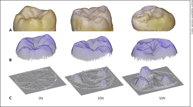
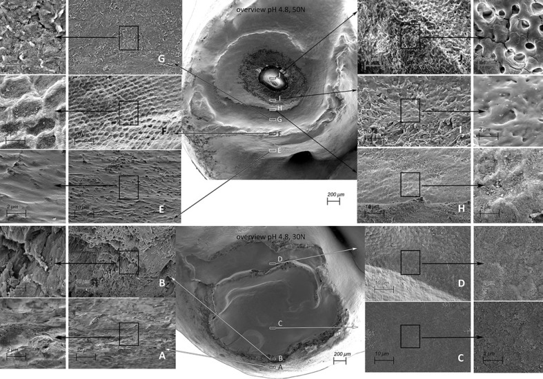
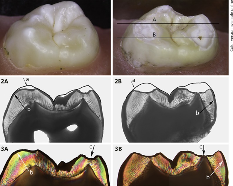
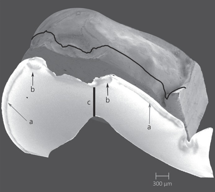
Methods: A total of 48 extracted human molar teeth were exposed to acidic aqueous solutions at pH of 4.8 and 5.5 in constant motion, in combination with different loading conditions: no load (0N group, control), 30 N (30N group) or 50 N (50N group) (n = 8 per group). Before and after 3 months of exposure (1,422,000 loading cycles), the samples were scanned using a non-contact profilometer. Pre- and post-exposure scans were subtracted and height loss and volume tissue loss were calculated. Representative samples with wear and cupping lesions were imaged using scanning electron microscopy, light microscopy and micro-computed tomography.

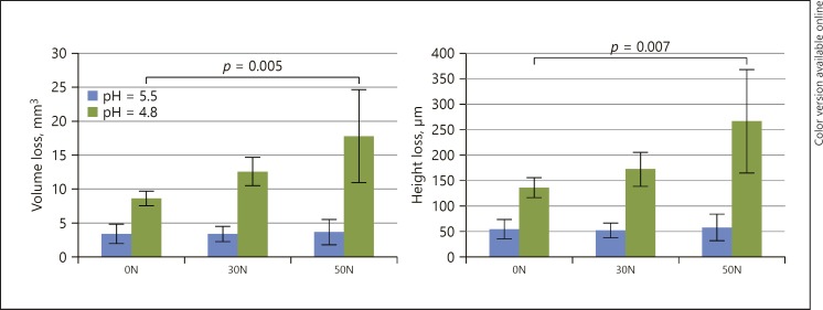
Results: Average height and volume tissue loss at pH 5.5 was 54 µm and 3.4 mm3 (0N), 52 µm and 3.4 mm3 (30N) and 58 µm and 3.7 mm3 (50N), respectively, with no statistically significant differences. Average height and volume loss at pH 4.8 were 135 µm and 8.7 mm3 (0N), 172 µm and 12.6 mm3 (30N) and 266 µm and 17.8 mm3 (50N), respectively, with a statistically significant difference between 0N and 50N (p < 0.002). Cup-shaped lesions had formed only at pH of 4.8, in the 30N and 50N groups.

Conclusion: The study showed that a cup can arise fully in enamel and that mechanical loading in addition to erosive challenges are required.

Keywords: Bite force; Profilometry; Tooth abrasion; Tooth erosion; Tooth wear.


Conflict of interest statement

The authors have no conflicts of interest to declare. They received no funding relevant to this study.

Figures

Fig. 1
Fig. 1
Mean volume loss (mm3) and height loss (µm) for all groups. Standard deviations are indicated by whiskers. Statistical differences between groups are indicated.
Fig. 2
Fig. 2
Images of a representative specimen from each loading group exposed to pH 4.8. A Stereomicroscopic image. B Non-contact profilometer scan. C Subtraction result. Minimal wear of the cusps can be observed at 0N, with cup-shaped lesions visible in the 30N and more distinctly in the 50N group. In the stereomicroscopic view of the 50N sample exposure of dentine at the bottom of one cup-shaped lesion can be observed.
Fig. 3
Fig. 3
SEM pictures of cup-shaped defects from a 50N specimen exposed to pH 4.8 (top, cup-shaped lesion extending into dentine) and a 30N specimen exposed to pH 4.8 (bottom, cup-shaped lesion limited to the enamel). Overview magnification, ×50. The frames indicated A–J show enlargements of areas in the cup-shaped lesion. Magnification, ×2,000 and ×5,000. In the overview cup shape lesions can be observed by multiple cavities and partly overlapping grooves, where on the surface a terrace structure can be observed with indented layering. A, E The enamel on the top of the cusp is slightly polished, the enamel is softened, and the solved components may exhibit a smearing action resulting in less friction and damage. B Enamel prism separated from each other. Separation of prisms will facilitate a deep penetration of acid. F “Polished” key-hole structures can be seen. C, D, G The damaged prisms are eroded/abraded and compressed, and a smear layer seems to arise. H, I In the lowest part of the formed cup-shaped lesion the surface is eroded/abraded and compressed, and a more pronounced smear layer is visible. J Exposed dentine.
Fig. 4
Fig. 4
Stereomicroscopic images in the top row show a sound specimen (left) and after exposure (right) of a specimen from the pH 4.8/50N group, with lines A and B indicating the location of the 100-µm sections imaged below. Sections assessed by transmitted light microscopy (2A, 2B) and polarized light (3A, 3B): (a) indicates the outline of the original shape of the cusps, (b) the demineralized top surface, and (c) the “deepest” point of the cup-shaped lesion at the location of the dentine “cusp” tip.
Fig. 5
Fig. 5
Representative specimen from the pH 4.8/50N group. Top view is an SEM image (magnification, ×50) and underneath a µCT image slice at the cross-section indicated by the line: (a) indicates surface demineralization, (b) damaged enamel near the cusp tip, and (c) the remaining enamel between the bottom of the cup-shaped lesion and the dentine “cusp” tip.

Similar articles

Cited by

References

    1. Abrahamsen TC. The worn dentition—pathognomonic patterns of abrasion and erosion. Int Dent J. 2005;55((4 Suppl 1)):268–76. - PubMed
    1. Bajaj D, Arola DD. On the R-curve behavior of human tooth enamel. Biomaterials. 2009 Aug;30((23-24)):4037–46. - PMC - PubMed
    1. Dawes C. What is the critical pH and why does a tooth dissolve in acid? J Can Dent Assoc. 2003 Dec;69((11)):722–4. - PubMed
    1. De Medeiros RC, Soares JD, De Sousa FB. Natural enamel caries in polarized light microscopy: differences in histopathological features derived from a qualitative versus a quantitative approach to interpret enamel birefringence. J Microsc. 2012 May;246((2)):177–89. - PubMed
    1. Eisenburger M, Addy M. Erosion and attrition of human enamel in vitro part I: interaction effects. J Dent. 2002 Sep-Nov;30((7-8)):341–7. - PubMed

Publication types