. 2019 Mar;62(2):175-182.
doi: 10.3340/jkns.2018.0048. Epub 2019 Feb 27.

Prevalence and Anatomy of Aberrant Right Subclavian Artery Evaluated by Computed Tomographic Angiography at a Single Institution in Korea

Affiliations

Prevalence and Anatomy of Aberrant Right Subclavian Artery Evaluated by Computed Tomographic Angiography at a Single Institution in Korea

Yunsuk Choi et al. J Korean Neurosurg Soc. 2019 Mar.

Abstract

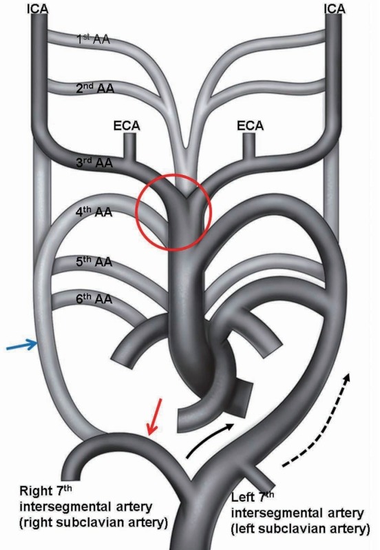
Objective: Aberrant right subclavian artery (ARSA) is a rare anatomical variant of the origin of the right subclavian artery. ARSA is defined as the right subclavian artery originating as the final branch of the aortic arch. The purpose of this study is to determine the prevalence and the anatomy of ARSA evaluated with computed tomography (CT) angiography.

Methods: CT angiography was performed in 3460 patients between March 1, 2014 and November 30, 2015 and the results were analyzed. The origin of the ARSA, course of the vessel, possible inadvertent ARSA puncture site during subclavian vein catheterization, Kommerell diverticula, and associated vascular anomalies were evaluated. We used the literature to review the clinical importance of ARSA.

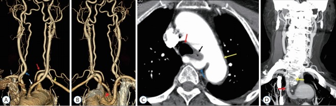
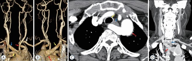
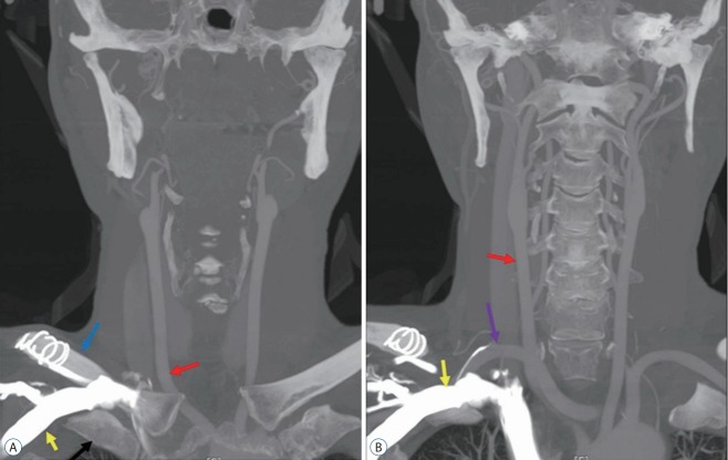
Results: Seventeen in 3460 patients had ARSA. All ARSAs in 17 patients originated from the posterior aspect of the aortic arch and traveled along a retroesophageal course to the right thoracic outlet. All 17 ARSAs were located in the anterior portion from first to fourth thoracic vertebral bodies and were located near the right subclavian vein at the medial third of the clavicle. Only one of 17 patients presented with dysphagia.

Conclusion: It is important to be aware ARSA before surgical approaches to upper thoracic vertebrae in order to avoid complications and effect proper treatment. In patients with a known ARSA, a right transradial approach for aortography or cerebral angiography should be changed to a left radial artery or transfemoral approach.

Keywords: Aberrant subclavian artery; Clinical; Computed tomography angiography.


Conflict of interest statement

No potential conflict of interest relevant to this article was reported.

Figures

Fig. 1.
Fig. 1.
A 72-year-old woman with an aberrant right subclavian artery (ARSA) with an inferoposterior origin (case 15). A : Anterior view of the aortic arch branch showing separate origin of both common carotid arteries (CCAs) and the left vertebral artery (VA) from the aorta (red arrow, left VA originating from the aortic arch; yellow arrow, left subclavian artery; white arrow, left CCA; blue arrow, right CCA; gray arrow, right subclavian artery). B : Posterior view of the aortic arch branch showing the ARSA (gray arrow, ARSA; red arrowhead, Kommerell diverticulum). C : Axial image showing the ARSA located anterior to the thoracic vertebrae (yellow arrow, aorta; red arrow, trachea; black arrow, esophagus; blue arrow, ARSA). D : Coronal image showing the ARSA with a horizontal course anterior to the vertebral body and an ascending course to the right side of the vertebral body (red arrow, ARSA; yellow arrow, second thoracic vertebral body).
Fig. 2.
Fig. 2.
A 60-year-old woman with an aberrant right subclavian artery (ARSA) with a superoposterior origin (case 11). A : Anterior view of the aortic arch branch showing the common origin of both common carotid arteries (CCAs) (red arrow, left CCA; yellow arrow, right CCA; white arrow, left subclavian artery; blue arrow, ARSA). B : Posterior view of the aortic arch branch showing the ARSA (yellow arrow, ARSA; red arrow, Kommerell diverticulum). C : Axial image showing the ARSA located anterior to the thoracic vertebrae (yellow arrow, trachea; red arrow, aorta; blue arrow, esophagus; gray arrow, ARSA). D : Coronal image showing the ARSA with an oblique course in the anterior portion of the thoracic vertebrae (red arrow, ARSA; blue arrow, first thoracic vertebral body).
Fig. 3.
Fig. 3.
A 56-year-old man with an aberrant right subclavian artery (ARSA) (case 7). A : Coronal image showing the subclavian vein (yellow arrow) located between the clavicle (blue arrow) and first rib (black arrow). Red arrow indicates right common carotid artery. B : Coronal image showing close proximity of the ARSA (violet arrow) and right subclavian vein (yellow arrow). Red arrow indicates right common carotid artery.
Fig. 4.
Fig. 4.
Abnormal embryonic development of the great vessels leading to the formation of an ARSA and common stem of both CCAs. Embryonic development of the AA takes place during the 4th and 8th week of fetal life. Normal embryonic development of the AA and great vessels begins as six paired AAs. The 1st and 2nd AAs regress. The paired 3rd arches form the 1st part of the ICA bilaterally. The proximal right 4th arch persists as the right subclavian artery at the origin of the internal mammary artery, whereas the distal right 4th arch regresses. The left 4th arch forms the anatomical basis of the subsequent fully formed AA. The 5th arch has not been formed completely. In abnormal embryonic development, involution of the right 4th AA and proximal right dorsal aorta leaves the right 7th intersegmental artery to arise from the left dorsal aorta, resulting in an ARSA. With further development, differential growth shifts the origin of the ARSA and the left subclavian artery cranially. Regression of the ventral aortic roots between the 3rd and 4th AAs in red circle of this figure result to common stem of both CCAs. Black half tone vessel indicates fully developed vessel in adult. Gray half tone vessel indicates obliterated vessel in development. Blue arrow indicates abnormally disappeared proximal right dorsal aorta, red arrow is persistent distal right dorsal aorta, and black and black dotted arrows are growth direction of both 7th intersegmental arteries. ICA : internal carotid artery, AA : aortic arch, ECA : external carotid artery, ARSA : aberrant right subclavian artery, CCA : common carotid artery.

References

    1. Babu CS, Sharma V. Two common trunks arising from arch of aorta: case report and literature review of a very rare variation. J Clin Diagn Res. 2015;9:AD05–AD07. - PMC - PubMed
    1. Backer CL, Ilbawi MN, Idriss FS, DeLeon SY. Vascular anomalies causing tracheoesophageal compression. Review of experience in children. J Thorac Cardiovasc Surg. 1989;97:725–731. - PubMed
    1. Bayford D. An account of singular case of obstructed deglutition. Mem Med Soc Lond. 1794;2:275–286.
    1. Berko NS, Jain VR, Godelman A, Stein EG, Ghosh S, Haramati LB. Variants and anomalies of thoracic vasculature on computed tomographic angiography in adults. J Comput Assist Tomogr. 2009;33:523–528. - PubMed
    1. Celikyay ZR, Koner AE, Celikyay F, Denız C, Acu B, Firat MM. Frequency and imaging findings of variations in human aortic arch anatomy based on multidetector computed tomography data. Clin Imaging. 2013;37:1011–1019. - PubMed

Grants and funding

LinkOut - more resources