. 2019 Mar 6;21(1):36.
doi: 10.1186/s13058-019-1123-2.

Separation of breast cancer and organ microenvironment transcriptomes in metastases

Affiliations

Separation of breast cancer and organ microenvironment transcriptomes in metastases

Mohammad A Alzubi et al. Breast Cancer Res. .

Abstract

Background: The seed and soil hypothesis was proposed over a century ago to describe why cancer cells (seeds) grow in certain organs (soil). Since then, the genetic properties that define the cancer cells have been heavily investigated; however, genomic mediators within the organ microenvironment that mediate successful metastatic growth are less understood. These studies sought to identify cancer- and organ-specific genomic programs that mediate metastasis.

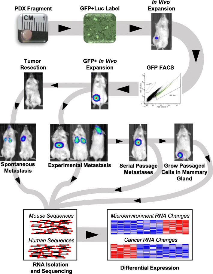
Methods: In these studies, a set of 14 human breast cancer patient-derived xenograft (PDX) metastasis models was developed and then tested for metastatic tropism with two approaches: spontaneous metastases from mammary tumors and intravenous injection of PDX cells. The transcriptomes of the cancer cells when growing as tumors or metastases were separated from the transcriptomes of the microenvironment via species-specific separation of the genomes. Drug treatment of PDX spheroids was performed to determine if genes activated in metastases may identify targetable mediators of viability.

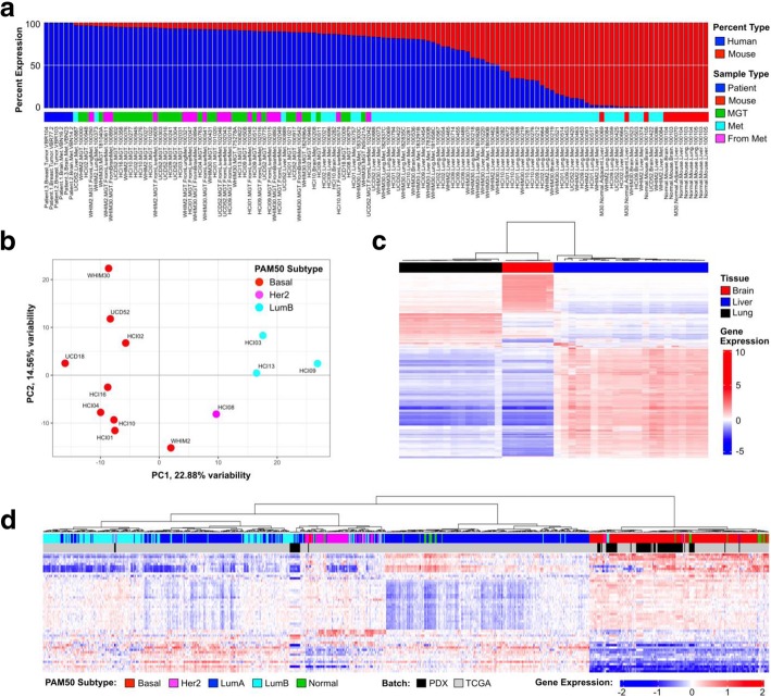
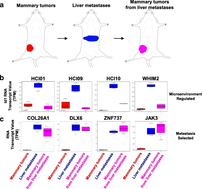
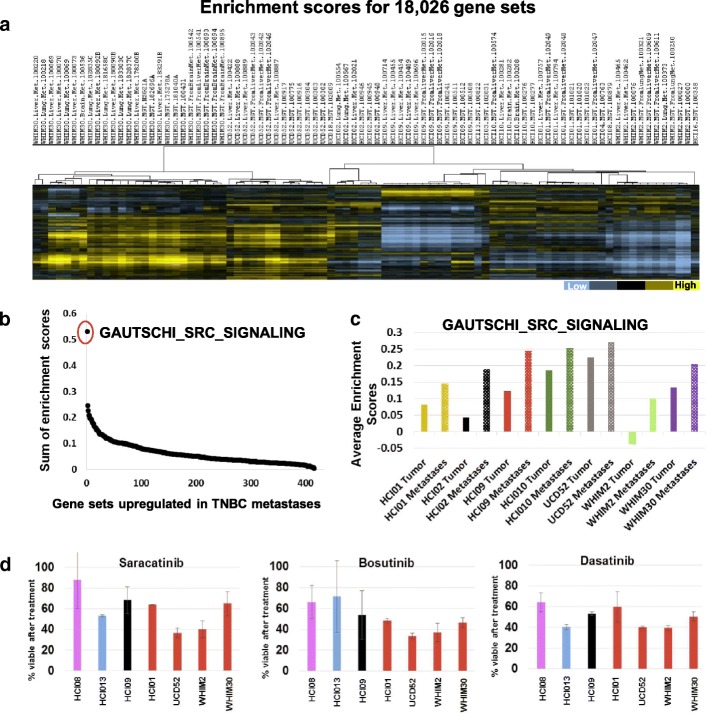
Results: The experimental approaches that generated metastases in PDX models were identified. RNA sequencing of 134 tumors, metastases, and normal non-metastatic organs identified cancer- and organ-specific genomic properties that mediated metastasis. A common genomic response of the liver microenvironment was found to occur in reaction to the invading PDX cells. Genes within the cancer cells were found to be either transiently regulated by the microenvironment or permanently altered due to clonal selection of metastatic sublines. Gene Set Enrichment Analyses identified more than 400 gene signatures that were commonly activated in metastases across basal-like PDXs. A Src signaling signature was found to be extensively upregulated in metastases, and Src inhibitors were found to be cytotoxic to PDX spheroids.

Conclusions: These studies identified that during the growth of breast cancer metastases, there were genomic changes that occurred within both the cancer cells and the organ microenvironment. We hypothesize that pathways upregulated in metastases are mediators of viability and that simultaneously targeting changes within different cancer cell pathways and/or different tissue compartments may be needed for inhibition of disease progression.

Keywords: Breast cancer; Luciferase; Metastasis; Microenvironment; Patient-derived xenograft; RNA sequencing.


Conflict of interest statement

Ethics approval

All animal procedures were approved by the Virginia Commonwealth University and University of North Carolina Institutional Animal Care and Use Committees.

Consent for publication

Non-identifiable patient data was utilized in these studies.

Competing interests

C.M.P is an equity stockholder, consultant, and Board of Director Member of BioClassifier LLC. C.M.P is also listed as an inventor on patent applications for the Breast PAM50 Subtyping assay. The other authors declare no potential conflicts of interest.

Publisher’s Note

Springer Nature remains neutral with regard to jurisdictional claims in published maps and institutional affiliations.

Figures

Fig. 1
Fig. 1
Schematic of experimental approach used to identify genomic mediators of patient-derived xenograft metastases. PDX tumors were grown in the mammary gland, extracted and digested into single-cell suspensions, and then labeled with lentivirus that encoded for green fluorescent protein and luciferase (GFP + Luc). After expansion in vivo, the cells with the brightest GFP were expanded in the orthotopic location, then used for metastasis studies. Paired-end RNA sequencing was then performed on mammary tumors, normal tissues, and brain, lung, and liver metastases. The sequencing data was then aligned to both the mouse and human reference genomes
Fig. 2
Fig. 2
Assessment of tumor growth rates and metastatic distributions of 14 breast cancer PDXs. a Cancer cells were injected into the abdominal mammary gland and measured weekly for tumor growth. ER+ and HER2+ PDXs are designated, all the others are TNBC. b Quantification of spontaneous metastases after surgical excision of primary tumors. c Quantification of metastases from after tail-vein injection of PDX cells
Fig. 3
Fig. 3
Virtual dissection of PDX mammary tumors and metastases into cancer (human) and microenvironment (mouse) gene expression datasets. The 134 sample RNA-sequencing dataset; PDX mammary tumors and metastases to the brain, lung, and liver; normal mouse brain, lung, and liver; 100% human breast tumors and brain metastases. a The transcripts from the 134 samples were segregated into human and mouse datasets; shown is the percent of each genome within each sample. b One mammary tumor from each PDX line was used in a Principal Component Analysis based on the 2000 most variable genes. c Mouse transcripts from metastases to the brain, lung, and liver, as well as normal mouse brain, lung, liver, were used to cluster the mouse genome based on the most variable top 2000 genes. d PDX tumors and metastases with more than ten million human mapped transcripts were combined with RNA-seq data from 817 tumors from The Cancer Genome Atlas. The PAM50 genes were used to hierarchical cluster the combined dataset
Fig. 4
Fig. 4
Validation of human and mouse-specific transcripts with immunofluorescence microscopy and immunohistochemistry. a Box and whisker plots showing human vimentin RNA expression levels in TNBC mammary tumors. b Immunofluorescence microscopy for pan-cytokeratin (green), vimentin (red), and DAPI (blue) in TNBC mammary tumors. c The mouse RNA-seq dataset was queried for genes that were differentially expressed in normal mouse liver as compared to mouse livers colonized by metastatic cells. Immunohistochemistry of liver metastases to validate the increased RNA expression observed in liver metastases. Asterisk denotes the location of cancer cells; arrows denote S100a9 cells in the peri-tumor area surrounding the cancer cells
Fig. 5
Fig. 5
Identification of genes within human metastasis signatures that originate from liver cells. a RNA-seq data from patient-matched breast tumors and liver metastases were downloaded from Siegel et al. [30], median centered, and hierarchically clustered. Highlighted in the purple box are 171 genes increased in each liver metastasis compared to the primary tumor it was derived from. b ANOVA was performed on the PDX mouse RNA-seq dataset for all 171 genes to identify the transcripts which were induced in the liver cell transcriptome during PDX metastasis compared to normal mouse liver. Twenty-four liver genes were identified as significantly upregulated (p < 0.05) in metastases compared to normal liver and were averaged to generate a signature value for each sample. PDX liver macro-metastases were used for the analyses that had human RNA content > 50% (n = 19), normal mouse liver (n = 3). c 27 liver relapses and 17 breast relapse fine-needle aspirates from Tobin et al. [31] were queried for the 27-gene liver microenvironment gene signature presented in Additional file 7c
Fig. 6
Fig. 6
Identification of microenvironment-regulated genes or metastasis-selected genes. a Approach used to compare gene expression profiles of mammary gland tumors (MGT), liver metastases, and MGT which were grown from liver metastases. b Human ID1 gene expression levels in MGT, metastases, and MGT grown from metastases. Most metastases have increased ID1 expression, which reverts to levels observed in parental MGT when the metastases are collected and regrown in the mammary gland. c Shown are examples of genes upregulated in UCD52 liver metastases, which maintained high expression when the liver metastases were collected and grown in the mammary gland
Fig. 7
Fig. 7
Gene Set Enrichment Analyses identify signatures upregulated in metastases. a Gene set enrichment scores for tumors and metastases were identified for 18,026 gene signatures and hierarchical clustered. b > 400 gene sets were found to be upregulated on average in metastases from all six basal-like PDXs, the sum of enrichment scores was identified by adding together the difference in enrichment scores of all 6 PDXs (metastases compared to tumors). c Average enrichment score values for one gene set across different tumors and metastases. d Effect of targeting the most upregulated pathway with three different Src inhibitors on viability of PDX spheroids in suspension culture

References

    1. Perou CM, Sorlie T, Eisen MB, van de Rijn M, Jeffrey SS, Rees CA, Pollack JR, Ross DT, Johnsen H, Akslen LA, et al. Molecular portraits of human breast tumours. Nature. 2000;406(6797):747–752. - PubMed
    1. Curtis C, Shah SP, Chin SF, Turashvili G, Rueda OM, Dunning MJ, Speed D, Lynch AG, Samarajiwa S, Yuan Y, et al. The genomic and transcriptomic architecture of 2,000 breast tumours reveals novel subgroups. Nature. 2012;486(7403):346–352. - PMC - PubMed
    1. Ciriello G, Gatza ML, Beck AH, Wilkerson MD, Rhie SK, Pastore A, Zhang H, McLellan M, Yau C, Kandoth C, et al. Comprehensive molecular portraits of invasive lobular breast cancer. Cell. 2015;163(2):506–519. - PMC - PubMed
    1. Cancer Genome Atlas N. Comprehensive molecular portraits of human breast tumours. Nature. 2012;490(7418):61–70. - PMC - PubMed
    1. Harrell JC, Prat A, Parker JS, Fan C, He X, Carey L, Anders C, Ewend M, Perou CM. Genomic analysis identifies unique signatures predictive of brain, lung, and liver relapse. Breast Cancer Res Treat. 2012;132(2):523–535. - PMC - PubMed

Publication types

MeSH terms