. 2019 Apr 1;216(4):772-785.
doi: 10.1084/jem.20181773. Epub 2019 Mar 6.

KAT8 selectively inhibits antiviral immunity by acetylating IRF3

Affiliations

KAT8 selectively inhibits antiviral immunity by acetylating IRF3

Wanwan Huai et al. J Exp Med. .

Erratum in

Abstract

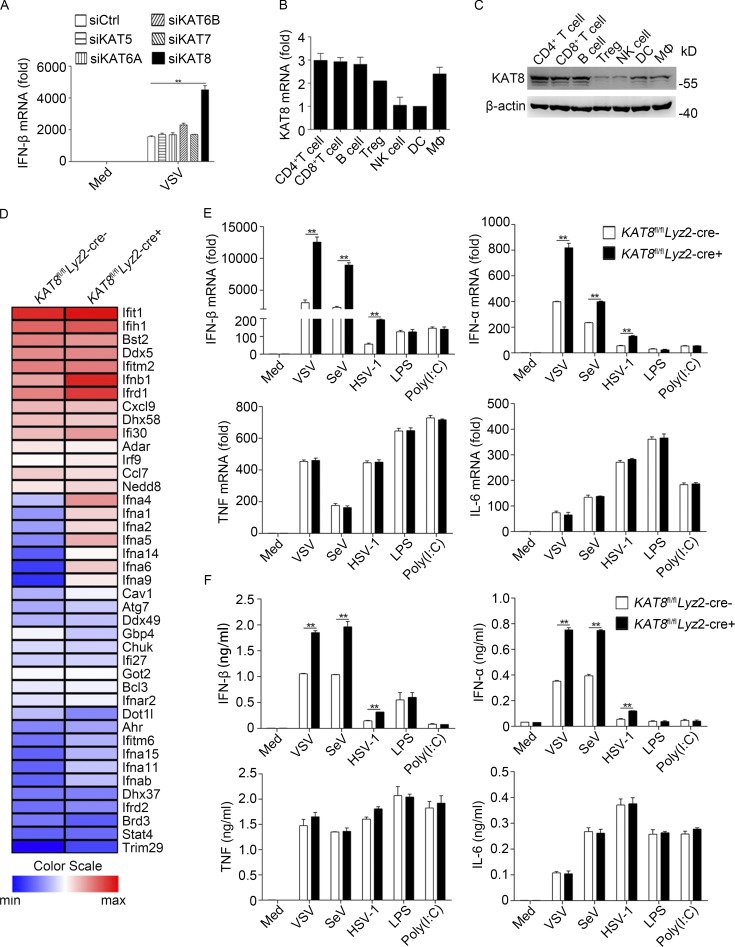
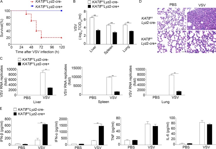
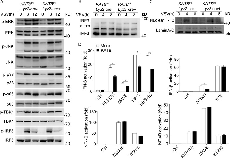
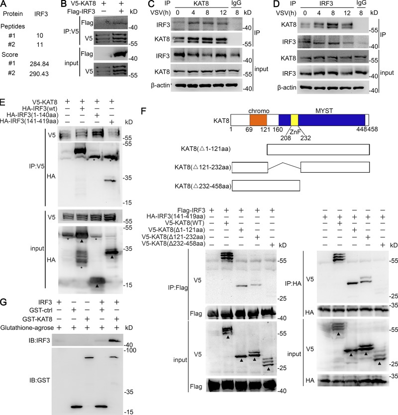
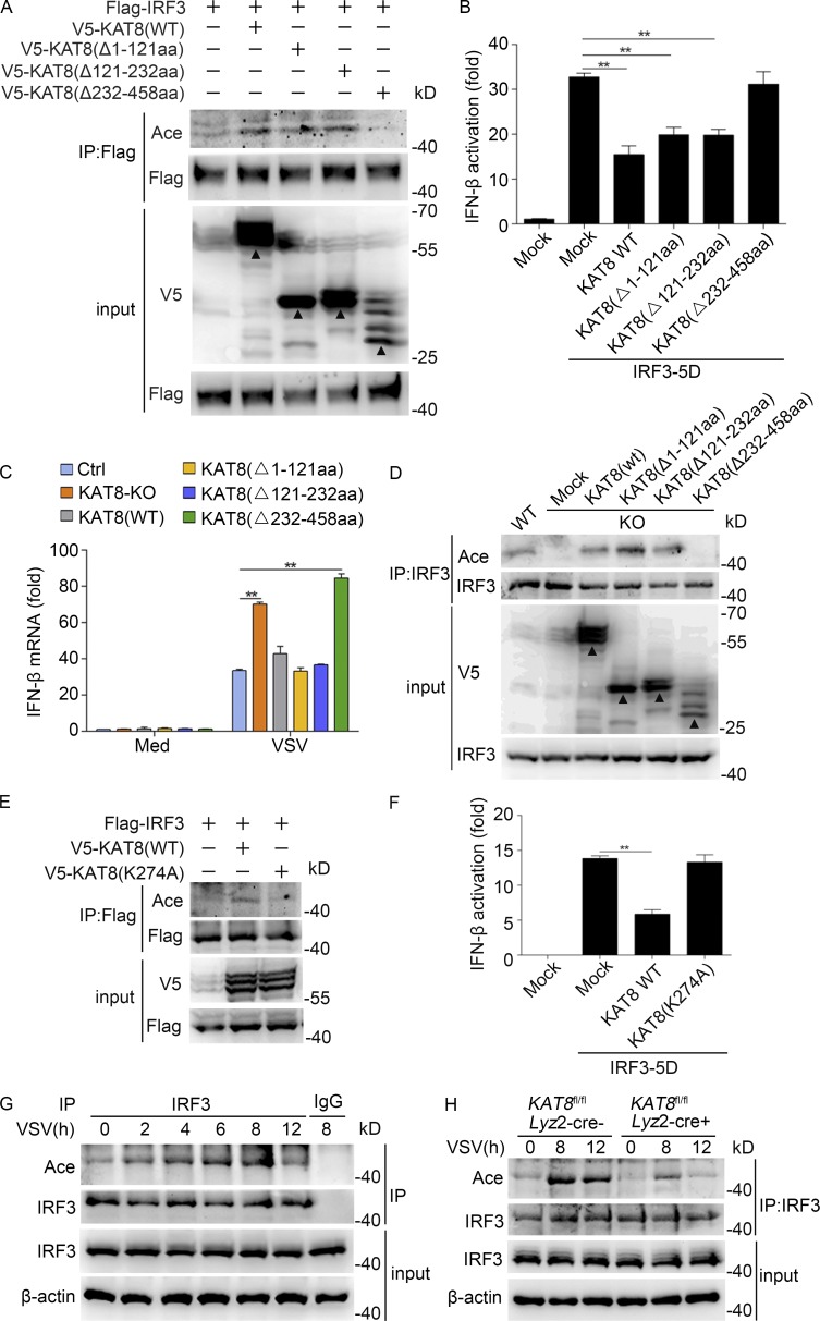
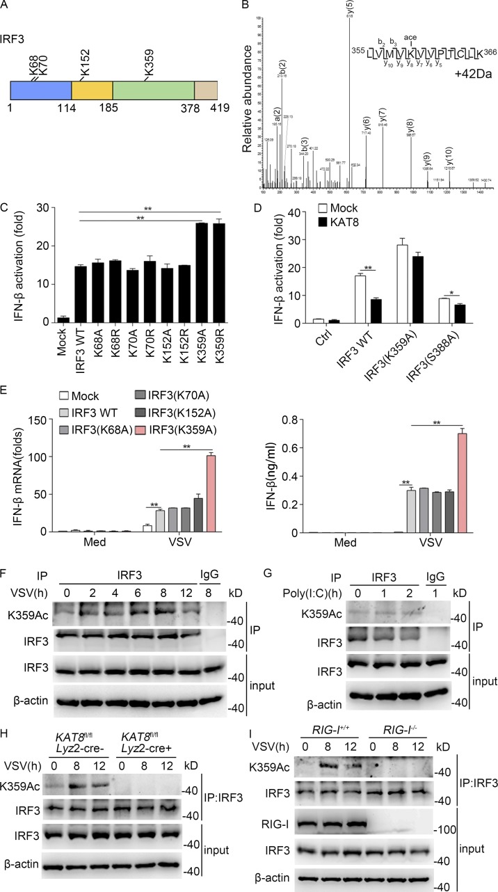
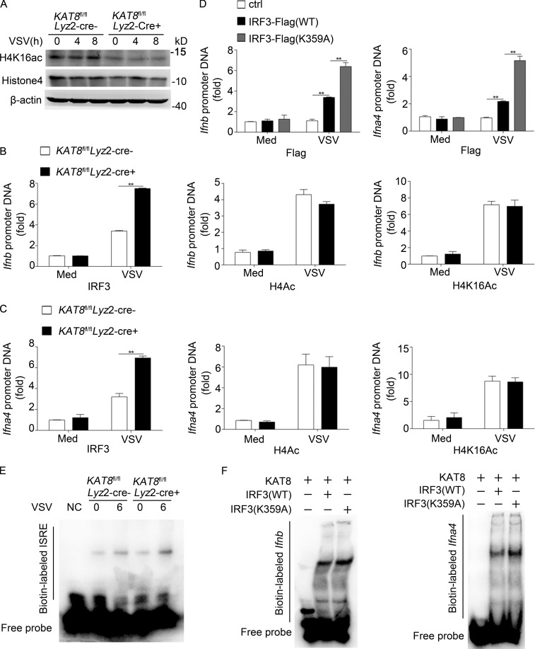
The transcription factor interferon regulatory factor 3 (IRF3) is essential for virus infection-triggered induction of type I interferons (IFN-I) and innate immune responses. IRF3 activity is tightly regulated by conventional posttranslational modifications (PTMs) such as phosphorylation and ubiquitination. Here, we identify an unconventional PTM of IRF3 that directly inhibits its transcriptional activity and attenuates antiviral immune response. We performed an RNA interference screen and found that lysine acetyltransferase 8 (KAT8), which is ubiquitously expressed in immune cells (particularly in macrophages), selectively inhibits RNA and DNA virus-triggered IFN-I production in macrophages and dendritic cells. KAT8 deficiency protects mice from viral challenge by enhancing IFN-I production. Mechanistically, KAT8 directly interacts with IRF3 and mediates IRF3 acetylation at lysine 359 via its MYST domain. KAT8 inhibits IRF3 recruitment to IFN-I gene promoters and decreases the transcriptional activity of IRF3. Our study reveals a critical role for KAT8 and IRF3 lysine acetylation in the suppression of antiviral innate immunity.


Figures

Figure 1.
Figure 1.
Deficiency in KAT8 selectively promotes the production of IFN-I. (A) Q-PCR analysis of IFN-β mRNA in peritoneal macrophages transfected with control siRNA (siCtrl) or specific siRNA targeting KAT5 (siKAT5), KAT6A (siKAT6A), KAT6B (siKAT6B), KAT7 (siKAT7), or KAT8 (siKAT8) and then infected with VSV (1 MOI) for 8 h. (B) Q-PCR analysis of KAT8 mRNA in mouse immune cells; results were normalized to β-actin mRNA. (C) Immunoblot analysis of KAT8 and β-actin in mouse immune cells. (D) RNA sequencing analysis of KAT8fl/flLyz2-Cre or KAT8fl/flLyz2-Cre+ peritoneal macrophages infected with VSV (1 MOI). (E) Q-PCR analysis of IFN-β, IFN-α, TNF, and IL-6 mRNA in KAT8fl/flLyz2-Cre or KAT8fl/flLyz2-Cre+ peritoneal macrophages left untreated (Med), infected with VSV (1 MOI), SeV (1 MOI), or HSV-1 (10 MOI) for 8 h, or stimulated with LPS (100 ng/ml) or poly(I:C) (10 µg/ml) for 3 h. (F) ELISA of IFN-β, IFN-α, TNF, and IL-6 in supernatants of KAT8fl/flLyz2-Cre or KAT8fl/flLyz2-Cre+ peritoneal macrophages infected with VSV (1 MOI), SeV (1 MOI), or HSV-1 (10 MOI) for 12 h or stimulated with LPS (100 ng/ml) or poly(I:C) (10 µg/ml) for 6 h. **, P < 0.01; two-tailed Student’s t test (A, B, E, and F). Data are representative of three independent experiments with similar results (C and D) or are from three independent experiments (A, B, E, and F; mean ± SEM).
Figure 2.
Figure 2.
Deficiency of KAT8 protects mice against viral infection. (A) Survival of KAT8fl/flLyz2-Cre or KAT8fl/flLyz2-Cre+ mice (n = 10 per group) after infection with VSV (5 × 10⁷ plaque-forming units per gram body weight; Wilcoxon test). (B) VSV load in the liver, spleen, and lung of KAT8fl/flLyz2-Cre or KAT8fl/flLyz2-Cre+ mice (n = 8 per group) 18 h after infection with VSV (as in A), assessed by endpoint-dilution assay and presented as 50% tissue culture infectious dose (TCID50). (C) Q-PCR analysis of VSV RNA in the liver, spleen, and lungs of KAT8fl/flLyz2-Cre or KAT8fl/flLyz2-Cre+ mice 18 h after intraperitoneal injection of PBS or VSV (as in A). (D) Hematoxylin and eosin staining of sections of lungs from mice as in B. Bars, 50 µm. (E) ELISA of cytokines in serum from mice as in C. **, P < 0.01; two-tailed Student’s t test (B, C, and E). Data are from three independent experiments (B, C, and E; mean ± SEM) or are representative of three independent experiments with similar results (D).
Figure 3.
Figure 3.
KAT8 targets IRF3. (A) Immunoblot analysis of phosphorylated (p-) or total ERK, JNK, p38, p65, TBK1, IRF3, or β-actin in KAT8fl/flLyz2-Cre or KAT8fl/flLyz2-Cre+ peritoneal macrophages infected for the indicated times with VSV. (B) Immunoblot analysis of IRF3 dimerization in KAT8fl/flLyz2-Cre or KAT8fl/flLyz2-Cre+ peritoneal macrophages infected for the indicated times with VSV. (C) Immunoblot analysis of IRF3 among nuclear proteins in KAT8fl/flLyz2-Cre or KAT8fl/flLyz2-Cre+ peritoneal macrophages infected for the indicated times with VSV. (D) Luciferase activity assay of an IFN-β reporter in HEK293T cells transfected with KAT8 or Mock together with control vector, RIG-I(N), MAVS, TBK1, IRF3-5D, STING, or TRIF or luciferase activity assay of an NF-κB reporter in HEK293T cells transfected with KAT8 or Mock together with control vector, MyD88, TRAF6, RIG-I(N), MAVS, or STING assessed by dual-luciferase assay. *, P < 0.05; **, P < 0.01; two-tailed Student’s t test (D). Data are representative of three independent experiments with similar results (A–C) or are from three independent experiments (D; mean ± SEM).
Figure 4.
Figure 4.
KAT8 interacts with IRF3 via the MYST domain. (A) MS analysis of KAT8-binding proteins in peritoneal macrophages infected with VSV (1 MOI). #1 and #2 indicates two independent experiments. (B) Immunoblot analysis of KAT8 or IRF3 in HEK293T cells transfected with V5-tagged KAT8 alone or together with Flag-tagged IRF3, assessed before (input) or after IP with antibody to V5 or Flag. (C) Immunoblot analysis of endogenous KAT8 or IRF3 in peritoneal macrophages infected with VSV for the indicated times, assessed before (input) or after IP with IgG (control) or antibody to KAT8. (D) Immunoblot analysis of endogenous KAT8 or IRF3 in peritoneal macrophages infected for the indicated times with VSV, assessed before (input) or after IP with IgG or antibody to IRF3. (E) Immunoblot analysis of HEK293T cells transiently transfected with HA-tagged WT or mutant IRF3 plus V5-tagged KAT8 and assessed 24 h later before (input) or after IP with antibody to V5. (F) Immunoblot analysis of HEK293T cells transiently transfected with V5-tagged WT or mutant KAT8 plus Flag-tagged IRF3 or HA-tagged mutant IRF3 (141–419 aa) and assessed 24 h later before (input) or after IP with antibody to Flag or HA. (G) Immunoblot (IB) analysis of IRF3 and KAT8 interaction in a GST pull-down assay. Data are representative of three independent experiments with similar results. *, nonspecific bands; arrowheads, specific bands.
Figure 5.
Figure 5.
KAT8 promotes IRF3 acetylation via its MYST domain. (A) Immunoblot analysis of acetylation in HEK293T cells transiently transfected with V5-tagged WT or mutant KAT8 plus Flag-tagged IRF3 and assessed 24 h later before (input) or after IP with antibody to Flag. (B) Luciferase activity of an IFN-β reporter in HEK293T cells transfected with WT KAT8 or KAT8 truncations together with IRF3-5D. (C) KAT8-KO RAW264.7 cells were transfected with WT KAT8 or KAT8 truncations and then infected 24 h later with VSV (1 MOI) for 8 h. The expression of IFN-β at the mRNA level was measured by Q-PCR. (D) Immunoblot analysis of IRF3 acetylation and total IRF3 in KAT8-KO RAW264.7 cells transiently transfected with V5-tagged WT or mutant KAT8 and then infected 24 h later with VSV (1 MOI) for 8 h, assessed before (input) or after IP with antibody to IRF3. (E) Immunoblot analysis of acetylation in HEK293T cells transiently transfected with V5-tagged WT or mutant KAT8 plus Flag-tagged IRF3 and assessed 24 h later before (input) or after IP with antibody to Flag. (F) Luciferase activity of an IFN-β reporter in HEK293T cells transfected with WT KAT8 or KAT8 mutant (K274A) together with IRF3-5D. (G) Immunoblot analysis of endogenous acetylation of IRF3 in peritoneal macrophages infected for the indicated times with VSV (1 MOI), assessed before (input) or after IP with IgG or antibody to IRF3. (H) Immunoblot analysis of IRF3 acetylation in KAT8fl/flLyz2-Cre or KAT8fl/flLyz2-Cre+ peritoneal macrophages infected for the indicated times with VSV (1 MOI), assessed before (input) or after IP with antibody to IRF3. **, P < 0.01 (one-way ANOVA; B and F); **, P < 0.01 (two-tailed Student’s t test; C). Data are representative of three independent experiments with similar results (A, D, E, G, and H) or are from three independent experiments (B, C, and F; mean ± SEM). Arrowheads, specific bands.
Figure 6.
Figure 6.
Acetylation of IRF3 at K359 in VSV-infected macrophages. (A) Illustration of acetylated lysine residues of IRF3 identified by an MS assay. (B) The tryptic peptide IRF3 (355–366 LVMVKVVPTCLK), consistent with acetylation, is characterized by a neutral addition of 42 D (neutral addition of H2O or NH3 omitted for clarity). (C) Luciferase activity of an IFN-β reporter in HEK293T cells transfected with control vector (Mock) or vectors encoding WT or mutant IRF3. (D) Luciferase activity of an IFN-β reporter in HEK293T cells transfected with control vector or vector encoding WT or mutant IRF3 (IRF3-K359A, and IRF3-S388A) along with vector encoding KAT8. (E) MEF cells from IRF3-deficient mice were transfected with WT or IRF3 mutants and then infected 24 h later with VSV (1 MOI) for 8 h. The mRNA level of IFN-β was measured by Q-PCR, and the production of IFN-β was measured by ELISA. (F) Immunoblot analysis of IRF3 acetylated at Lys359 (K359Ac) and total IRF3 in peritoneal macrophages infected for the indicated times with VSV (1 MOI), assessed before (input) or after IP with IgG or antibody to IRF3. (G) Immunoblot analysis of IRF3 acetylated at Lys359 (K359Ac) and total IRF3 in peritoneal macrophages stimulated for the indicated times with poly(I:C) (10 µg/ml), assessed before (input) or after IP with IgG or antibody to IRF3. (H) Immunoblot analysis of IRF3 acetylated at Lys359 (K359Ac) and total IRF3 in KAT8fl/flLyz2-Cre or KAT8fl/flLyz2-Cre+ peritoneal macrophages infected with VSV (1 MOI), followed by IP with antibody to IRF3. (I) Immunoblot analysis of IRF3 acetylated at Lys359 (K359Ac) and total IRF3 in RIG-I–sufficient or RIG-I–deficient peritoneal macrophages infected with VSV (1 MOI), followed by IP with antibody to IRF3. **, P < 0.01 (one-way ANOVA; C); *, P < 0.05; **, P < 0.01 (two-tailed Student’s t test; D and E). Data are representative of three independent experiments with similar results (F–I) or are from three independent experiments (C–E; mean ± SEM).
Figure 7.
Figure 7.
KAT8 abrogates the abundance of IRF3 at IFN-I promoters. (A) Immunoblot analysis of H4K16ac, histone 4, or β-actin in KAT8fl/flLyz2-Cre or KAT8fl/flLyz2-Cre+ peritoneal macrophages infected with VSV (1 MOI) for the indicated times. (B) ChIP analysis of IRF3, H4Ac, or H4K16Ac at the Ifnb promoter in KAT8fl/flLyz2-Cre or KAT8fl/flLyz2-Cre+ peritoneal macrophages infected for 6 h with VSV (1 MOI) or left untreated. (C) ChIP analysis of IRF3, H4Ac, or H4K16Ac at Ifna4 promoter in KAT8fl/flLyz2-Cre or KAT8fl/flLyz2-Cre+ peritoneal macrophages infected for 6 h with VSV (1 MOI) or left untreated. (D) ChIP analysis of IRF3 at Ifnb or Ifna4 promoter in MEF cells from IRF3-deficient mice overexpressed with WT or mutant IRF3 for 24 h and then infected with VSV (1 MOI) for 6 h. (E) EMSA of nuclear extract from KAT8fl/flLyz2-Cre or KAT8fl/flLyz2-Cre+ peritoneal macrophages infected with VSV (1 MOI) for 6 h. The ISRE motif was biotin labeled. (F) Analysis of recombinant IRF3 WT or IRF3 mutant and IFN interaction in an EMSA assay. The Ifnb and Ifna motifs were biotin labeled. **, P < 0.01; two-tailed Student’s t test (B–D). Data are representative of three independent experiments with similar results (A, E, and F) or are from three independent experiments (B–D; mean ± SEM).

References

    1. Ageta-Ishihara N., Miyata T., Ohshima C., Watanabe M., Sato Y., Hamamura Y., Higashiyama T., Mazitschek R., Bito H., and Kinoshita M.. 2013. Septins promote dendrite and axon development by negatively regulating microtubule stability via HDAC6-mediated deacetylation. Nat. Commun. 4:2532 10.1038/ncomms3532 - DOI - PMC - PubMed
    1. Akhtar A., Zink D., and Becker P.B.. 2000. Chromodomains are protein-RNA interaction modules. Nature. 407:405–409. 10.1038/35030169 - DOI - PubMed
    1. Allfrey V.G., Faulkner R., and Mirsky A.E.. 1964. Acetylation and methylation of histones and their possible role in the regulation of rna synthesis. Proc. Natl. Acad. Sci. USA. 51:786–794. 10.1073/pnas.51.5.786 - DOI - PMC - PubMed
    1. Beltrao P., Albanèse V., Kenner L.R., Swaney D.L., Burlingame A., Villén J., Lim W.A., Fraser J.S., Frydman J., and Krogan N.J.. 2012. Systematic functional prioritization of protein posttranslational modifications. Cell. 150:413–425. 10.1016/j.cell.2012.05.036 - DOI - PMC - PubMed
    1. Cao X. 2016. Self-regulation and cross-regulation of pattern-recognition receptor signalling in health and disease. Nat. Rev. Immunol. 16:35–50. 10.1038/nri.2015.8 - DOI - PubMed

Publication types

MeSH terms