Review
. 2019 Mar 6;11(482):eaao0935.
doi: 10.1126/scitranslmed.aao0935.

Delivery of therapeutics to the inner ear: The challenge of the blood-labyrinth barrier

Affiliations
Review

Delivery of therapeutics to the inner ear: The challenge of the blood-labyrinth barrier

Sophie Nyberg et al. Sci Transl Med. .

Abstract

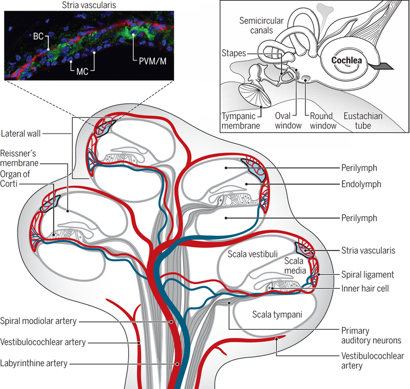
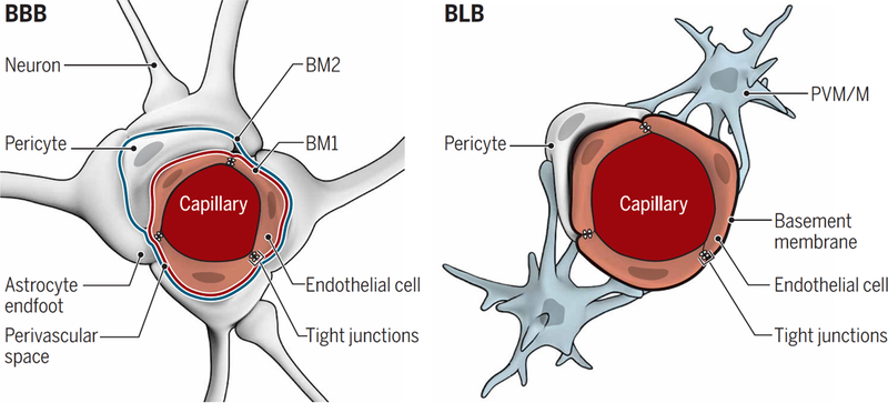
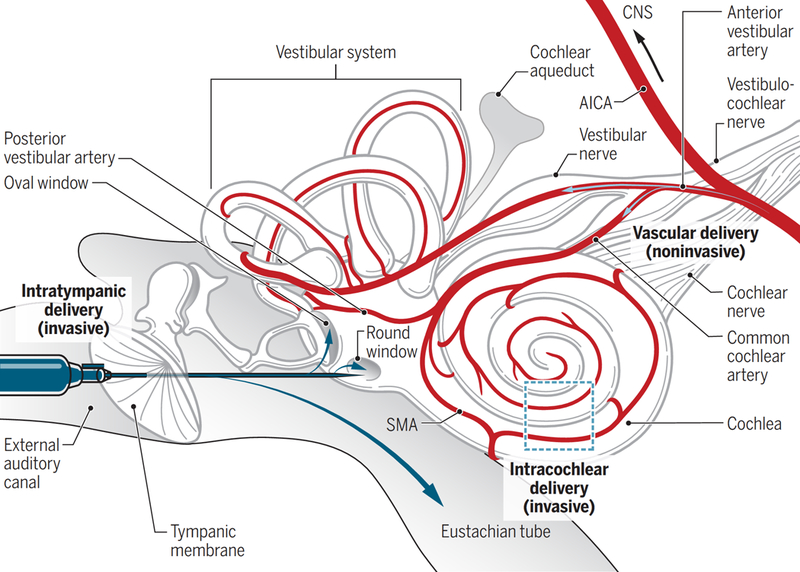
Permanent hearing loss affects more than 5% of the world's population, yet there are no nondevice therapies that can protect or restore hearing. Delivery of therapeutics to the cochlea and vestibular system of the inner ear is complicated by their inaccessible location. Drug delivery to the inner ear via the vasculature is an attractive noninvasive strategy, yet the blood-labyrinth barrier at the luminal surface of inner ear capillaries restricts entry of most blood-borne compounds into inner ear tissues. Here, we compare the blood-labyrinth barrier to the blood-brain barrier, discuss invasive intratympanic and intracochlear drug delivery methods, and evaluate noninvasive strategies for drug delivery to the inner ear.


Conflict of interest statement

Competing interests: The authors declare that they have no competing interests.

Figures

Fig. 1
Fig. 1. Structure and blood supply of the cochlea.
Inset: (top left) Fluorescence micrograph of the mouse stria vascularis showing three cell layers comprising basal cells (BC), marginal cells (MC), and an intermediate cell layer of perivascular-resident macrophage-like melanocytes (PVM/Ms). Basal cells are immunostained for actin (red), and PVM/ Ms are immunostained for the K+ channel Kir4.1 (green); nuclei are stained with DAPI (4′,6-diamidino-2-phenylindole) counterstain (blue). Inset: (top right) Structure of the middle ear containing the tympanic membrane, oval window, round window, and auditory ossicles. Also shown are the cochlea and semicircular canals (vestibular system) of the inner ear. Main figure: The blood supply to the cochlea (shown in cross section) comes from the common cochlear artery, which divides into the spiral modiolar artery and the vestibulocochlear artery. The spiral modiolar artery supplies the apical turns of the cochlea, and the cochlear branch of the vestibulocochlear artery supplies the basal turns of the cochlea. The spiral modiolar artery supplies the organ of Corti and primary auditory neurons of the modiolus and forms the capillaries of the spiral ligament and stria vascularis in the cochlear lateral wall. The blue vessels represent the venous return route of deoxygenated blood. Image credit: Sophie Nyberg, University of Toronto.
Fig. 2
Fig. 2. Comparison of the BBB and BLB.
Shown are cross sections of the capillaries and cell types forming the BBB and BLB. (Left) The BBB endothelium is surrounded by a basement membrane that splits to accommodate pericytes so that the basement membrane (BM1) is shared between endothelial cells and pericytes. A second basement membrane (BM2) is formed by the endfeet of astrocytes, generating a perivascular space between the two basement membranes. Within the CNS, most of the outer basement membrane encapsulating BBB capillaries is covered by astrocytic endfeet. (Right) The BLB endothelium is similarly surrounded by a basement membrane with pericytes wrapping around the basement membrane enclosing the capillaries, but there is no outer basement membrane as in the BBB. PVM/Ms are wrapped around the basement membrane surrounding the BLB endothelium. In both the BBB and BLB, tight junctions are present between endothelial cells and are composed of similar proteins.
Fig. 3
Fig. 3. Invasive and noninvasive delivery of drugs to the cochlea.
Intratympanic delivery involves using a syringe needle to penetrate the tympanic membrane and to inject the therapeutic into the middle ear. Blue arrows indicate the potential fates of water-soluble therapeutics after injection into the middle ear, including uptake by the oval window or round window epithelial membranes or clearance by the Eustachian tube. Intracochlear delivery can be performed after a cochleostomy through the temporal bone of the skull and can be used to deliver drugs via injections or catheters directly to the cochlea. This is usually only performed in conjunction with surgery to implant a cochlear device. Water-soluble therapeutics delivered through the vasculature enter via the anterior cerebellar inferior artery branching into the cochlear and vestibular blood vessels. Only arteries and arterioles are shown for the blood supply. SMA, spiral modiolar artery; AICA, anterior inferior cerebellar artery.

References

    1. Wu Y-X, Zhu G-X, Liu X-Q, Sun F, Zhou K, Wang S, Wang C-M, Jia J-W, Song J-T, Lu L-J, Noise alters guinea pig’s blood-labyrinth barrier ultrastructure and permeability along with a decrease of cochlear Claudin-5 and Occludin. BMC Neurosci 15, 136 (2014). - PMC - PubMed
    1. Bhandare N, Antonelli PJ, Morris CG, Malayapa RS, Mendenhall WM, Ototoxicity after radiotherapy for head and neck tumors. Int. J. Radiat. Oncol. Biol. Phys 67, 469–479 (2007). - PubMed
    1. Schell MJ, McHaney VA, Green AA, Kun LE, Hayes FA, Horowitz M, Meyer WH, Hearing loss in children and young adults receiving cisplatin with or without prior cranial irradiation. J. Clin. Oncol 7, 754–760 (1989). - PubMed
    1. Sone M, Schachern PA, Paparella MM, Loss of spiral ganglion cells as primary manifestation of aminoglycoside ototoxicity. Hear. Res 115, 217–223 (1998). - PubMed
    1. Müller U, Barr-Gillespie PG, New treatment options for hearing loss. Nat. Rev. Drug Discov 14, 346–365 (2015). - PubMed

Publication types

Substances