. 2019 Mar 6;11(482):eaav1648.
doi: 10.1126/scitranslmed.aav1648.

Sustained B cell depletion by CD19-targeted CAR T cells is a highly effective treatment for murine lupus

Affiliations

Sustained B cell depletion by CD19-targeted CAR T cells is a highly effective treatment for murine lupus

Rita Kansal et al. Sci Transl Med. .

Abstract

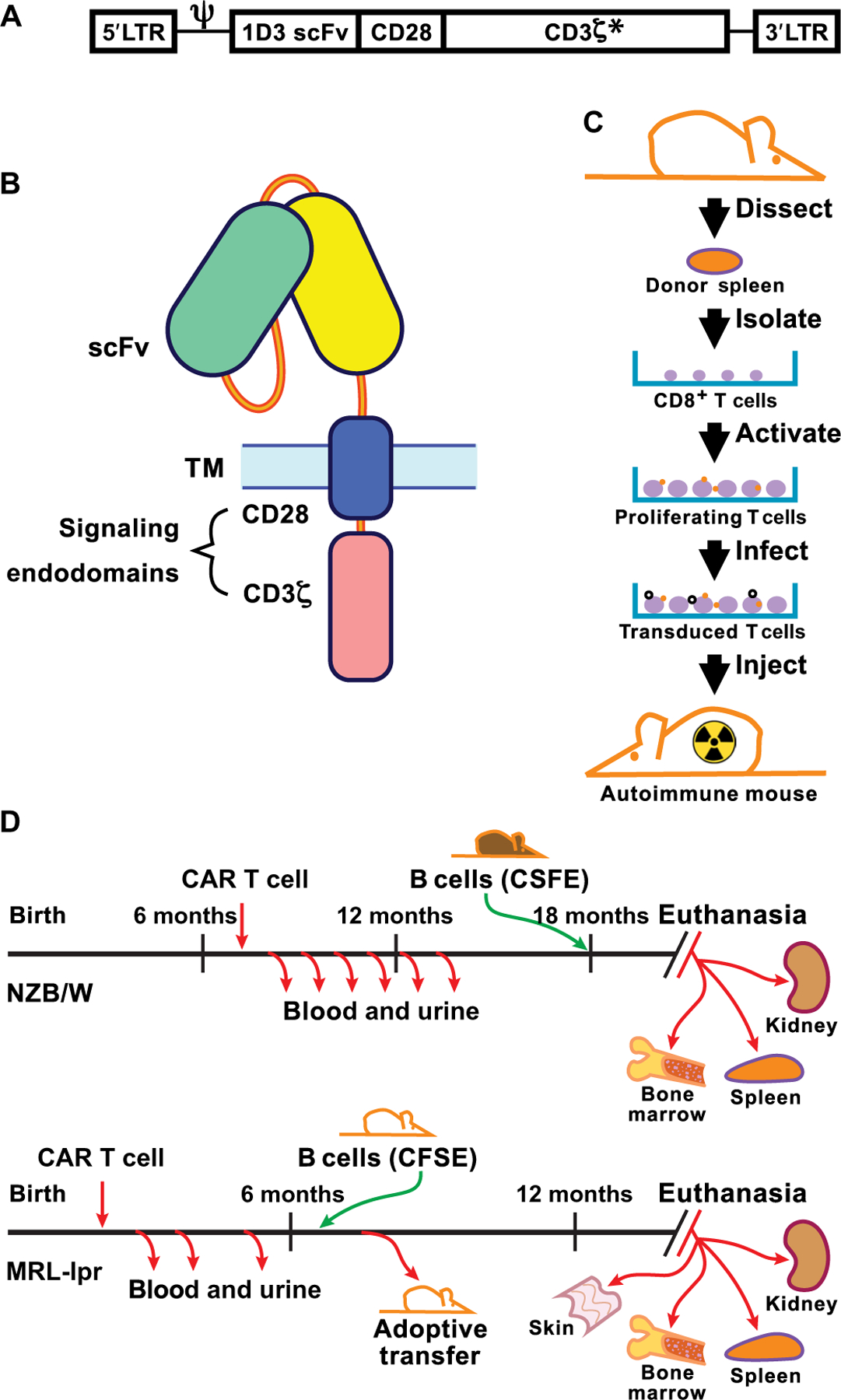
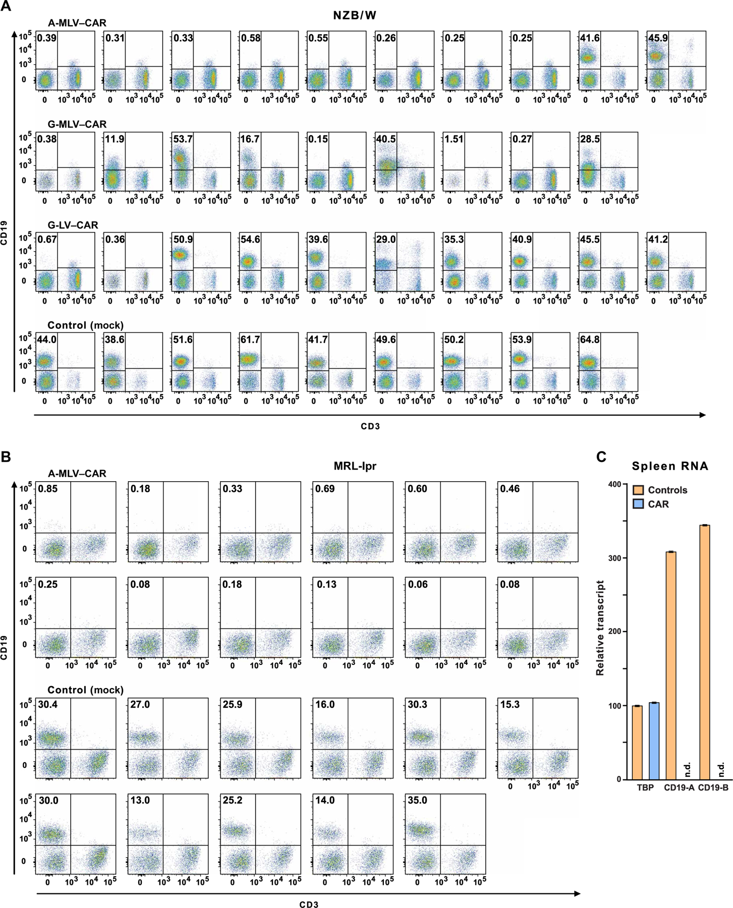
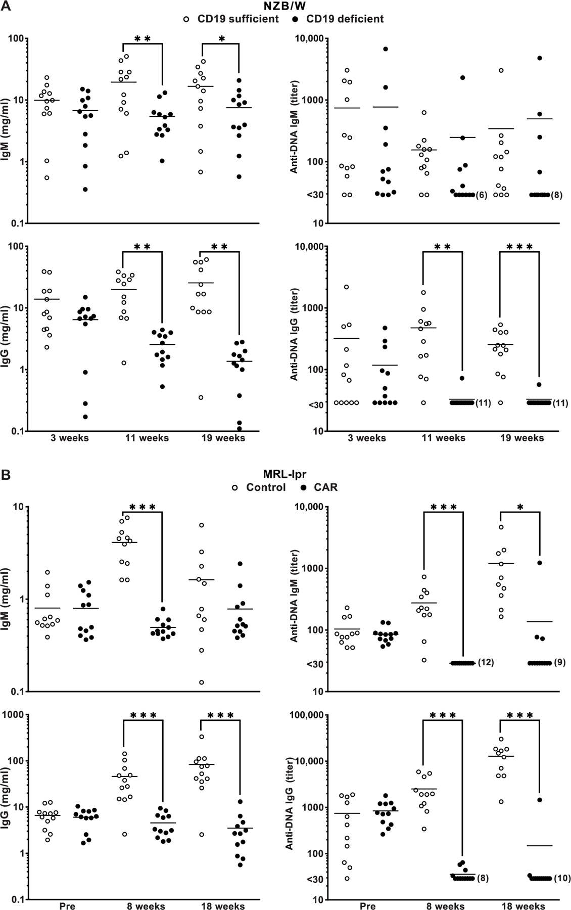
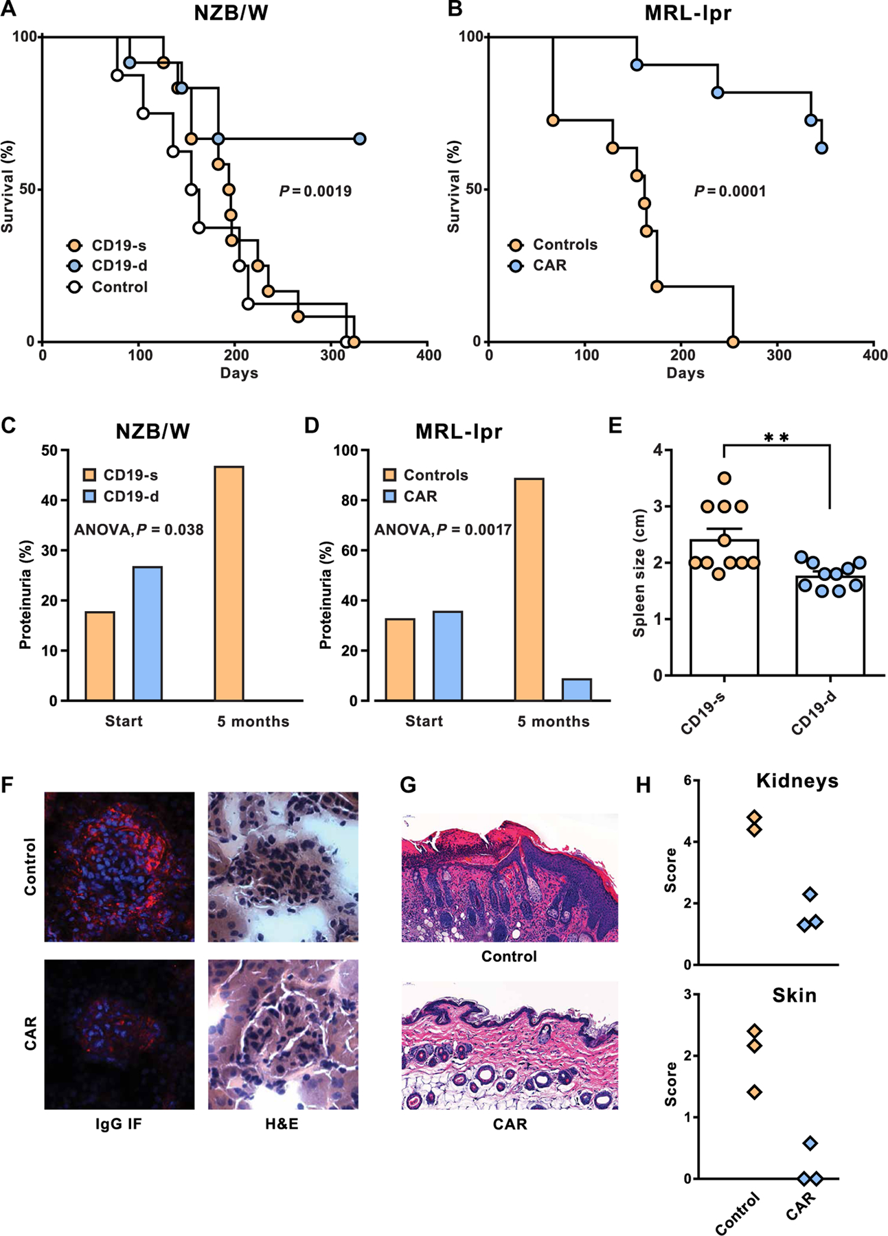
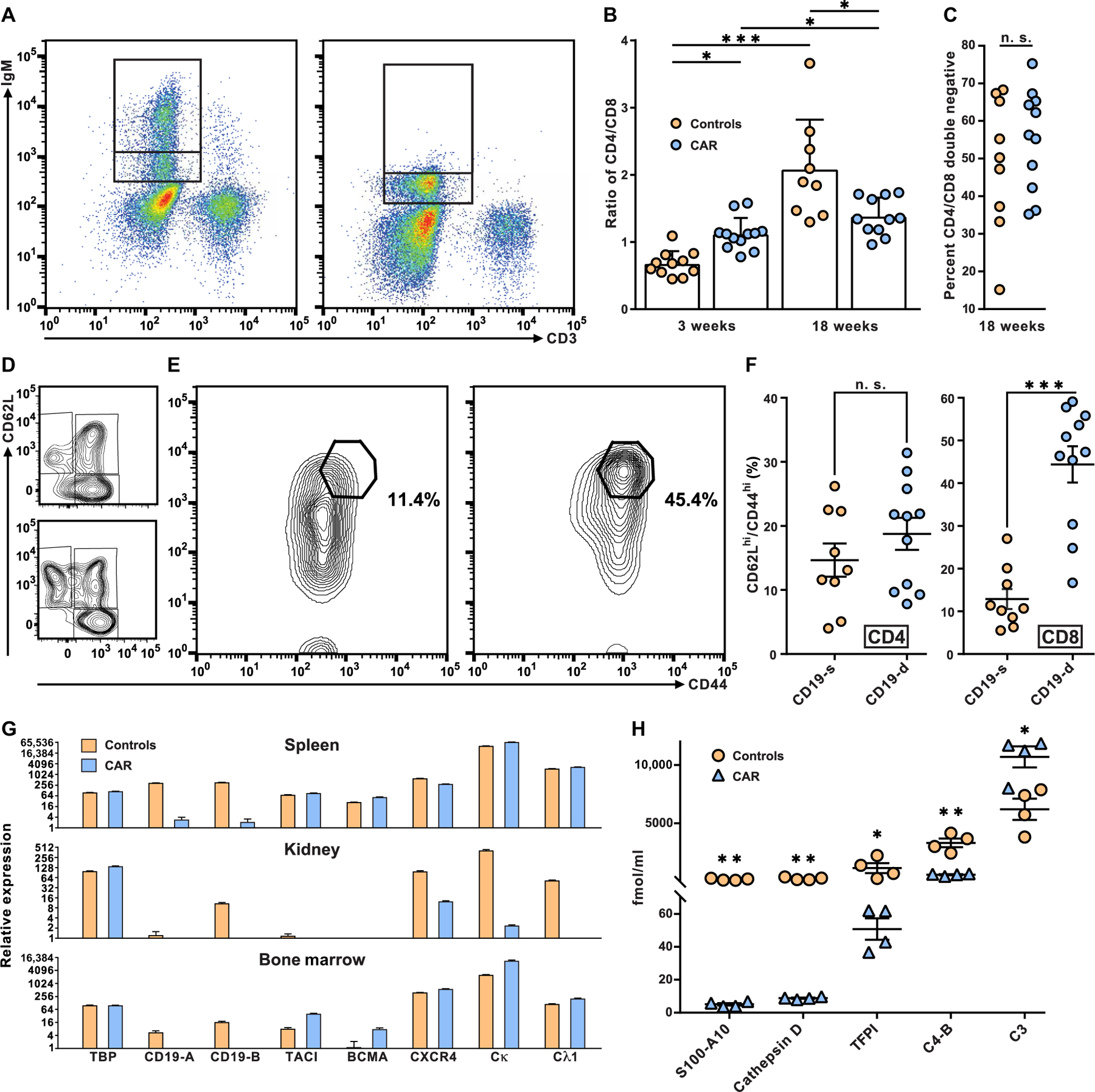
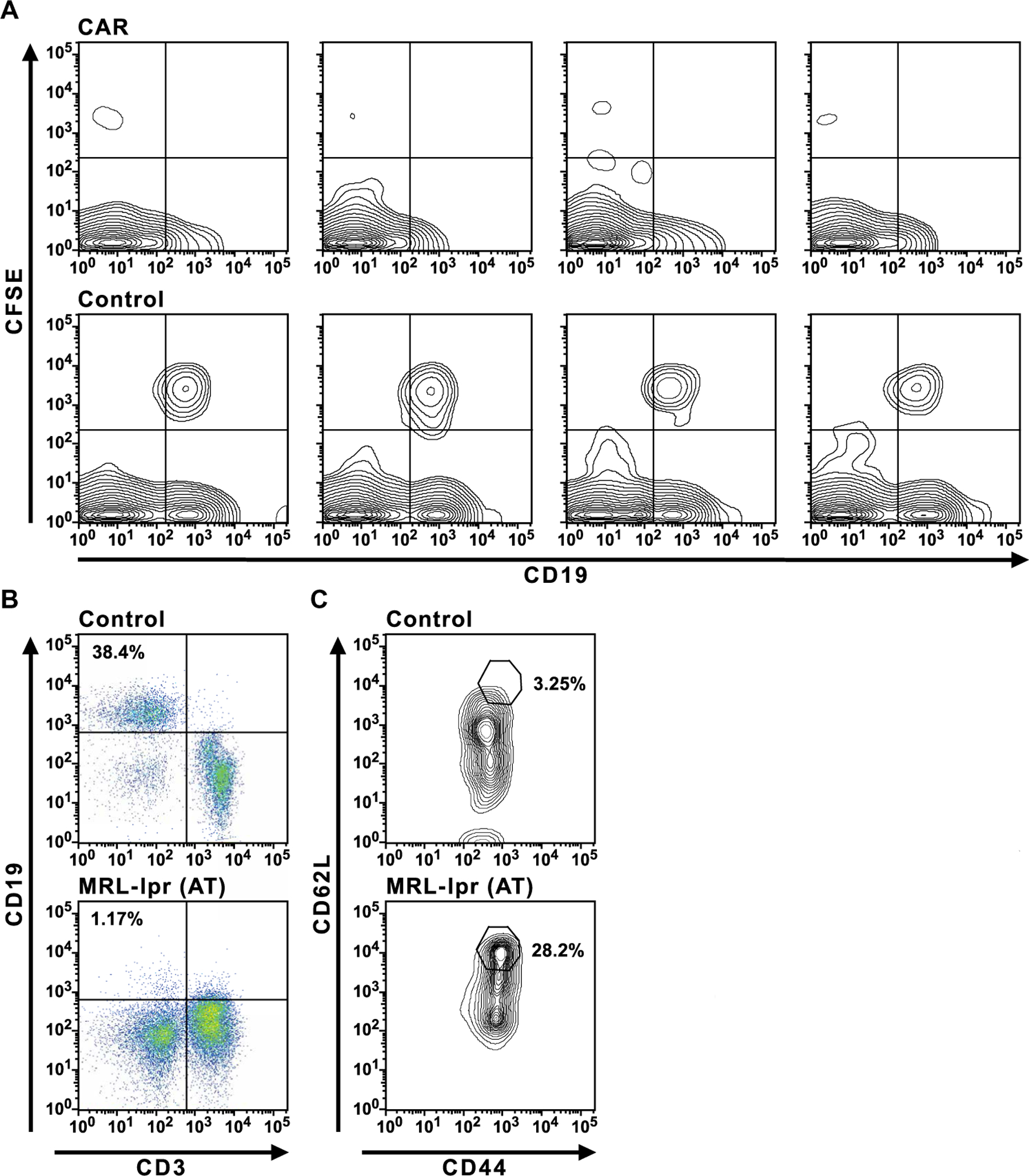
The failure of anti-CD20 antibody (Rituximab) as therapy for lupus may be attributed to the transient and incomplete B cell depletion achieved in clinical trials. Here, using an alternative approach, we report that complete and sustained CD19+ B cell depletion is a highly effective therapy in lupus models. CD8+ T cells expressing CD19-targeted chimeric antigen receptors (CARs) persistently depleted CD19+ B cells, eliminated autoantibody production, reversed disease manifestations in target organs, and extended life spans well beyond normal in the (NZB × NZW) F1 and MRL fas/fas mouse models of lupus. CAR T cells were active for 1 year in vivo and were enriched in the CD44+CD62L+ T cell subset. Adoptively transferred splenic T cells from CAR T cell-treated mice depleted CD19+ B cells and reduced disease in naive autoimmune mice, indicating that disease control was cell-mediated. Sustained B cell depletion with CD19-targeted CAR T cell immunotherapy is a stable and effective strategy to treat murine lupus, and its effectiveness should be explored in clinical trials for lupus.


Conflict of interest statement

Competing interests: UTHSC filed a patent application, no. 65022–274929, titled “Use of Anti-CD19 CAR T Cells in Lupus.” (M.R., inventor). J.N.K. has patent applications for CARs targeting human CD19.

Figures

Fig. 1.
Fig. 1.. CAR design and transduction.
(A) The structural gene for 1D3–28Z.1–3 is shown downstream of the MSGV 5′ long terminal repeat (LTR) and splice acceptor (Ψ). The asterisk indicates that the first and third ITAMs in CD3ζ have tyrosine to alanine replacement mutations. (B) Diagram of CAR protein in the cell membrane showing the extracellular single-chain Fv (scFv) domain and the transmembrane (TM) and signaling domains that combine CD28 and CD3ζ C termini. (C) Steps involved in the isolation, activation, and transduction of CD8+ splenocytes with virus before infusion into a recipient mouse that was irradiated ahead of T cell transfer to achieve transient myeloablation. (D) Timeline of CAR T cell treatment and sample collection in NZB/W and MRL-lpr mice. CFSE, carboxyfluorescein diacetate succinimidyl ester.
Fig. 2.
Fig. 2.. Flow cytometry of blood lymphocytes from CAR T cell–treated mice and controls.
Mice were administered CD19-targeted CAR CD8+ T cells transduced by one of three different viruses, as indicated above the plots. CD19+ B cell depletion was assessed 2 months after treatment. Pseudocolor plots indicate CD19 and CD3 expression on lymphocytes, as defined by forward and side scatter in blood from individual mice. Frequencies of CD19+ B cells are shown in the top left quadrant. (A) Fourteen NZB/W mice in the three transduction groups that had less than 1% CD19+ lymphocytes were designated as CD19-d, whereas control mice and 15 of the CAR T cell–treated mice that had 12 to 60% CD19+ B cells were combined into the CD19-sufficient (CD19-s) experimental group. (B) MRL-lpr mice were injected with A-MLV–CAR–transduced CD8+ T cells or with nontransduced CD8+ T cells (control), and frequencies of CD19+ lymphocytes were determined. (C) Total RNA from the spleens of four control and four CAR T cell–treated mice was purified to measure transcripts coding for CD19 and TATA binding protein (TBP). CD19 message, as measured by reverse transcription polymerase chain reaction (RT-PCR) at two exon-intron boundaries (CD19-A or CD19-B; see table S2 for details), was determined in CAR T cell–treated mice and controls. n.d., nondetectable.
Fig. 3.
Fig. 3.. Serologic analysis of IgM and IgG concentrations and anti-DNA reactivities at different times after CAR T cell infusion.
(A) NZB/W and (B) MRL-lpr plasma were tested using ELISA for total IgM and IgG concentration and IgM and IgG anti-DNA titers. Values for CD19-d and CD19-s and control mice are indicated. IgM and IgG standards were used to generate standard curves for estimating IgM and IgG concentrations. Times of plasma collection for the relevant groups are indicated. Horizontal lines are means. Differences between measurements taken at the indicated time points were compared by one-tailed t test, and significance is indicated by *P < 0.05, **P < 0.01, and ***P < 0.001.
Fig. 4.
Fig. 4.. Impact of CD19-targeted CAR T cell treatment on lupus pathogenesis and survival.
(A) Survival curves of NZB/W mice after CAR T cell infusion into 7-month-old recipients. Survival of CD19-d mice (n = 12) was compared relative to CD19-s mice (n = 12), and significance was determined by log rank (Mantel-Cox; P = 0.0019). For comparison, the survival of control mice (n = 8) is plotted. (B) Survival curves of CAR T cell-treated and control MRL-lpr mice. CAR T cell–treated mice (n = 11) were compared to control MRL-lpr mice (n = 11), and survival of mice in the two groups was evaluated by long rank (Mantel-Cox; P = 0.0001). (C and D) High-grade proteinuria (1 mg/ml and higher) was determined before treatment (start) and at 5 months after CAR T cell treatment. (C) Proteinuria in CD19-s (n = 12) and CD19-d (n = 11) NZB/W mice was analyzed by single-factor analysis of variance (ANOVA) (P = 0.038). (D) High-grade proteinuria in CAR T cell–treated MRL-lpr mice (n = 11) and controls (n = 9) was similarly compared (P = 0.0017). (E) Lengths of spleens in CD19-d and CD19-s NZB/W mice were measured at euthanasia (P < 0.005). Bars are means, and horizontal lines are SEMs. (F) Kidneys from NZB/W mice were sectioned and analyzed for cellularity and morphology by H&E staining and for mouse IgG deposits by immunofluorescence (IF). (G) MRL-lpr skin sections were stained by H&E to compare the epidermis of control and CAR T cell–treated mice. (H) Pathology scores of MRL-lpr kidney and skin sections were determined as described in Materials and Methods.
Fig. 5.
Fig. 5.. Effect of CD19-targeted CAR T cell treatment on lymphocyte phenotype and plasma proteome.
(A) The expression of surface IgM on peripheral blood B lymphocytes from control MRL-lpr mice (left) was used to define an intermediate and a high IgM+ population (indicated by stacked gates). These gates were used to assess expression of IgM on B cells from CAR T cell–treated MRL-lpr mice (right). (B) The ratio of CD4 to CD8 T cells was determined in 18-week-old MRL-lpr mice treated with CAR T cells and controls. Each symbol represents data from a single mouse, bars are means, and horizontal lines are SDs. Differences between CAR T cell–treated and control mice were examined by two-tailed t test and noted by *P < 0.05 and ***P < 0.001. (C) Proportions of CD4CD8 DN T cells as the percentage of CD3+ cells from CAR T cell–treated and control MRL-lpr mice were plotted (n. s., no significant differences). (D) Effector memory (CD62LloCD44hi), central memory (CD62LhiCD44hi), and naïve (CD62LhiCD44lo) CD4+ T cells from representative control (top) and CAR T cell–treated MRL-lpr mice (bottom) were determined (for complete CD4+ T cell phenotype assessment, see table S3). (E) The CD62LhiCD44hi population of CD8+ T cells from a representative control mouse (left) and CAR T cell–treated MRL-lpr mouse (right) are shown, and the percentage of CD8+ T cells in the CD62LhiCD44hi gates was displayed. (F) Overall proportions of CD62LhiCD44hi cells among CD8+ or CD4+ T cells in CAR T cell–treated and control mice were determined and plotted. Each symbol represents data from a single mouse, horizontal lines are means, and offset lines are SDs. Differences between CAR T cell–treated and control mice were examined by two-tailed t test and marked by ***P < 0.001. (G) Relative RNA expression in spleens, kidneys, and bone marrow from four CAR T cell–treated and four control NZB/W mice were measured by RT-PCR for transcripts originating from TBP, a house keeping gene, two regions of CD19, TACI, BCMA, CXCR4, and the light-chain constant regions of Igκ and λ1 (see Materials and Methods and table S2). Values were plotted using ln2. Means and SE are displayed. (H) Plasma was prepared from CAR T cell–treated and control MRL-lpr mice, and plasma proteins were analyzed by mass spectrometry. Horizontal lines are means, and offset lines are SDs. Concentrations were compared by two-tailed t test, and significance was indicated by *P < 0.05, **P < 0.01.
Fig. 6.
Fig. 6.. Tests of CAR T cell function in recipient mice.
(A) Injections of CFSE-labeled B cells into control or CAR T cell–treated MRL-lpr mice were performed 4 months after CAR T cell administration. Six days after injection into control or CAR T cell–treated mice, peripheral blood was assayed for labeled B cells by flow cytometry. (B and C) Adoptive transfer (AT) of purified splenic CD8+ T cells from an MRL-lpr mouse that was treated 7 months earlier with CD19-targeted CAR T cells into a second group of previously untreated, 2-month-old MRL-lpr mice. The secondary MRL-lpr recipients (MRL-lpr AT) and control MRL-lpr mice were assayed for CD19+ B cells (B) in peripheral blood 4 months after transfer and CD44hiCD62Lhi CD8+ T cells (C). The data in (B) and (C) are representative of four MRL-lpr AT mice observed in two experiments.

Comment in

References

    1. Rahman A, Isenberg DA, Systemic lupus erythematosus. N. Engl. J. Med 358, 929–939 (2008). - PubMed
    1. Shlomchik MJ, Activating systemic autoimmunity: B’s, T’s, and tolls. Curr. Opin. Immunol 21, 626–633 (2009). - PMC - PubMed
    1. Sang A, Zheng Y-Y, Morel L, Contributions of B cells to lupus pathogenesis. Mol. Immunol 62, 329–338 (2014). - PMC - PubMed
    1. Li Y, Chen F, Putt M, Koo YK, Madaio M, Cambier JC, Cohen PL, Eisenberg RA, B cell depletion with anti-CD79 mAbs ameliorates autoimmune disease in MRL/lpr mice. J. Immunol 181, 2961–2972 (2008). - PMC - PubMed
    1. Gallagher S, Yusuf I, McCaughtry TM, Turman S, Sun H, Kolbeck R, Herbst R, Wang Y, MEDI-551 treatment effectively depletes B cells and reduces serum titers of autoantibodies in mice transgenic for Sle1 and human CD19. Arthritis Rheumatol 68, 965–976 (2016). - PubMed

Publication types