. 2019 May 1;93(10):e00308-19.
doi: 10.1128/JVI.00308-19. Print 2019 May 15.

Quantitative Proteomic Analysis Reveals Unfolded-Protein Response Involved in Severe Fever with Thrombocytopenia Syndrome Virus Infection

Affiliations

Quantitative Proteomic Analysis Reveals Unfolded-Protein Response Involved in Severe Fever with Thrombocytopenia Syndrome Virus Infection

Lei-Ke Zhang et al. J Virol. .

Abstract

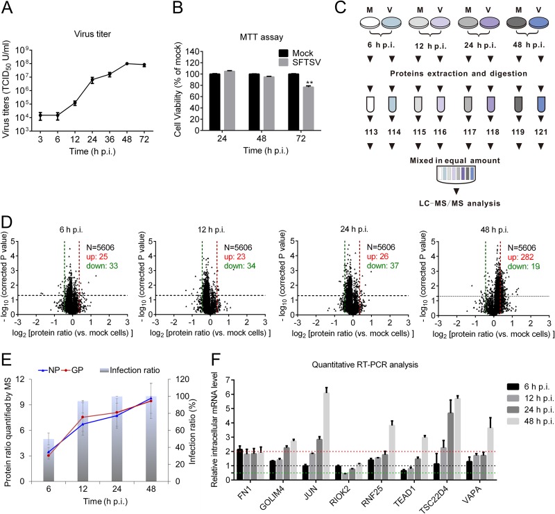
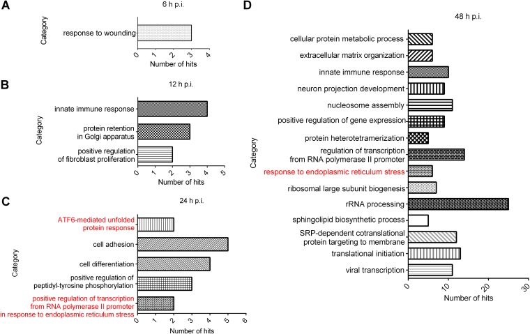
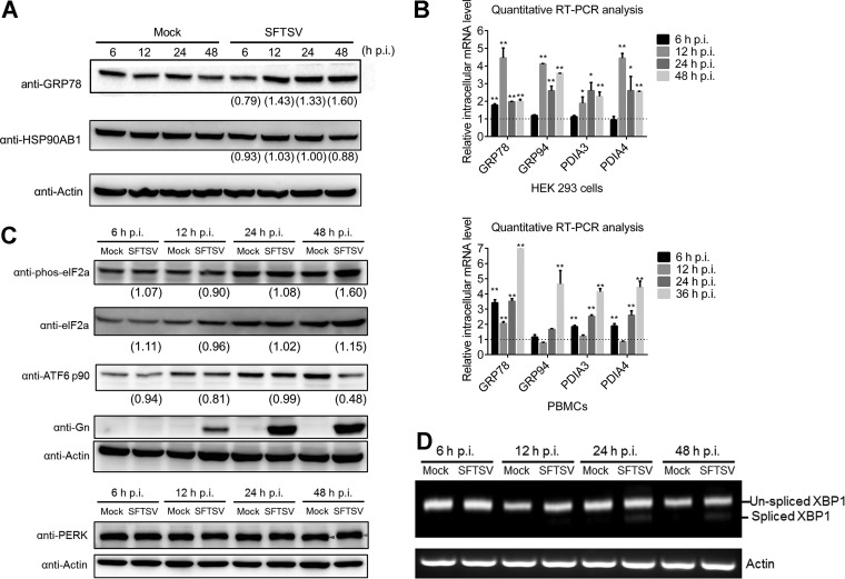
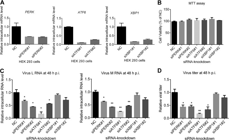
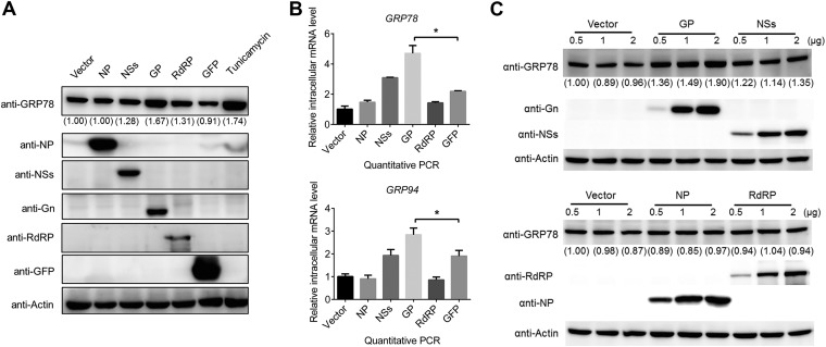
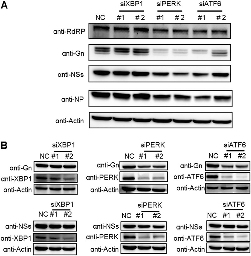
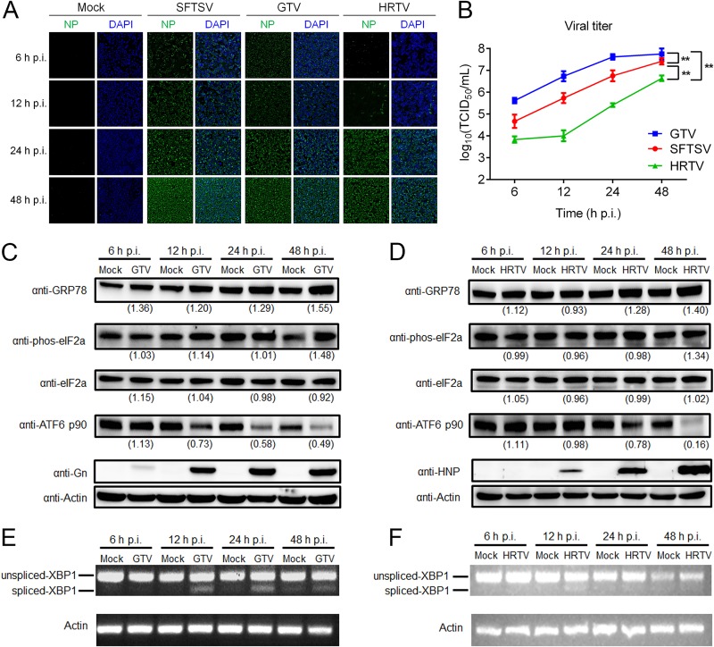
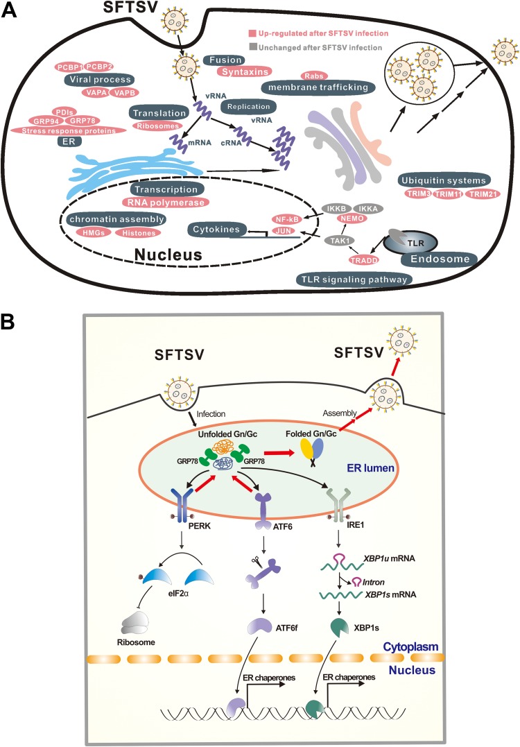
Severe fever with thrombocytopenia syndrome (SFTS) is an emerging, highly pathogenic, infectious disease caused by infection with a newly discovered tick-borne phlebovirus, SFTS virus (SFTSV). Limited information on the molecular mechanism of SFTSV infection and pathogenesis impedes the development of effective vaccines and drugs for SFTS prevention and treatment. In this study, an isobaric tag for relative and absolute quantification (iTRAQ)-based quantitative proteomic analysis of SFTSV-infected HEK 293 cells was performed to explore dynamic host cellular protein responses toward SFTSV infection. A total of 433 of 5,606 host proteins involved in different biological processes were differentially regulated by SFTSV infection. The proteomic results highlighted a potential role of endoplasmic reticular stress-triggered unfolded-protein response (UPR) in SFTSV infection. Further functional studies confirmed that all three major branches of the UPR, including the PKR-like endoplasmic reticulum kinase (PERK), the activating transcription factor-6 (ATF6), and the inositol-requiring protein-1 (IRE1)/X-box-binding protein 1 (XBP1) pathways, were activated by SFTSV. However, only the former two pathways play a crucial role in SFTSV infection. Furthermore, expression of SFTSV glycoprotein (GP) alone was sufficient to stimulate the UPR, whereas suppression of PERK and ATF6 notably decreased GP expression. Interestingly, two other newly discovered phleboviruses, Heartland virus and Guertu virus, also stimulated the UPR, suggesting a common mechanism shared by these genetically related phleboviruses. This study provides a global view to our knowledge on how host cells respond to SFTSV infection and highlights that host cell UPR plays an important role in phlebovirus infection.IMPORTANCE Severe fever with thrombocytopenia syndrome virus (SFTSV) is an emerging tick-borne bunyavirus that causes severe fever with thrombocytopenia syndrome in humans, with a mortality rate reaching up to 30% in some outbreaks. There are currently no U.S. Food and Drug Administration-approved vaccines or specific antivirals available against SFTSV. To comprehensively understand the molecular interactions occurring between SFTSV and the host cell, we exploit quantitative proteomic approach to investigate the dynamic host cellular responses to SFTSV infection. The results highlight multiple biological processes being regulated by SFTSV infection. Among these, we focused on exploration of the mechanism of how SFTSV infection stimulates the host cell's unfolded-protein response (UPR) and identified the UPR as a common feature shared by SFTSV-related new emerging phleboviruses. This study, for the first time to our knowledge, provides a global map for host cellular responses to SFTSV infection and highlighted potential host targets for further research.

Keywords: ATF6; ER stress; PERK; SFTSV; XBP1; quantitative proteomics; unfolded-protein response; virus-host interaction.


Figures

FIG 1
FIG 1
Quantitative proteomics analysis of SFTSV-infected HEK 293 cells. (A) Kinetics of SFTSV replication in HEK 293 cells. HEK 293 cells were infected with SFTSV at an MOI of 5, the supernatants were harvested at the indicated time points, and the virus titers were measured by determining the TCID50. All experiments were performed at least three times, and values represent means ± the SDs from three replicates. (B) HEK 293 cells were infected with SFTSV at an MOI of 5, and at the indicated time points HEK 293 cells were harvested and subjected to MTT assay to measure cell viability. (C) Workflow for iTRAQ-based quantitative proteomic analysis of SFTSV-infected HEK 293 cells. (D) Volcano plot showing log2-fold change plotted against the –log2-adjusted P value for SFTSV-infected cells versus mock-treated cells at different times p.i. (E) Kinetics of the viral protein ratio and infection ratio of SFTSV-infected HEK 293 cells. The viral protein ratio was measured by MS. The infection ratio was measured by detecting NP-positive cells versus all cells detected. (F) Validation of MS results using quantitative RT-PCR. HEK 293 cells were infected with SFTSV at an MOI of 5 or mock infected. At indicated time intervals, cells were harvested, and intracellular mRNAs were extracted and subjected to reverse transcription. The intracellular RNA levels of the corresponding proteins were measured with quantitative RT-PCR. Glyceraldehyde-3-phosphate dehydrogenase (GAPDH), tubulin, and actin were chosen as internal controls. Intracellular RNA levels at each time point of SFTSV infection were normalized to those in the mock-infected cells. The experiments were repeated twice. Bars in panels A and D represent the SD. M, mock treated; V, SFTSV infected; N, number of proteins quantified; up, upregulated protein; down, downregulated protein; FN1, fibronectin; GOLIM4, Golgi integral membrane protein 4; JUN, transcription factor AP-1; RIOK2, serine/threonine protein kinase RIO2; RNF25, E3 ubiquitin-protein ligase RNF25; TEAD1, transcriptional enhancer factor TEF-1; TSC22D4, TSC22 domain family protein 4; VAPA, vesicle-associated membrane protein-associated protein A.
FIG 2
FIG 2
GO analysis of regulated proteins based on biological processes. Differentially regulated proteins at each time point were subjected to DAVID, respectively. Regulated proteins were grouped based on their roles in biological processes, and a statistical overrepresentation test was performed to determine which biological process was overrepresented by differentially regulated proteins. Only biological processes overrepresented by differentially regulated proteins at 6 h p.i. (A), 12 h p.i. (B), 24 h p.i. (C), and 48 h p.i. (D) were considered to be regulated by SFTSV infection. The categories labeled in red in panels C and D are UPR-related pathways.
FIG 3
FIG 3
SFTSV infection activates all three branches of the UPR. (A) SFTSV-infected HEK 293 cells were collected at the indicated time intervals, and total proteins were extracted and subjected to Western blot analysis for GRP78/94. (B) RNA samples from the above cells were extracted and subjected to reverse transcription. The relative mRNA levels of the indicated proteins were measured using quantitative RT-PCR. GAPDH, tubulin, and actin were chosen as internal controls. Intracellular RNA levels at each time point of SFTSV infection were normalized to those in the mock-infected cells. All experiments were performed at least three times, and values represent means ± the SDs from three replicates. *, P < 0.05; **, P < 0.01 (Student t test). (C) SFTSV-infected HEK 293 cells were collected at the indicated time intervals, and total proteins were extracted and subjected to Western blot analysis for ATF6 p90, phos-eIF2α (Ser51), total eIF2α, PERK, Gn, and the internal control actin. Red arrow, phosphorylated PERK; purple arrow, PERK. (D) RNA samples were also analyzed for spliced XBP1 mRNA by using reverse transcription-PCR. The intensity of protein band was measured by ImageJ_v1.8.0. For each time point, the protein intensity was first normalized to actin and then normalized to the corresponding mock group.
FIG 4
FIG 4
Effects of UPR on SFTSV production. (A and B) Knockdown of targeted proteins by using RNA interference. HEK 293 cells were transfected with siRNAs against targeted host genes or scrambled siRNA (NC). Cells were collected at 48 h p.t., and total cellular RNA was extracted and subjected to reverse transcription. Intracellular RNA levels of ATF6, XBP1, and PERK were measured with quantitative RT-PCR (A). MTT analysis was performed to determine the CPE of the siRNAs. (B). (C and D) Effects of knockdown of host proteins on SFTSV production and replication. HEK 293 cells were transfected with siRNAs against targeted genes or scrambled siRNA (NC), and at 48 h p.t. the cells were superinfected with SFTSV at an MOI of 1. (C) At 48 h p.i., total cellular RNA was extracted, and SFTSV genomic RNA levels were measured with quantitative RT-PCR. (D) The cell supernatant was collected, and the viral titer was measured by EPDAs. All experiments were performed in triplicate, and values represent means ± the SDs from three replicates. *, P < 0.05; **, P < 0.01 (Student t test).
FIG 5
FIG 5
Expression of SFTSV G proteins, but not other viral proteins, induces the cellular UPR. (A) HEK 293 cells were transfected with plasmids expressing viral proteins, or with GFP or empty vector as controls, and at 48 h p.t. the cells were collected, and the intracellular protein levels of GRP94, GRP78, viral proteins, and the loading control actin were detected with Western blots. Tunicamycin (Tm), a reported inducer of the UPR, was used as a positive control. (B) HEK 293 cells were transfected with plasmids expressing viral proteins or GFP, and the intracellular RNA levels of GRP94 and GRP78 were detected with quantitative RT-PCR at 48 h p.t. (C) HEK 293 cells were transfected with increasing amounts of SFTSV protein-expressing plasmids and empty vector as a control. At 48 h p.t., the cells were collected, and the intracellular protein levels of GRP78, viral proteins, and actin as a loading control were detected by Western blotting. *, P < 0.05; **, P < 0.01 (Student t test). The intensity of protein band was measured by ImageJ_v1.8.0. Protein intensity was first normalized to actin and then further normalized to that of the empty vector-transfected cells.
FIG 6
FIG 6
Knockdown of PERK and ATF6 reduces intracellular levels of SFTSV GP. (A) HEK 293 cells were transfected with siRNAs against targeted host genes or scrambled siRNA (NC). At 48 h p.t., cells were superinfected with SFTSV at an MOI of 1 and then collected at 48 h p.i. Viral protein levels were analyzed by Western blotting. (B) HEK 293 cells were cotransfected with plasmids expressing NSs or G proteins and siRNAs against targeted host genes or scrambled siRNA (NC). At 48 h p.t., the cells were collected and viral/host proteins were subjected to Western blot analyses.
FIG 7
FIG 7
GTV and HRTV infection activate three branches of the UPR. (A) HEK 293 cells were infected by GTV, HRTV, or SFTSV, and then, at the indicated time intervals, the cells were fixed, and the intracellular level of NP was monitored by using immunofluorescence. (B) The supernatants of infected cells were also collected, and viral titers were measured by determining the TCID50. All experiments were performed at least three times, and values represent means ± the SDs from three replicates. ***, P < 0.001 (Fisher LSD tests). GTV (C)- or HRTV (E)-infected HEK 293 cells were collected at the indicated time intervals, and total proteins were extracted and subjected to Western blot analyses for GRP78, ATF6 p90, phos-eIF2α (Ser51), total eIF2α, Gn, NP, and the internal control actin. Anti-SFTSV Gn (anti-Gn) and anti-HRTV NP (anti-HNP) were used to detect GTV GP and HRTV NP, respectively. (D and F) RNA samples from the cells described above were also analyzed for spliced XBP1 mRNA by using reverse transcription-PCR. The intensity of protein band was measured by ImageJ_v1.8.0. For each time point, the protein intensity was first normalized to actin and then normalized to the corresponding mock-treated group.
FIG 8
FIG 8
Proposed model for the UPR and other cellular responses regulated by SFTSV infection. (A) Global host cellular protein responses to SFTSV infection. According to the results of the quantitative proteomic analysis, proteins or protein complexes specifically regulated by SFTSV infection are sorted and aligned according to their biological functions. (B) Proposed interaction model between host UPR and SFTSV infection. SFTSV infection produces large amounts of unfolded GP in the ER, which activates all three main branches of the UPR. Among these, the ATF6 and PERK pathways facilitate proper folding of GP and thus favor SFTSV replication.

References

    1. Cyranoski D. 2018. East Asia braces for surge in deadly tick-borne virus. Nature 556:282–283. doi:10.1038/d41586-018-04486-6. - DOI - PubMed
    1. Yu XJ, Liang MF, Zhang SY, Liu Y, Li JD, Sun YL, Zhang L, Zhang QF, Popov VL, Li C, Qu J, Li Q, Zhang YP, Hai R, Wu W, Wang Q, Zhan FX, Wang XJ, Kan B, Wang SW, Wan KL, Jing HQ, Lu JX, Yin WW, Zhou H, Guan XH, Liu JF, Bi ZQ, Liu GH, Ren J, Wang H, Zhao Z, Song JD, He JR, Wan T, Zhang JS, Fu XP, Sun LN, Dong XP, Feng ZJ, Yang WZ, Hong T, Zhang Y, Walker DH, Wang Y, Li DX. 2011. Fever with thrombocytopenia associated with a novel bunyavirus in China. N Engl J Med 364:1523–1532. doi:10.1056/NEJMoa1010095. - DOI - PMC - PubMed
    1. Kim KH, Yi J, Kim G, Choi SJ, Jun KI, Kim NH, Choe PG, Kim NJ, Lee JK, Oh MD. 2013. Severe fever with thrombocytopenia syndrome, South Korea, 2012. Emerg Infect Dis 19:1892–1894. doi:10.3201/eid1911.130792. - DOI - PMC - PubMed
    1. Takahashi T, Maeda K, Suzuki T, Ishido A, Shigeoka T, Tominaga T, Kamei T, Honda M, Ninomiya D, Sakai T, Senba T, Kaneyuki S, Sakaguchi S, Satoh A, Hosokawa T, Kawabe Y, Kurihara S, Izumikawa K, Kohno S, Azuma T, Suemori K, Yasukawa M, Mizutani T, Omatsu T, Katayama Y, Miyahara M, Ijuin M, Doi K, Okuda M, Umeki K, Saito T, Fukushima K, Nakajima K, Yoshikawa T, Tani H, Fukushi S, Fukuma A, Ogata M, Shimojima M, Nakajima N, Nagata N, Katano H, Fukumoto H, Sato Y, Hasegawa H, Yamagishi T, Oishi K, Kurane I, Morikawa S, Saijo M. 2014. The first identification and retrospective study of severe fever with thrombocytopenia syndrome in Japan. J Infect Dis 209:816–827. doi:10.1093/infdis/jit603. - DOI - PMC - PubMed
    1. McMullan LK, Folk SM, Kelly AJ, MacNeil A, Goldsmith CS, Metcalfe MG, Batten BC, Albarino CG, Zaki SR, Rollin PE, Nicholson WL, Nichol ST. 2012. A new phlebovirus associated with severe febrile illness in Missouri. N Engl J Med 367:834–841. doi:10.1056/NEJMoa1203378. - DOI - PubMed

Publication types

MeSH terms

Supplementary concepts