. 2019 Mar 6;9(1):3684.
doi: 10.1038/s41598-019-39812-z.

"Hit the missing stimulus". A simultaneous EEG-fMRI study to localize the generators of endogenous ERPs in an omitted target paradigm

Affiliations

"Hit the missing stimulus". A simultaneous EEG-fMRI study to localize the generators of endogenous ERPs in an omitted target paradigm

Aldo Ragazzoni et al. Sci Rep. .

Abstract

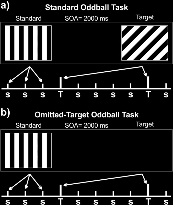
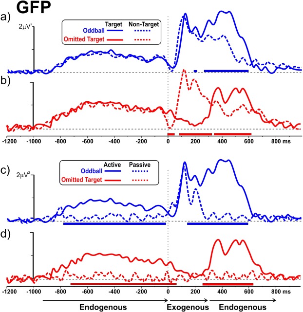
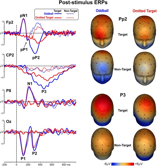
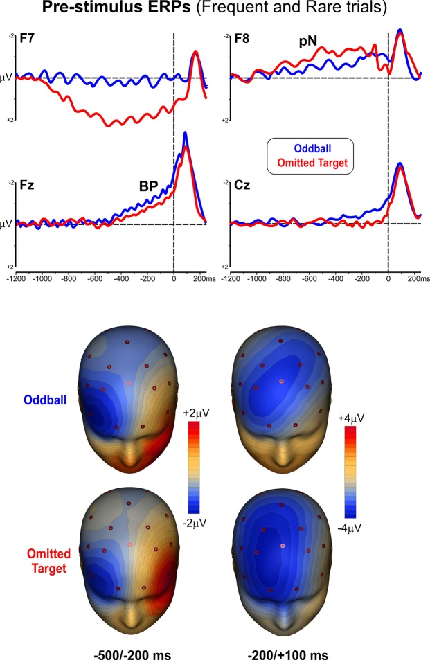
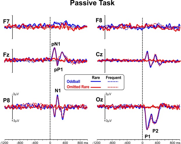
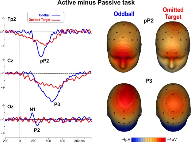
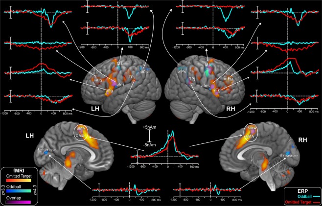
Event-Related Potentials (ERPs) occurring independently from any stimulus are purely endogenous (emitted potentials) and their neural generators can be unequivocally linked with cognitive processes. In the present study, the subjects performed two similar visual counting tasks: a standard two-stimulus oddball, and an omitted-target oddball task, characterized by the physical absence of the target stimulus. Our investigation aimed at localizing the neural sources of the scalp-recorded endogenous/emitted ERPs. To optimize the source localization, the high temporal resolution of electrophysiology was combined with the fine spatial information provided by the simultaneous recording of functional magnetic resonance (fMRI). Both tasks identified two endogenous ERP components in the 300 to 520 ms interval. An earlier component, pP2, showed a bilateral generator in the anterior Insula. A later P3 component (P3b) was generated bilaterally in the temporal-parietal junction, the premotor and motor area and the anterior intraparietal sulcus (this latter one only in the standard oddball). Anticipatory slow waves (beginning 900 to 500 ms pre-stimulus), also of endogenous nature, were produced by the inferior and middle frontal gyrus and the supplementary and cingulate motor areas. Our protocol disentangled pre- from post-stimulus fMRI activations and provided original clues to the psychophysiological interpretation of emitted/endogenous ERPs.


Conflict of interest statement

The authors declare no competing interests.

Figures

Figure 1
Figure 1
Schematic representation of the two experimental tasks: (a) Standard visual Oddball; (b) Omitted-Target Oddball. s: standard-frequent stimuli. T: target-rare stimuli. SOA: Stimulus-Onset Asynchrony.
Figure 2
Figure 2
Global Field Power (GFP) of ERPs preceding and following the stimulus in the different experimental conditions. The GFP time course associated with Frequent/Non-Target and Rare/Target stimuli are presented for the Oddball Task (panel a, blue plot) and for the Omitted Target Task (panel b, red plot). GFP associated with Rare/Target stimuli in the passive and the active conditions are provided for the Oddball Task (panel c, blue plot) and Omitted Target Task (panel d, red plot). The horizontal thick blue and red lines signal epochs in which the difference between the GFP of the two ERPs was statistically significant. The temporal extents of Exogenous and Endogenous components are highlighted by the black thin horizontal arrows below the ERP traces.
Figure 3
Figure 3
Left: ERP waveforms of the post-stimulus responses to non-target and target stimuli in the two active tasks. Right: scalp topography of the pP2 and P3b components.
Figure 4
Figure 4
ERP waveforms (top) and scalp topography (bottom) of the pre-stimulus activities (pN, BP) in the two active tasks (oddball and omitted target). Maps represent the prefrontal lateral activity between −500/−200 ms (left) and the medial frontal activity between −200/+100 ms (right).
Figure 5
Figure 5
ERP waveforms in the two passive tasks for both frequent and rare stimuli.
Figure 6
Figure 6
Left: post-stimulus ERP differential waveforms (active minus passive) following the rare stimuli. Right: scalp topographical distribution of the pP2 and P3 differential components.
Figure 7
Figure 7
Spatiotemporal mapping obtained by the combination of ERP and fMRI data. fMRI activations resulting in the active > passive contrast for target stimuli for both tasks and their overlap from mesial and lateral views. LH, left hemisphere; RH, right hemisphere. Waveforms correspond to the ERP-based time-courses of the neural sources obtained from the fMRI-seeded dipoles. LH: left hemisphere. RH: right hemisphere. aIns: anterior Insula. aIPs: anterior IntraParietal sulcus. CMA: Cingulate Motor Area. iFg: inferior Frontal gyrus. mFg: middle Frontal gyrus. M1: motor cortex. SMA: Supplementary Motor Area. TPj: Temporal-Parietal junction.

References

    1. Sutton S, Tueting P, Zubin J, John ER. Information delivery and the sensory evoked potential. Science. 1967;155:1436–1439. - PubMed
    1. O’Connell RG, Dockree PM, Kelly SP. A supramodal accumulation-to-bound signal that determines perceptual decisions in humans. Nature Neuroscience. 2012;15:1729–1735. - PubMed
    1. Picton TW, et al. Guidelines for using human event-related potentials to study cognition. Psychophysiology. 2000;37:127–152. - PubMed
    1. Duncan CC, et al. Event-related potentials in clinical research: Guidelines for eliciting, recording, and quantifying mismatch negativity, P300 and N400. Clin. Neurophysiol. 2009;120:1883–1908. - PubMed
    1. Polich J. Updating P300: an integrative theory of P3a and P3b. Clin. Neurophysiol. 2007;118:2128–2148. - PMC - PubMed

Publication types