. 2019 Mar 6;9(1):3716.
doi: 10.1038/s41598-019-39884-x.

L1cam-mediated developmental processes of the nervous system are differentially regulated by proteolytic processing

Affiliations

L1cam-mediated developmental processes of the nervous system are differentially regulated by proteolytic processing

Cecilie Linneberg et al. Sci Rep. .

Abstract

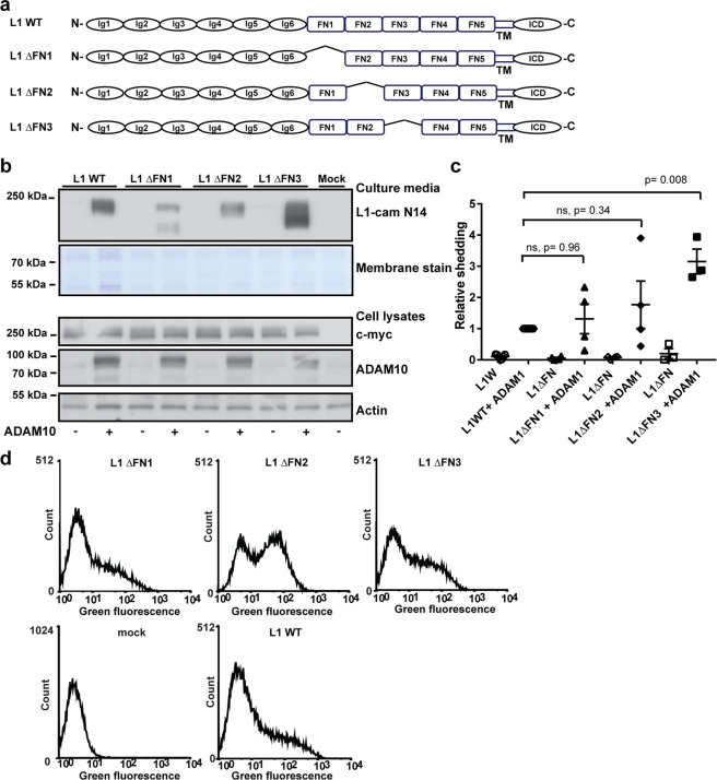
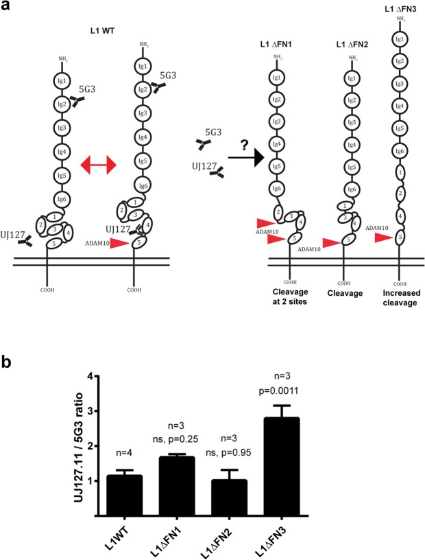
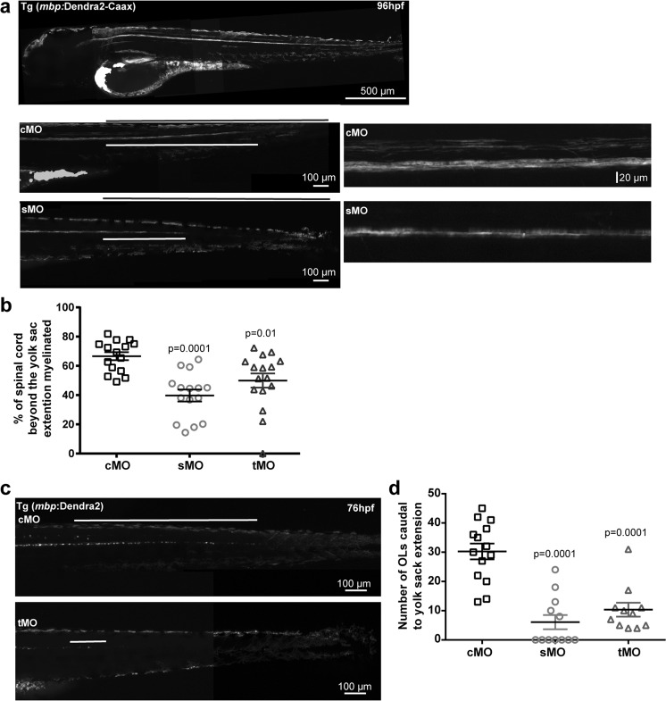
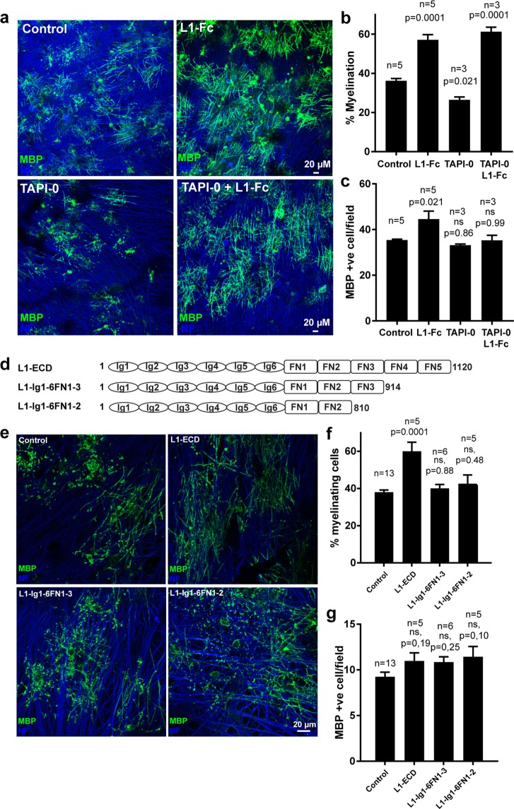
Normal brain development depends on tight temporal and spatial regulation of connections between cells. Mutations in L1cam, a member of the immunoglobulin (Ig) superfamily that mediate cell-cell contacts through homo- and heterophilic interactions, are associated with several developmental abnormalities of the nervous system, including mental retardation, limb spasticity, hydrocephalus, and corpus callosum aplasia. L1cam has been reported to be shed from the cell surface, but the significance of this during different phases of brain development is unknown. We here show that ADAM10-mediated shedding of L1cam is regulated by its fibronectin type III (FNIII) domains. Specifically, the third FNIII domain is important for maintaining a conformation where access to a membrane proximal cleavage site is restricted. To define the role of ADAM10/17/BACE1-mediated shedding of L1cam during brain development, we used a zebrafish model system. Knockdown of the zebrafish, l1camb, caused hydrocephalus, defects in axonal outgrowth, and myelination abnormalities. Rescue experiments with proteinase-resistant and soluble L1cam variants showed that proteolytic cleavage is not required for normal axonal outgrowth and development of the ventricular system. In contrast, metalloproteinase-mediated shedding is required for efficient myelination, and only specific fragments are able to mediate this stimulatory function of the shedded L1cam.


Conflict of interest statement

The authors declare no competing interests.

Figures

Figure 1
Figure 1
Deletion of the two membrane-proximal FNIII domains in L1cam is required to generate a proteinase resistant variant. (a) Diagram of WT L1cam and mutants with the two membrane proximal fibronectin type III (FN) domains deleted alone or in combination as indicated (b) HEK293T cells were transfected with plasmids encoding WT L1cam or deletion variants alone or in combination with ADAM10 as indicated. The extend of shedding of soluble L1cam into cell culture media was assessed by Western blotting using an antibody targeting the N-terminal of L1cam, membrane staining for total protein was used as loading control (2 top lanes). Expression of L1cam and ADAM10 in cell lysates was assessed by Western blotting, using antibodies targeting the C-terminal c-myc tag (L1cam), or ADAM10. Actin was used as loading control (3 bottom lanes). (c) The amount of soluble fragments in the culture medium was quantified and displayed relative to the amount of soluble fragments from cells co-transfected with WT-L1cam and ADAM10. Mean values +/− SEM for three independent experiments are plotted (Supplementary Fig. 11). Statistical significance was assessed by one-way ANOVA followed by Dunnett’s multiple comparison test. (d) HEK293T cells were transfected with plasmids encoding WT L1cam, deletion variants or an empty vector as indicated. The expression of the different variants on the cell surface was assessed by flow cytometry using an antibody targeting the N-terminal of L1cam. Example histograms are displayed. Mean fluorescence intensities for three independent experiments are displayed in Supplementary Fig. 1.
Figure 2
Figure 2
The first and third FNIII domain regulate ADAM10 mediated shedding of L1cam. (a) Diagram of WT L1cam and mutants with individual deletions of the first three fibronectin type III (FN) as indicated (b) HEK293T cells were transfected with plasmids encoding WT L1cam or deletion variants alone or in combination with ADAM10 as indicated. The extend of shedding of soluble L1cam into cell culture media was assessed by Western blotting using an antibody targeting the N-terminal of L1cam, membrane staining for total protein was used as loading control (2 top lanes). Expression of L1cam and ADAM10 in cell lysates was assessed by Western blotting, using antibodies targeting the C-terminal c-myc tag (L1cam), or ADAM10. Actin was used as loading control (3 bottom lanes). (c) The amount of shedded fragments in the culture medium was quantified and displayed relative to the amount of soluble fragments from cells co-transfected with WT-L1cam and ADAM10. For L1ΔFNIII1, both cleavage products were included for quantification. Mean values +/− SEM of at least three independent experiments are plotted (Supplementary Fig. 13). Statistical significance was assessed by one-way ANOVA followed by Dunnett’s multiple comparison test. (d) HEK293T cells were transfected with plasmids encoding WT L1cam, deletion variants or an empty vector as indicated. The expression of the different variants on the cell surface was assessed by flow cytometry using an antibody targeting the N-terminal of L1cam. Example histograms are displayed. Mean fluorescence intensities for three independent experiments are displayed in Supplementary Fig. 1.
Figure 3
Figure 3
Deletion of the third FNIII domain increases the accessibility to the fourth FNIII domain. (a) Model for control of L1 proteolysis by conformational changes within the FNIII domains. (b) The accessibility of an antibody targeting the membrane proximal fourth FNIII domain for deletion mutants of L1cam in which the first three FNIII domains were deleted individually was analyzed by flow cytometry. The ratio of the mean fluorescence intensity for cells stained with an antibody targeting the fourth FNIII domain (UJ127-11) was given relative to the mean fluorescence intensity obtained for an antibody targeting the second Ig-domain in the N-terminal (5G3). Mean values +/− SEM for at least three independent experiments are plotted. Example histograms and dot-plots are displayed in Supplemental Fig. 4. Statistical significance was assessed by one-way ANOVA followed by Dunnett’s multiple comparison test.
Figure 4
Figure 4
L1cam proteolysis is not required for axonal outgrowth from motor neurons, or development of the ventricular system. (a) Fertilized eggs from wild-type AB zebrafish, were injected with control morpholino (cMo), or morpholinos targeting splicing (sMo) or translation (tMo) of l1camb. The overall morphology of the larvae was assessed at 24 (left panel) and 48 (central panel) hpf. Enlarged view of the area of the mid- and hindbrain at 48 hpf (right panel). (b) Quantification of the penetration of the hydrocephalus phenotype, the percentage of larvae displaying an enlargement of the fourth ventricle was calculated for at least three independent injections. Mean values +/− SEM are plotted. The total number of larvae assessed in each group was cMo (149), sMo (215) and tMo (50). (c) Axonal outgrowth from motor neurons was assessed in fertilized embryos injected with morpholinos as in a, and following immunostaining for znp-1. (d) Quantification of axonal outgrowth from motor neurons. The number of motor neurons with visible axonal outgrowth at 24 hpf was counted. Mean values +/− SEM are displayed. Measurements are from at least three independent injections. (e) Fertilized eggs from wild-type AB zebrafish were injected with control morpholino (cMo) or a morpholino targeting splicing of l1camb alone or in combination with mRNA encoding wild-type (WT), proteinase-resistant (L1ΔFN45), or soluble (L1ECD) L1cam. The extend of hydrocephalus was evaluated at 48 hpf and the penetration of the hydrocephalus phenotype, the percentage of larvae displaying an enlargement of the fourth ventricle was calculated for at least three independent injections. Mean values +/− SEM are plotted. The total number of larvae assessed in each group was cMo (161), sMo (205), sMo- L1ECD (60), sMo-L1 WT (48), and sMo-L1ΔFN45 (51). Example pictures are displayed in Supplemental Fig. 6(f) Axonal outgrowth from motor neurons was assessed in fertilized embryos injected with morpholinos and mRNA as in e, following immune-staining with znp-1.The number of motor neurons with visible axonal outgrowth at 24 hpf was counted. Mean values +/− SEM are displayed. Measurements are from at least three independent injections. Statistical significance was assessed by one-way ANOVA followed by Dunnett’s multiple comparison test.
Figure 5
Figure 5
Knockdown of L1camb decreases the number of mature oligodendrocytes and inhibits myelination. (a) Fertilized eggs from Tg(mbp:Dendra2-CAAX) zebrafish, expressing a membrane targeted version of the fluorescent protein Dendra2 under control of the mbpa promoter, were injected with control morpholino (cMo), or morpholinos targeting splicing (sMo) or translation (tMo) of l1camb. The extent of myelination was assessed at 96 hpf. Micrographs of the posterior part of the fish are displayed (left panels) with the total length from the end of the yolk sac extension to the tail marked by the black line and the extend of myelinated fibers by the white line. Enlarged views of the area of the spinal cord surrounding the yolk sac extension for embryos injected with cMo, or sMo are displayed in the right panels. Note the reduced extent of myelinated fibers and the reduced myelination in both the ventral and dorsal tract of the spinal cord in larvae from l1camb_sMo injections compared to larvae from cMo injections. (b) The extent of myelinated fibers was quantified by measuring the percentage of the caudal spinal cord containing myelinated fibers and the mean values +/− SEM are plotted. The analyzed larvae were from at least three independent injections. (c) Fertilized eggs from a transgenic zebrafish line, Tg(mbp:Dendra2), expressing the fluorescent protein Dendra2 under control of the mbpa promoter, were injected with control morpholinos (cMo), morpholinos targeting splicing (sMo) or translation (tMo) of l1camb. The posterior part of the spinal cord caudal to the yolk sac extension at 76 hpf is displayed. (d) Quantification of the number of Dendra2-positive cells caudal to the yolk sac extension at 76 hpf. Mean values +/− SEM for analyzed larvae from at least three independent injections are plotted. Statistical significance was assessed by one-way ANOVA followed by Dunnett’s multiple comparison test.
Figure 6
Figure 6
Metalloproteinase mediated cell surface shedding is required for efficient myelination. (a) Oligodendrocyte precursor cells (OPCs) were added to Dorsal Root Ganglion (DRG) neurons and co-cultured in the absence or presence of the metalloproteinase inhibitor TAPI-0 and/or soluble L1-Fc. The degree of myelination was analyzed following staining for MBP (marker of mature oligodendrocytes, green), and neurofilament (NF, neuronal marker, blue). Quantification is shown in the following two panels. Mean values +/− SEM are plotted (n values is the total number of coverslips analyzed in three independent culture sets). (b) Percentage of the MBP-positive cells forming at least two internode-like structures and therefore scored as myelinating. (c) The number of MBP-positive cells/10x field. (d) Schematic view of truncation variants of soluble L1. (e) OPCs and DRG neurons were co-cultured in the absence or presence of soluble L1 variants, L1-ECD, L1-Ig1-6FN1-3, or L1-Ig1-6FN1-4 as indicated. The degree of myelination was assessed following staining for MBP (green), and NF (blue). Quantification is shown in the following two panels. Mean values +/− SEM are plotted (n values is the total number of coverslips analyzed in three independent culture sets). (f) Percentage of the MBP-positive cells forming at least two internode-like structures and therefore scored as myelinating. (g) The number of MBP-positive cells/20x field. Statistical significance was assessed by one-way ANOVA followed by Dunnett’s multiple comparison test.

References

    1. Fransen E, et al. CRASH Syndrome: Clinical Spectrum of Corpus Callosum Hypoplasia, Retardation, Adducted Thumbs, Spastic Paraparesis and Hydrocephalus Due to Mutations in One Single Gene, L1. European Journal of Human Genetics: EJHG. 1995;3:273–284. doi: 10.1159/000472311. - DOI - PubMed
    1. Stumpel, C. & Vos, Y. J. In GeneReVIEws((R)) (eds Adam, M. P. et al.) (1993).
    1. Dahme M, et al. Disruption of The Mouse L1 Gene Leads to Malformations of the Nervous System. Nature Genetics. 1997;17:346–349. doi: 10.1038/ng1197-346. - DOI - PubMed
    1. Demyanenko GP, Tsai AY, Maness PF. Abnormalities in Neuronal Process Extension, Hippocampal Development, and the Ventricular System of L1 Knockout Mice. The Journal of Neuroscience: The Official Journal of the Society for Neuroscience. 1999;19:4907–4920. doi: 10.1523/JNEUROSCI.19-12-04907.1999. - DOI - PMC - PubMed
    1. Fransen E, et al. L1 Knockout Mice Show Dilated Ventricles, Vermis Hypoplasia and Impaired Exploration Patterns. Human Molecular Genetics. 1998;7:999–1009. doi: 10.1093/hmg/7.6.999. - DOI - PubMed

Publication types

MeSH terms