. 2019 Mar 6;9(1):3622.
doi: 10.1038/s41598-019-40392-1.

Ischaemic stroke in mice induces lung inflammation but not acute lung injury

Affiliations

Ischaemic stroke in mice induces lung inflammation but not acute lung injury

Victoria Austin et al. Sci Rep. .

Abstract

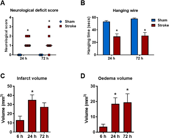
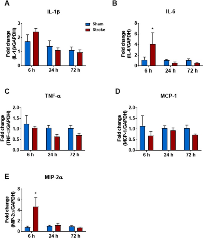
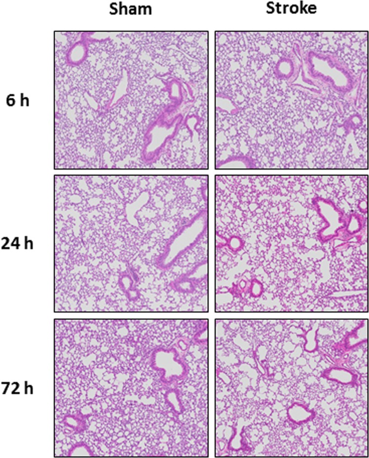
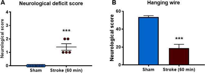
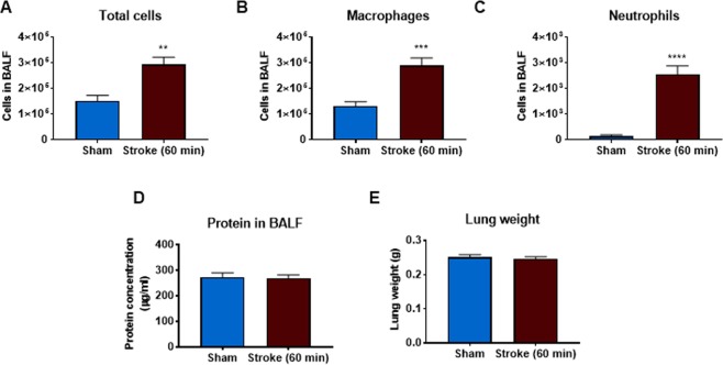
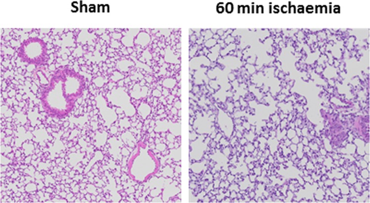
Stroke is a major cause of death worldwide and ischemic stroke is the most common subtype accounting for approximately 80% of all cases. Pulmonary complications occur in the first few days to weeks following ischemic stroke and are a major contributor to morbidity and mortality. Acute lung injury (ALI) occurs in up to 30% of patients with subarachnoid haemorrhage but the incidence of ALI after ischemic stroke is unclear. As ischemic stroke is the most common subtype of stroke, it is important to understand the development of ALI following the initial ischemic injury to the brain. Therefore, this study investigated whether focal ischemic stroke causes lung inflammation and ALI in mice. Ischemic stroke caused a significant increase in bronchoalveolar lavage fluid (BALF) macrophages and neutrophils and whole lung tissue proinflammatory IL-1β mRNA expression but this did not translate into histologically evident ALI. Thus, it appears that lung inflammation, but not ALI, occurs after experimental ischemic stroke in mice. This has significant implications for organ donors as the lungs from patient's dying of ischemic stroke are not severely damaged and could thus be used for transplantation in people awaiting this life-saving therapy.


Conflict of interest statement

The authors declare no competing interests.

Figures

Figure 1
Figure 1
Stroke outcomes at 6 h, 24 h and 72 h after 50 min ischaemia. Neurological deficit scores and hanging wire tests (A,B; n = 10–15), infarct and oedema volumes (C,D; n = 6–11). Data are expressed as mean ± SEM. Neurological deficit score data expressed as median (*P < 0.05 vs sham, Mann-Whitney test). Hanging wire test was analysed by two-way ANOVA (*P < 0.05 vs sham). Infarct and oedema data was analysed by one-way ANOVA followed by Sidak post-hoc test (*P < 0.05).
Figure 2
Figure 2
Effect of 50 min ischaemia on inflammatory cell counts in BALF, lung weight and BALF protein concentration. BALF cellularity is shown as (A) the total number of cells, (B) macrophages and (C) neutrophils (n = 10–15). Protein concentration in BALF (D) and lung weight (E) data are also shown (n = 9–13). Data are expressed as mean ± SEM. Two-way ANOVA followed by Sidak post-hoc test (*P < 0.05).
Figure 3
Figure 3
Effect of 50 min ischaemia on mRNA expression of proinflammatory cytokines and chemokines in whole lung tissue. Data are expressed as mean ± SEM. Two-way ANOVA followed by Sidak post-hoc test (*P < 0.05; n = 6–12).
Figure 4
Figure 4
Hematoxylin and eosin stained lung tissue section from mice 6, 24 and 72 h post stroke (50 min ischaemia) or sham surgery.
Figure 5
Figure 5
Stroke outcomes at 24 h after 60 min ischaemia. Neurological deficit scores (A) expressed as median (***P < 0.0005 vs sham, Mann-Whitney test; n = 5–11). Hanging wire test (B) expressed as mean ± SEM (***P < 0.0001 vs sham, Student’s t-test; n = 5–11).
Figure 6
Figure 6
Effect of 60 min ischaemia on inflammatory cell counts in BALF, lung weight and BALF protein concentration. BALF cellularity is shown as (A) the total number of cells, (B) macrophages and (C) neutrophils. Protein concentration in BALF (D) and lung weight (E) data are also shown (n = 5–10). Data are expressed as mean ± SEM. Student’s t-test was performed to assess statistical significance (*P < 0.05 vs sham, **P < 0.005 vs sham, ***P < 0.0005 vs sham).
Figure 7
Figure 7
Effect of 60 min ischaemia on mRNA expression of proinflammatory cytokines and chemokines in whole lung tissue. Data are expressed as mean ± SEM. Student’s t-test was performed to assess statistical significance (*P < 0.05; n = 5–10).
Figure 8
Figure 8
Hematoxylin and eosin stained lung tissue sections from mice 24 h post 60 min post stroke (60 min ischaemia) or sham surgery.

References

    1. Feigin, V., Norrving, B. & Mensah, G. Global Burden of Stroke. Vol. 120 (2017). - PubMed
    1. Krishnamurthi RV, et al. Stroke Incidence by Major Pathological Type and Ischemic Subtypes in the Auckland Regional Community Stroke Studies. Stroke. 2018;49:3. doi: 10.1161/STROKEAHA.117.019358. - DOI - PubMed
    1. Johnston KC, et al. Medical and Neurological Complications of Ischemic. Stroke. Stroke. 1998;29:447. doi: 10.1161/01.STR.29.2.447. - DOI - PubMed
    1. Silver FL, Norris JW, Lewis AJ, Hachinski VC. Early mortality following stroke: a prospective review. Stroke. 1984;15:492–496. doi: 10.1161/01.STR.15.3.492. - DOI - PubMed
    1. Kumar S, Selim MH, Caplan LR. Medical complications after stroke. The Lancet Neurol. 2010;9:105–118. doi: 10.1016/S1474-4422(09)70266-2. - DOI - PubMed

Publication types