. 2019 Mar;567(7748):394-398.
doi: 10.1038/s41586-019-1000-2. Epub 2019 Mar 6.

Structural basis of STING binding with and phosphorylation by TBK1

Affiliations

Structural basis of STING binding with and phosphorylation by TBK1

Conggang Zhang et al. Nature. 2019 Mar.

Abstract

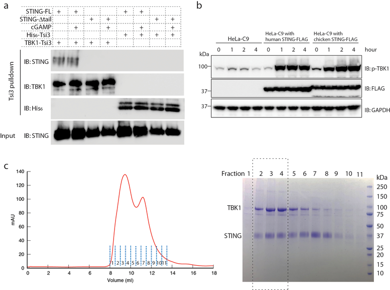
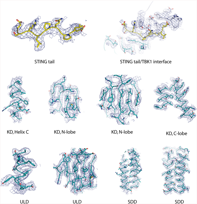
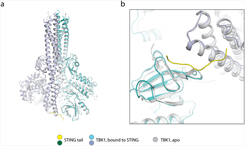
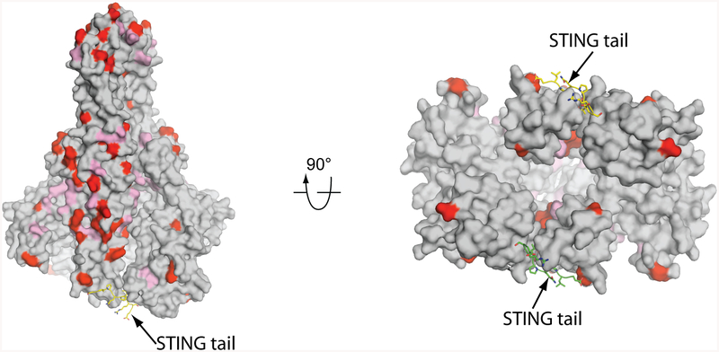
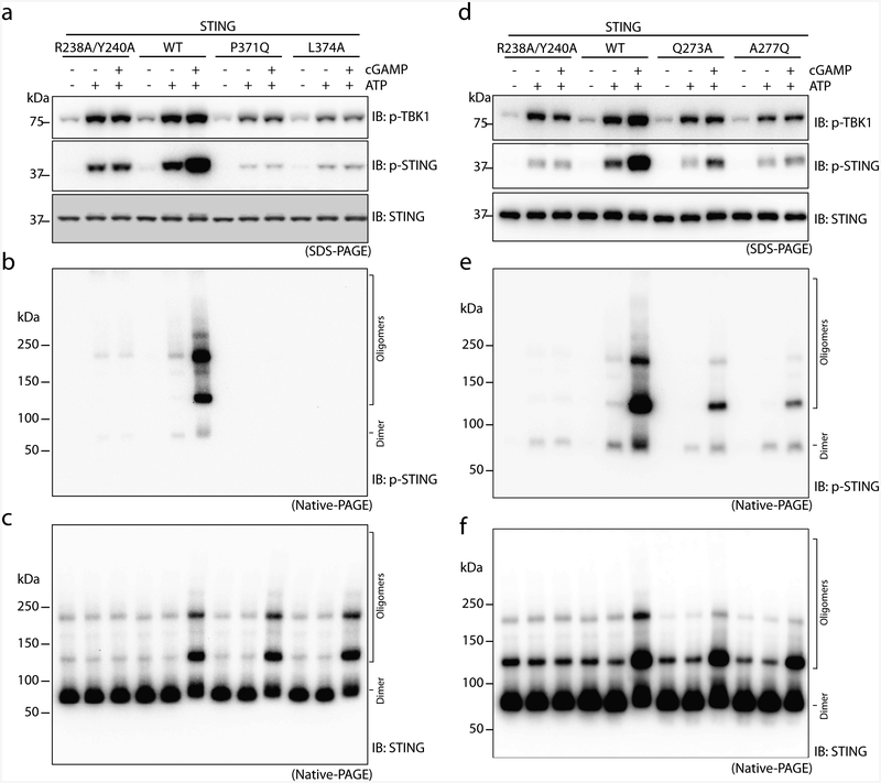
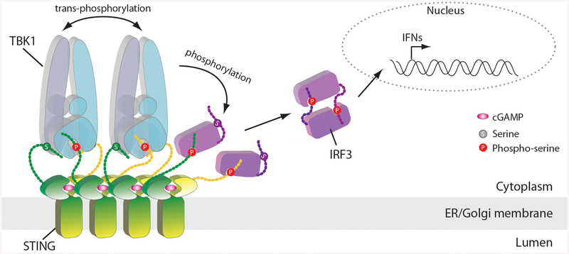
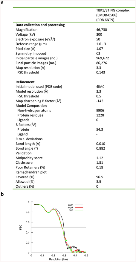
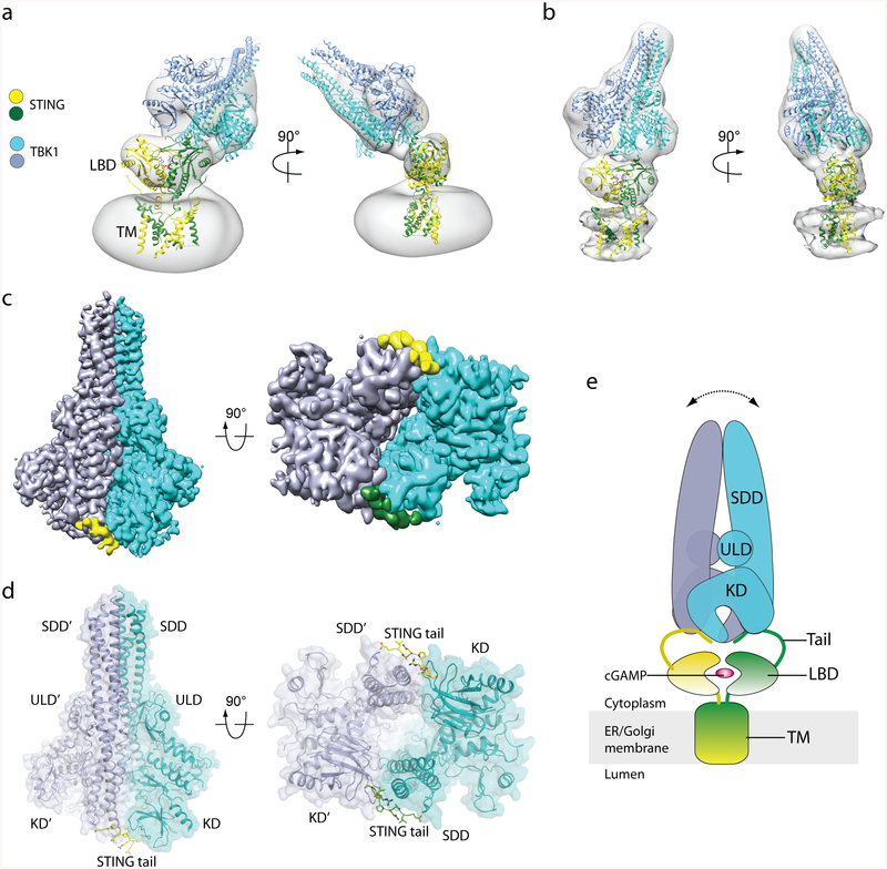
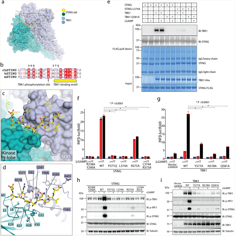
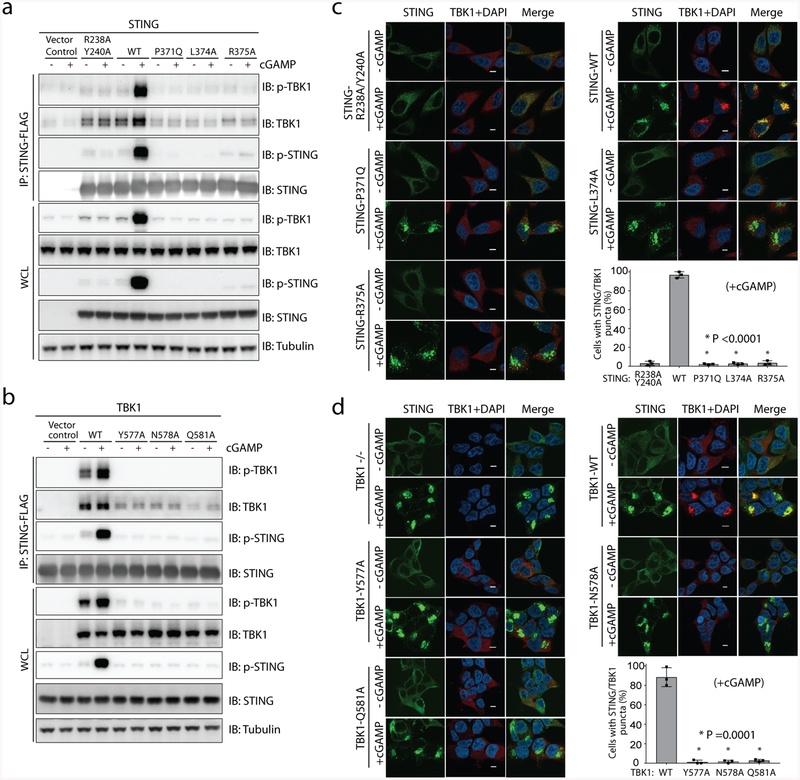
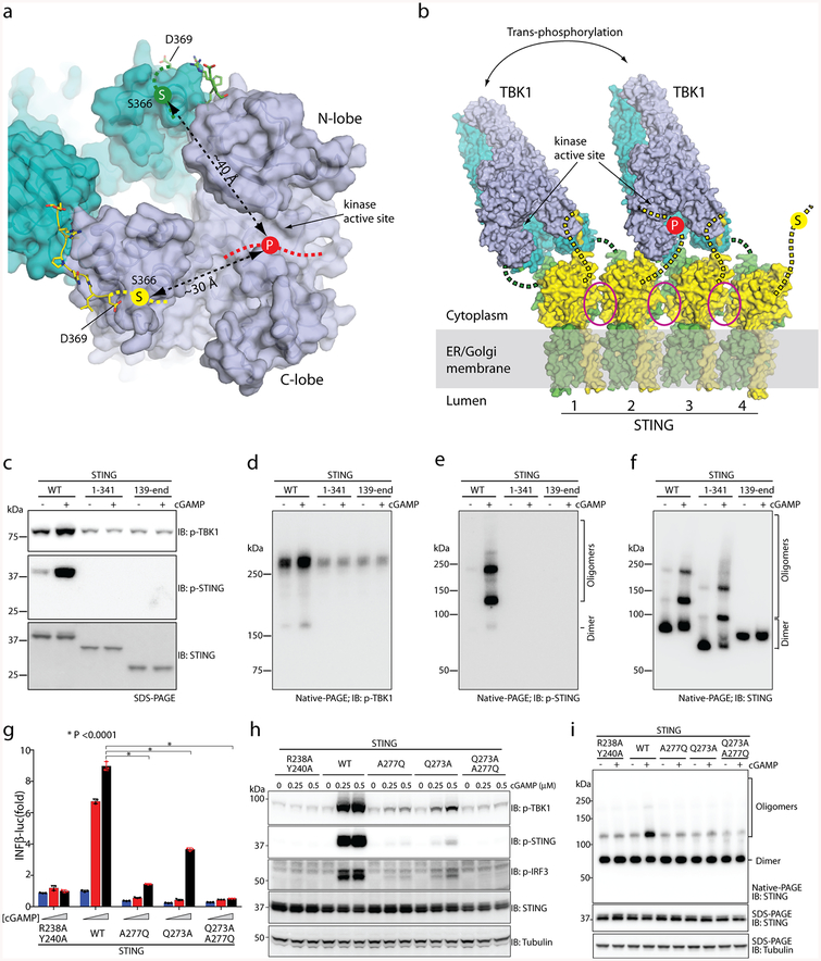
The invasion of mammalian cytoplasm by microbial DNA from infectious pathogens or by self DNA from the nucleus or mitochondria represents a danger signal that alerts the host immune system1. Cyclic GMP-AMP synthase (cGAS) is a sensor of cytoplasmic DNA that activates the type-I interferon pathway2. On binding to DNA, cGAS is activated to catalyse the synthesis of cyclic GMP-AMP (cGAMP) from GTP and ATP3. cGAMP functions as a second messenger that binds to and activates stimulator of interferon genes (STING)3-9. STING then recruits and activates tank-binding kinase 1 (TBK1), which phosphorylates STING and the transcription factor IRF3 to induce type-I interferons and other cytokines10,11. However, how cGAMP-bound STING activates TBK1 and IRF3 is not understood. Here we present the cryo-electron microscopy structure of human TBK1 in complex with cGAMP-bound, full-length chicken STING. The structure reveals that the C-terminal tail of STING adopts a β-strand-like conformation and inserts into a groove between the kinase domain of one TBK1 subunit and the scaffold and dimerization domain of the second subunit in the TBK1 dimer. In this binding mode, the phosphorylation site Ser366 in the STING tail cannot reach the kinase-domain active site of bound TBK1, which suggests that STING phosphorylation by TBK1 requires the oligomerization of both proteins. Mutational analyses validate the interaction mode between TBK1 and STING and support a model in which high-order oligomerization of STING and TBK1, induced by cGAMP, leads to STING phosphorylation by TBK1.


Conflict of interest statement

Competing interests: The authors declare no competing interests.

Figures

Extended Data Fig. 1 |
Extended Data Fig. 1 |. Purification of STING and TBK1, and characterization of their interaction.
a, Binding between purified human STING and TBK1. Tsi3-tagged TBK1 was captured by Tse3-conjugated beads. Pull down of STING by TBK1 was assessed by western blot. STING-Δtail, STING(1–343). b, Both human and chicken STING are able to induce phosphorylation of human TBK1 in cells. HeLa-C9 cells with undetectable endogenous STING were used to generate cell lines that stably express human STING–Flag or chicken STING–Flag. Cells were stimulated with cGAMP (1 μM) and analysed for TBK1 phosphorylation by immunoblotting. c, Gel filtration chromatography of the hybrid complex between chicken STING and human TBK1. Data are representative of two independent experiments.
Extended Data Figure 2 |
Extended Data Figure 2 |. Flow chart of cryo-EM image processing for the complex between chicken STING and human TBK1.
a, Representative micrograph. b, Representative 2D classes of the intact complex. c, Representative 2D classes from TBK1-focused image processing. n > 3. d, f, Final reconstructions of the intact STING–TBK1 complex (d) and from the TBK1-focused refinement (f), with colours based on local resolution. e, g, Gold-standard FSC curves of the final 3D reconstructions of the intact complex and from the TBK1-focused refinement. h, Image processing procedure.
Extended Data Fig. 3 |
Extended Data Fig. 3 |. Sample density maps.
Sample density maps are shown for the C-terminal tail of chicken STING and various parts of human TBK1.
Extended Data Fig. 4 |
Extended Data Fig. 4 |. Structural comparison of apo TBK1 and TBK1 bound to STING.
The structure of apo TBK1 is from PDB code 4IM0.
Extended Data Fig. 5 |
Extended Data Fig. 5 |. Sequence conservation of TBK1 from human and chicken.
Residues that are identical in the TBK1 of both species are coloured grey. Non-conserved residues are coloured red; non-identical, but similar, residues are coloured pink.
Extended Data Fig. 6 |
Extended Data Fig. 6 |. The binding and phosphorylation of STING by TBK1 relies on the interface between TBK1 and the STING C-terminal tail, and on the oligomerization of STING.
a, Mutations of TBK1-binding residues in the STING tail diminish cGAMP-induced phosphorylation of both TBK1 and STING. The S1 post-nuclear supernatant from HEK293T cells that express either the STING wild type or mutants was incubated with ATP in the presence or absence of cGAMP, and subjected to immunoblotting analyses for pTBK1, pSTING and STING. b, c, Mutations of TBK1-binding residues in the STING tail diminish cGAMP-induced STING phosphorylation (b) but not STING oligomerization (c). The same samples as in a were resolved by native gels, and analysed by immunoblotting. df, Mutations at the oligomerization interface of STING reduce cGAMP-induced oligomerization of STING, as well as phosphorylation of TBK1 and STING. The mutants are based on the accompanying paper on the structures of full-length STING. The analyses in d, e and f were conducted in the same manner as in a, b and c, respectively. Data shown here are representative of at least three independent biological replicates.
Extended Data Fig. 7 |
Extended Data Fig. 7 |. Cartoon model of STING-mediated activation of TBK1 and the downstream signalling pathway.
The cGAMP-induced oligomerization of STING leads to TBK1 clustering and trans-autophosphorylation. Activated TBK1 phosphorylates STING C-terminal tails that are not bound to the SDD–kinase domain groove in TBK1. Phosphorylated tails of STING recruit IRF3, which is phosphorylated by TBK1. Phosphorylated IRF3 forms a dimer and translocates to the nucleus to initiate the transcription of IFN genes.
Extended Data Fig. 8 |
Extended Data Fig. 8 |. Data collection and model statistics.
a, Data collection and model refinement statistics. b, FSC curves between the maps and model.
Fig. 1 |
Fig. 1 |. Structure of the complex of chicken STING and human TBK1.
a, b, Three-dimensional reconstructions of two 3D classes of the complex. The atomic models of dimeric full-length chicken STING bound to cGAMP (from ref.12) and the human TBK1 dimer (RCSB Protein Data Bank (PDB) code 4IM0) were fit into the maps (grey) through rigid-body docking. The extra density that surrounds the transmembrane (TM) domain of STING in a is from the detergent micelle. The protein density in b is stronger and therefore shown at a higher threshold, which led to partial cut-off of the detergent micelle density. LBD, ligand-binding domain. c, High-resolution 3D reconstruction from focused refinement on TBK1 with C2 symmetry. The densities for the two protomers of the TBK1 dimer are coloured either cyan or blue. The densities for the two STING tails are coloured either yellow or green. d, Atomic model of TBK1 bound to the C-terminal tail of STING. The colour scheme is the same as that of the atomic models in a. KD, kinase domain; ULD, ubiquitin-like domain. e, Cartoon model of the STING–TBK1 complex. The double-headed arrow indicates the wobble between TBK1 and STING, owing to flexibility in the STING tail. ER, endoplasmic reticulum.
Fig. 2 |
Fig. 2 |. Binding interface between human TBK1 and the chicken STING C-terminal tail.
a, Overview of the binding interface. The rectangle denotes the region shown in detail in c and d. b, Sequence alignment of the C-terminal tail of STING from chicken, human and mouse (denote by ch-, h- and m-prefixes, respectively). c, d, Detailed views of the binding interface. Potential hydrogen bonds are indicated with dotted lines. Numbers are inter-atom distances. N-lobe, N-terminal lobe. e, Mutants of interface residues in either STING or TBK1 abolish the STING/TBK1 interaction in vitro. Data are representative of two independent biological experiments. f, Mutants of interface residues in human STING abolish cGAMP-stimulated IFNβ expression. HEK293T cells that stably express IFNβ–luciferase (IFNβ–luc) were transfected with the STING–Flag wild type (WT) or mutants. The R238A/Y240A mutant (which does not bind cGAMP) served as a negative control. Cells were stimulated with increasing concentrations of cGAMP (0, 0.3 and 1.4 μM). Data are mean ± s.d. n = 3. g, Mutants of interface residues in human TBK1 abolish cGAMP-stimulated IFNβ expression. HEK293T TBK1-null cells that stably express human STING–Flag were reconstituted with the TBK1 wild type or mutants, using lentiviral infection. Cells were transiently transfected with the IFNβ–luciferase reporter and stimulated with increasing concentrations of cGAMP (0, 0.3 and 1.4 μM). Luciferase activity was determined 24 h after transfection. Data are mean ± s.d. h, i, Mutants of interface residues in either STING (h) or TBK1 (i) abolish cGAMP-stimulated phosphorylation (p) of TBK1, STING and IRF3. Cells used in h and i are similar to those in f and g, respectively, except the cells in h and i do not have the IFNβ–luciferase reporter. Cells were treated with cGAMP (1 μM) for 3 h, and subjected to immunoblotting analyses. Data in fi are representative of three independent biological replicates.
Fig. 3 |
Fig. 3 |. The interface between TBK1 and the STING C-terminal tail is required for TBK1 binding and colocalization with STING in cells.
a, b, Mutations of interface residues in either STING (a) or TBK1 (b) abolish cGAMP-induced interaction between STING and TBK1, as well as the phosphorylation of both proteins, as shown by immunoblotting. Cells used in a, b are the same as those used in Fig. 2h, i, respectively. After cGAMP stimulation, immunoprecipitation was carried out to examine the interaction between STING and TBK1. Data are representative of three independent biological replicates. WCL, whole-cell lysate. c, Mutants of interface residues in STING diminish colocalization of TBK1 with cGAMP-induced puncta of STING in cells. HeLa cells deficient in cyclic GMP–AMP synthase and expressing the wild type or mutant constructs of STING were stimulated with cGAMP (1 μM) for 1 h, and then subjected to immunostaining for STING (green, 488 nm), TBK1 (red, 568 nm) and DAPI. Representative confocal images of cells with or without cGAMP stimulation are shown. Scale bars, 5 μm. d, Mutants of interface residues in TBK1 diminish colocalization of TBK1 with cGAMP-induced puncta of STING in cells. HEK293T cell lines that stably express wild-type or mutant TBK1 (as in b) were stimulated with cGAMP (1 μM) for 1 h and subjected to immunostaining (as in c). Scale bars, 5 μm. In both c, d, the percentage of cells with STING and TBK1 colocalization was quantified from at least 150 cells for each group. Data are mean ± s.d. and representative of three independent biological replicates.
Fig. 4 |
Fig. 4 |. TBK1 activation and STING phosphorylation depend on STING oligomerization.
a, The STING C-terminal tail bound to TBK1 cannot reach the kinase active site for phosphorylation of Ser366 (‘S’ in yellow circle). ‘P’ on the red dotted line indicates the location of the TBK1 kinase active site, at which phosphorylation occurs. b, Model of TBK1 activation and STING phosphorylation upon STING oligomerization on the endoplasmic reticulum–Golgi membrane. The C-terminal tails in STING dimers 1 and 3 are bound to TBK1, whereas the tails in STING dimers 2 and 4 could enter the active site of TBK1 for phosphorylation. The two TBK1 dimers could activate one another by trans-autophosphorylation. Magenta ovals highlight the oligomer interface mediated by the STING ligand-binding domain, which in human STING involves Gln273 and Ala277. c, Phosphorylation of both TBK1 and STING requires the C-terminal tail and transmembrane region of STING. The S1 post-nuclear supernatant from HEK293T cells that express either the STING wild type or mutants was stimulated with cGAMP in the presence of ATP, resolved by sodium dodecyl sulfate polyacrylamide gel electrophoresis (SDS–PAGE), and subjected to immunoblotting analyses for pTBK1, pSTING and STING. df, Effects of the STING truncations on the phosphorylation and oligomerization of TBK1 and STING. The same samples as in c were resolved by native PAGE and subjected to immunoblotting to assess both oligomerization and phosphorylation of STING. g, Effects of mutations in the tetramer interface of human STING on cGAMP-stimulated IFNβ expression. The assays were conducted as in Fig. 2f. Data are mean ± s.d. and representative of three independent biological replicates. h, Effects of STING mutations on phosphorylation of STING, TBK1 and IRF3. HeLa cells that stably express either STING wild type or mutants were stimulated with cGAMP for 2 h and subjected to immunoblot analyses. i, Effects of the mutations in the tetramer interface on cGAMP-induced STING oligomerization. Lysates from cells stimulated with cGAMP (1 μM) for 1 h were resolved by native PAGE or SDS–PAGE, followed by immunoblotting. The immunoblotting results are representative of three independent biological replicates.

Comment in

References

    1. Li T & Chen ZJ The cGAS–cGAMP–STING pathway connects DNA damage to inflammation, senescence, and cancer. J. Exp. Med 215, 1287–1299 (2018). - PMC - PubMed
    1. Sun L, Wu J, Du F, Chen X & Chen ZJ Cyclic GMP–AMP synthase is a cytosolic DNA sensor that activates the type I interferon pathway. Science 339, 786–791 (2013). - PMC - PubMed
    1. Wu J et al. Cyclic GMP–AMP is an endogenous second messenger in innate immune signaling by cytosolic DNA. Science 339, 826–830 (2013). - PMC - PubMed
    1. Ishikawa H & Barber GN STING is an endoplasmic reticulum adaptor that facilitates innate immune signalling. Nature 455, 674–678 (2008). - PMC - PubMed
    1. Zhong B et al. The adaptor protein MITA links virus-sensing receptors to IRF3 transcription factor activation. Immunity 29, 538–550 (2008). - PubMed

Publication types

MeSH terms

Substances