. 2019 Mar;567(7747):262-266.
doi: 10.1038/s41586-019-1006-9. Epub 2019 Mar 6.

Autophagy induction via STING trafficking is a primordial function of the cGAS pathway

Affiliations

Autophagy induction via STING trafficking is a primordial function of the cGAS pathway

Xiang Gui et al. Nature. 2019 Mar.

Abstract

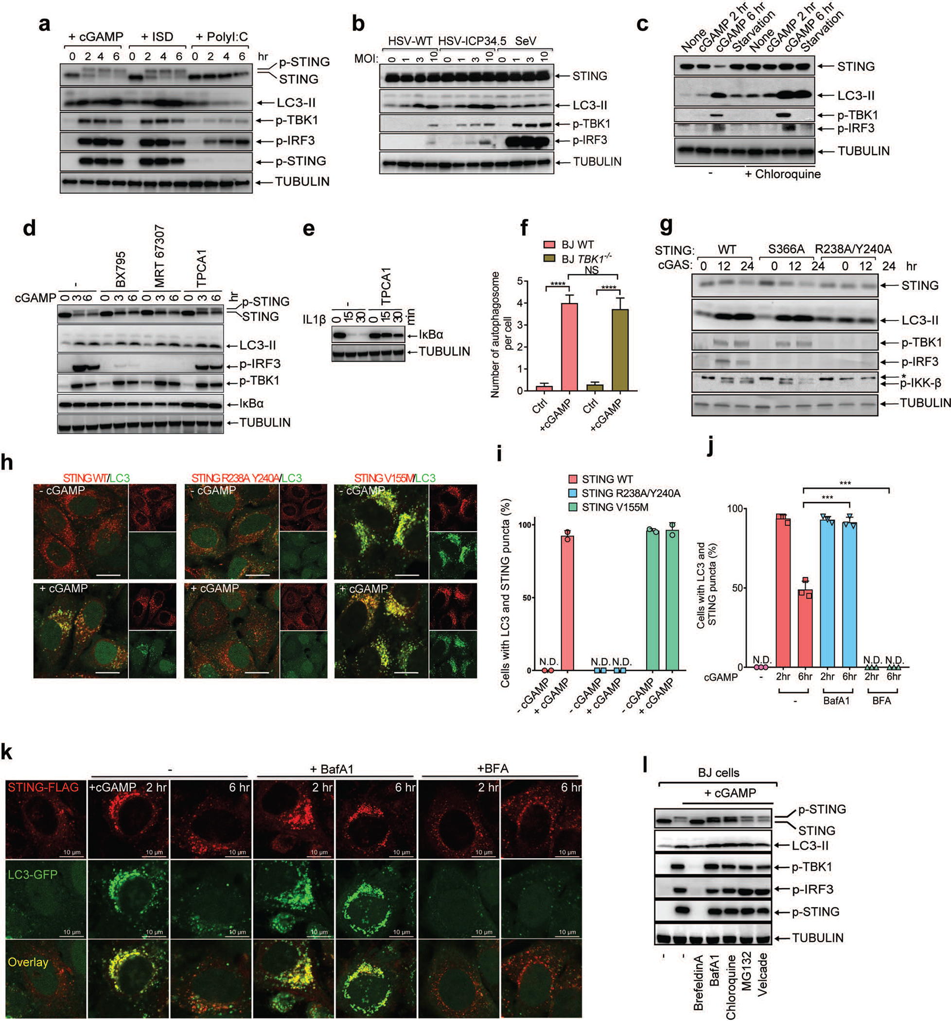
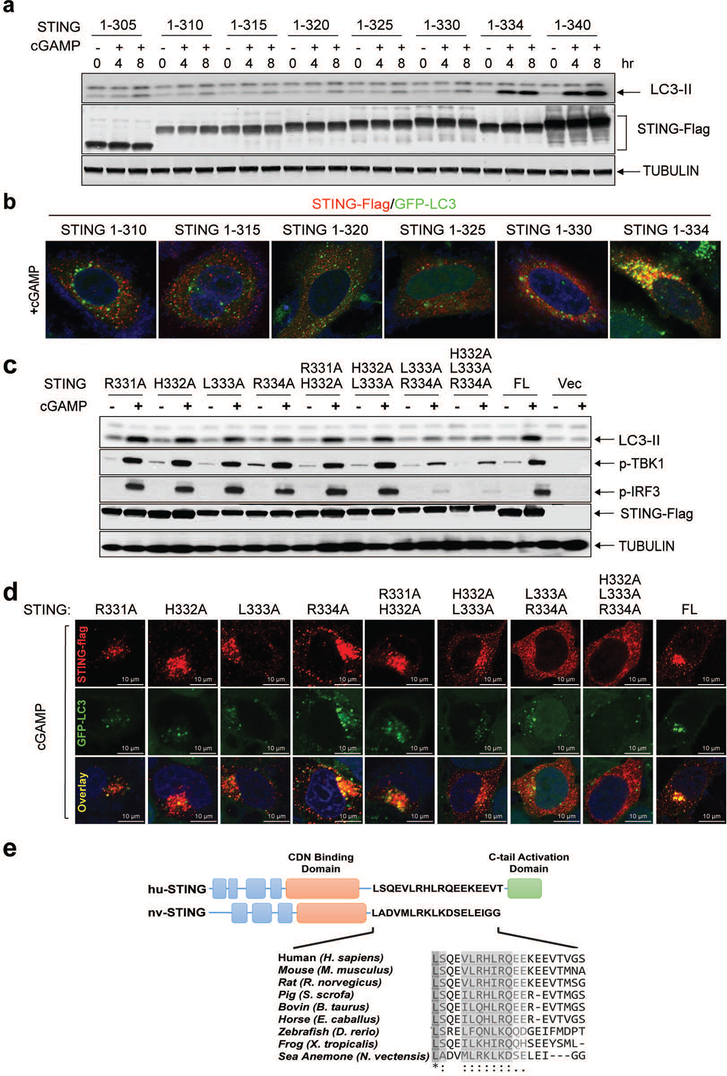
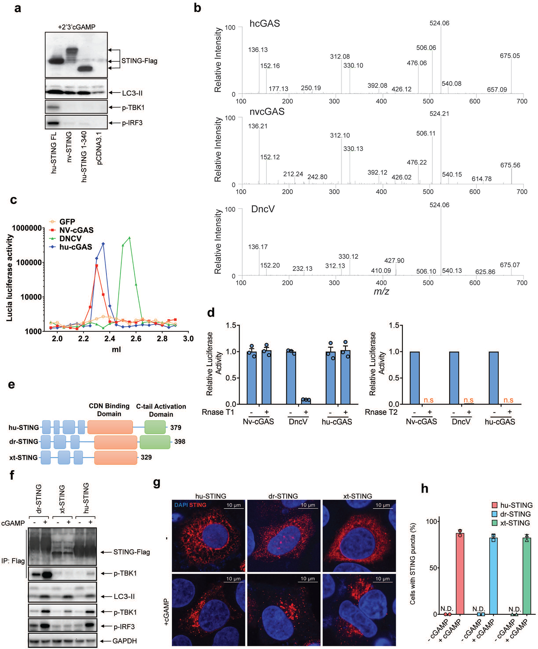
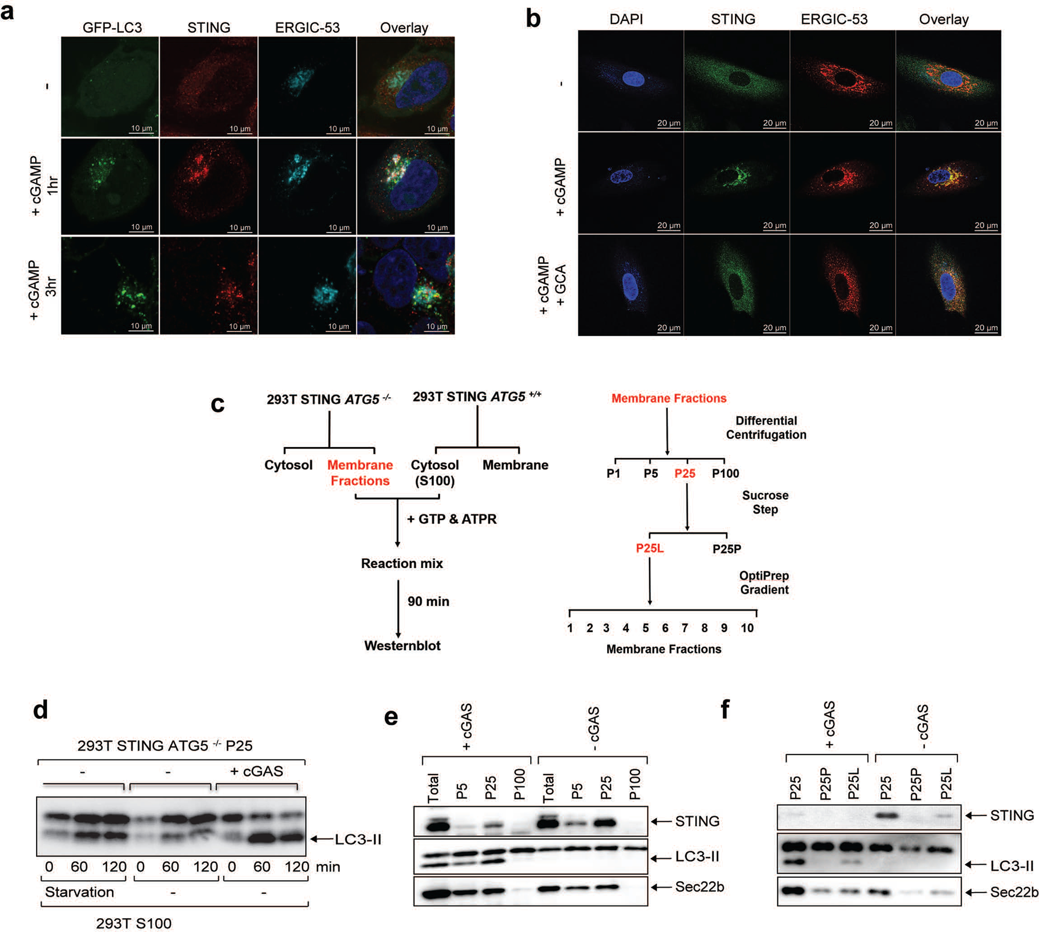
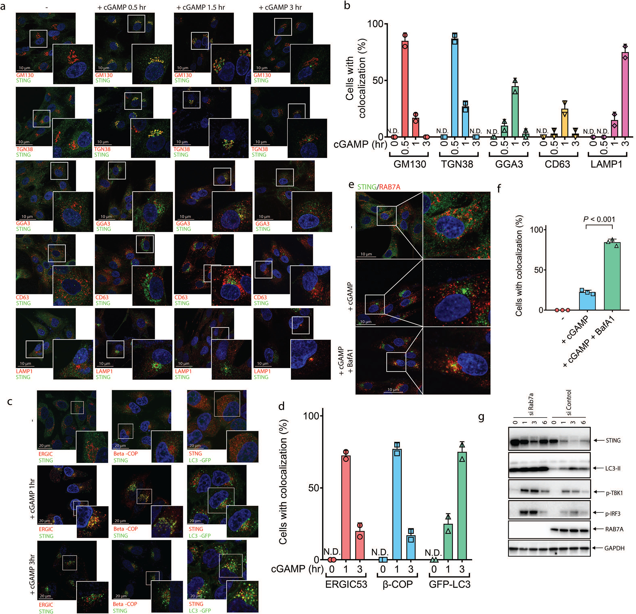
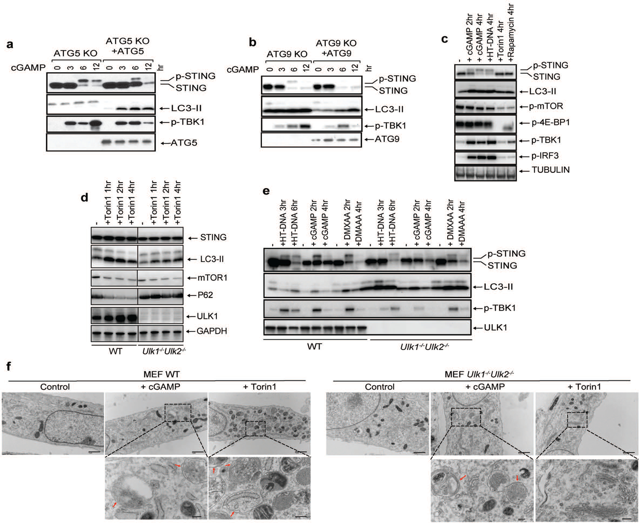
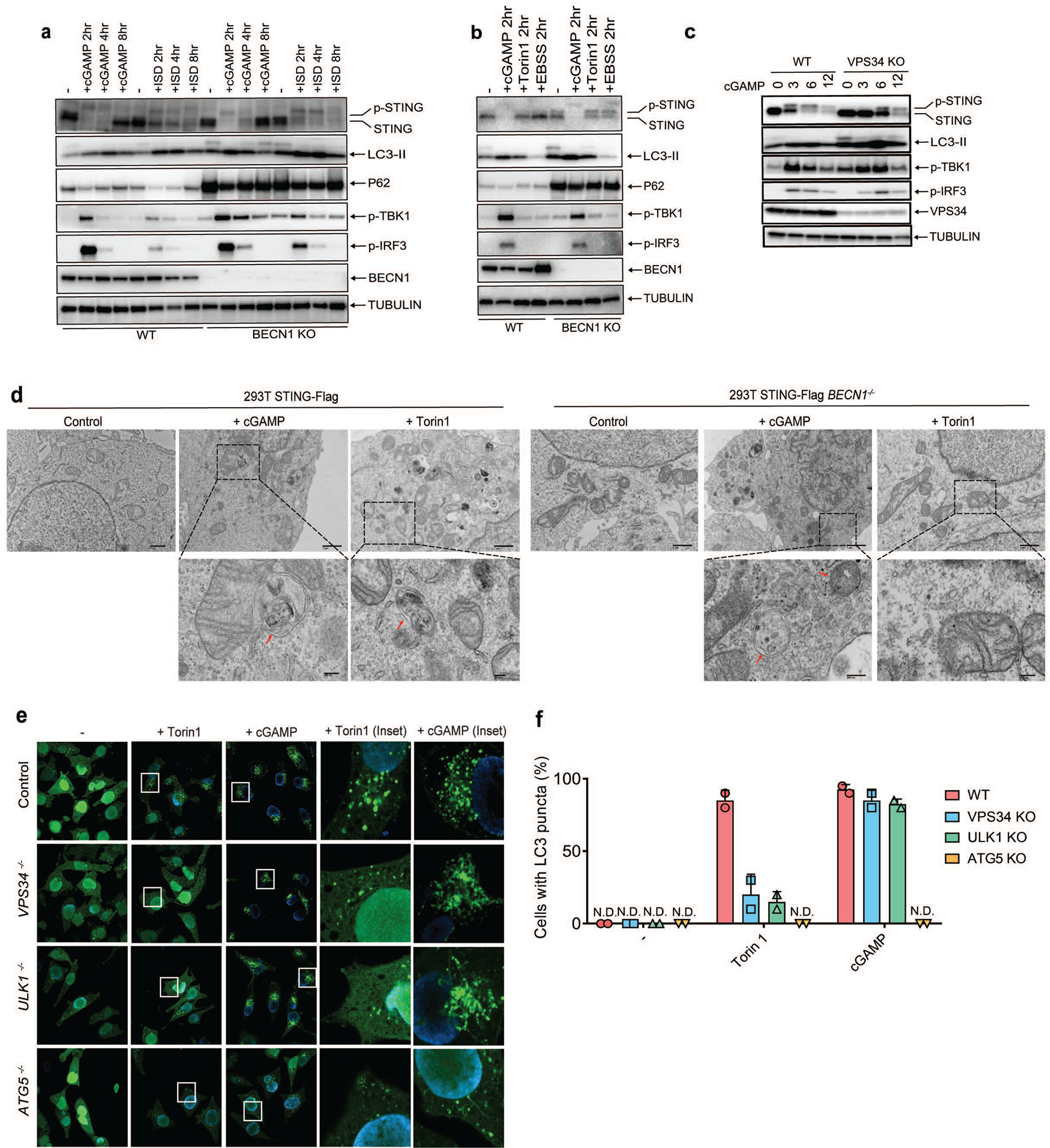
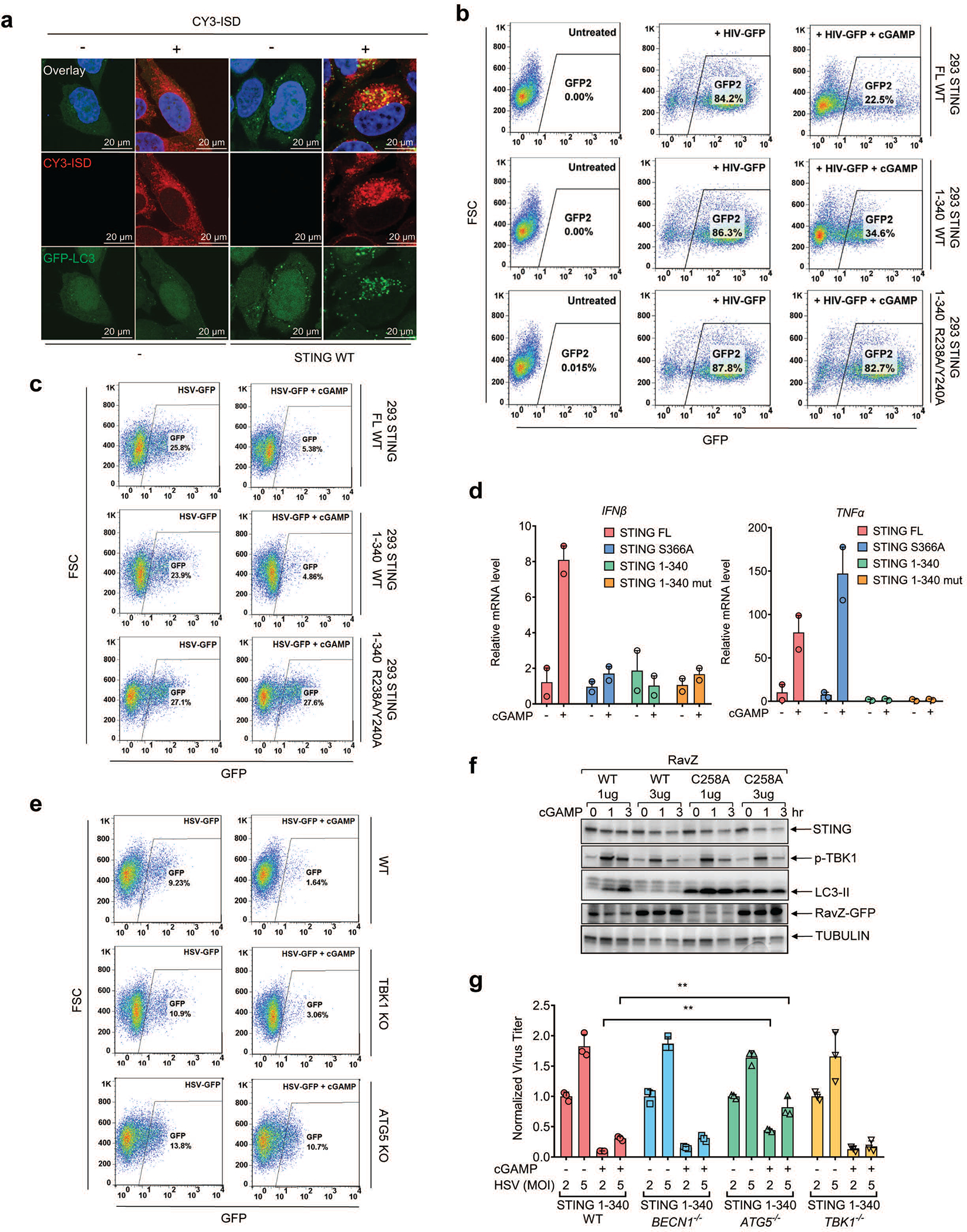
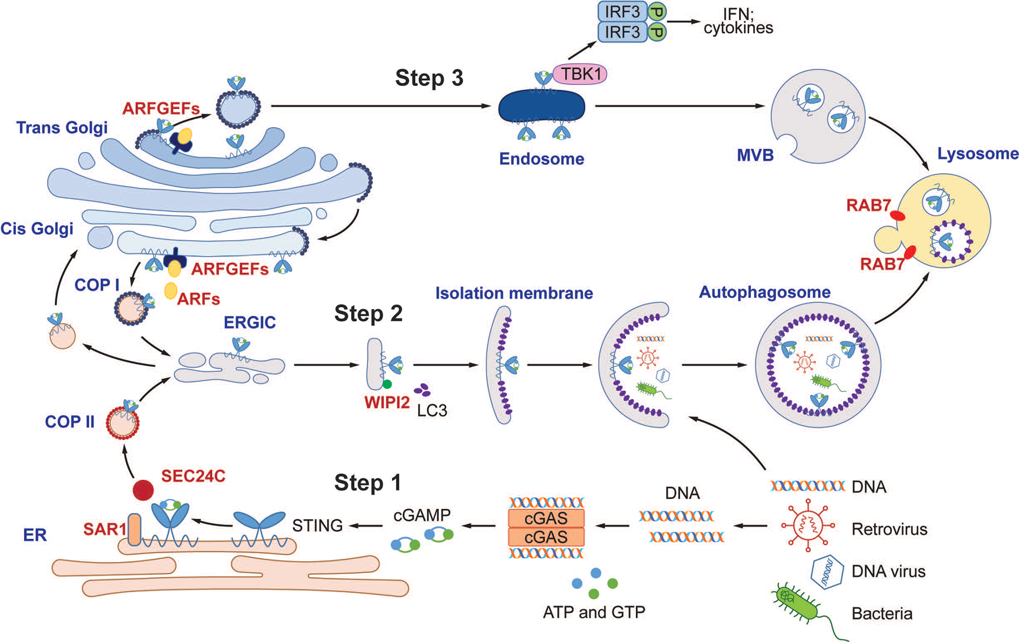
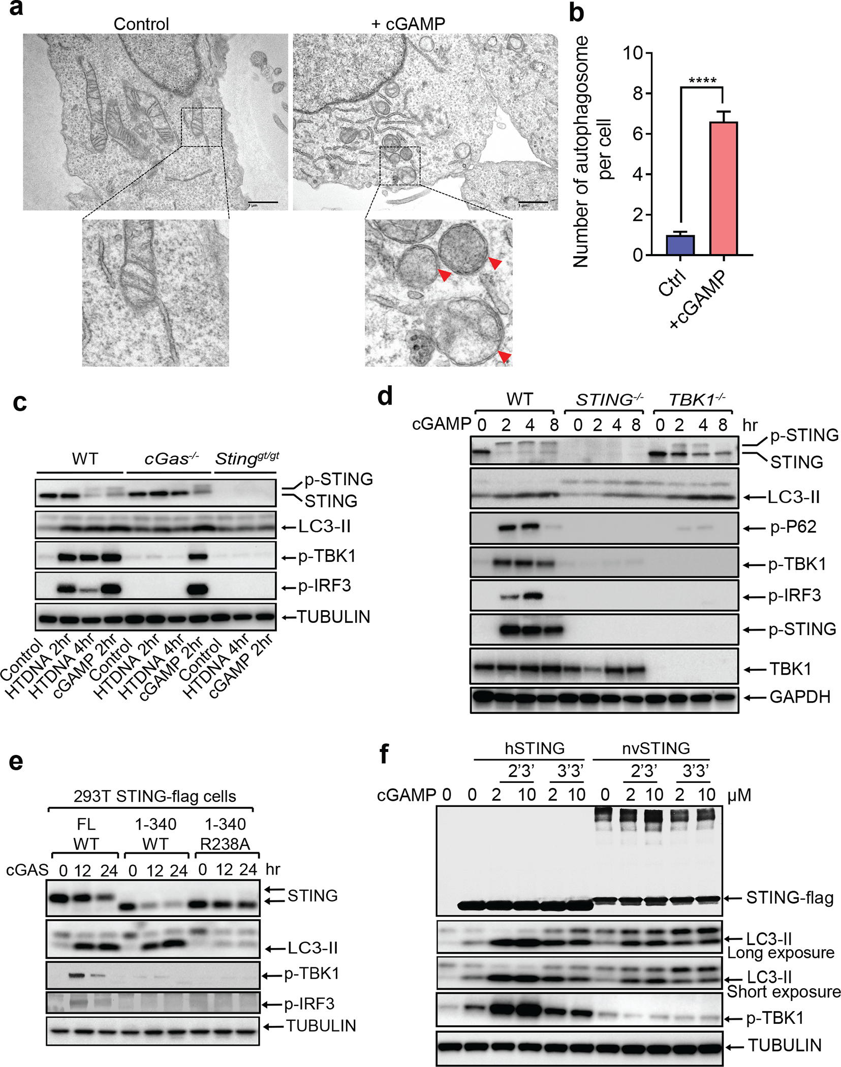
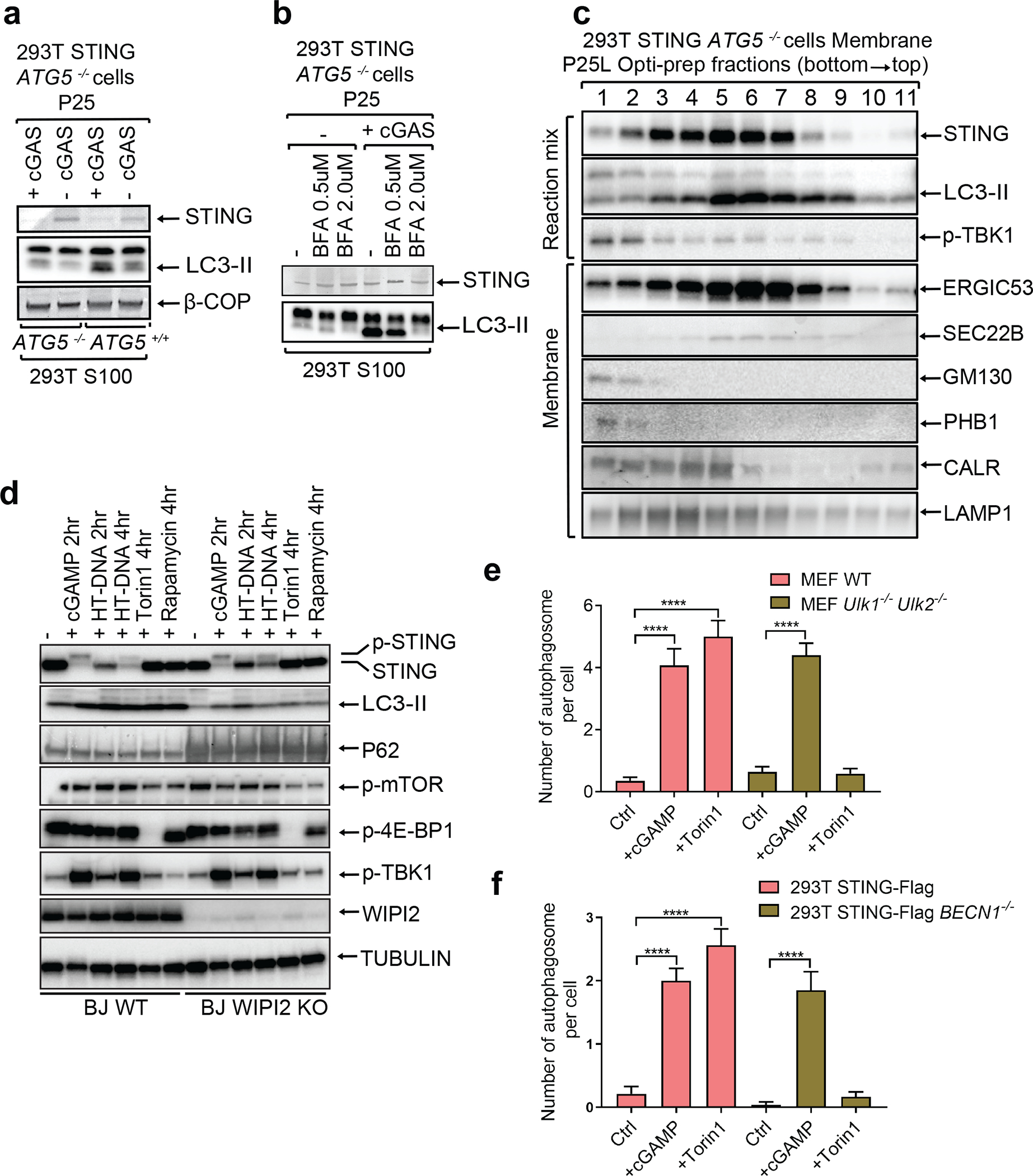
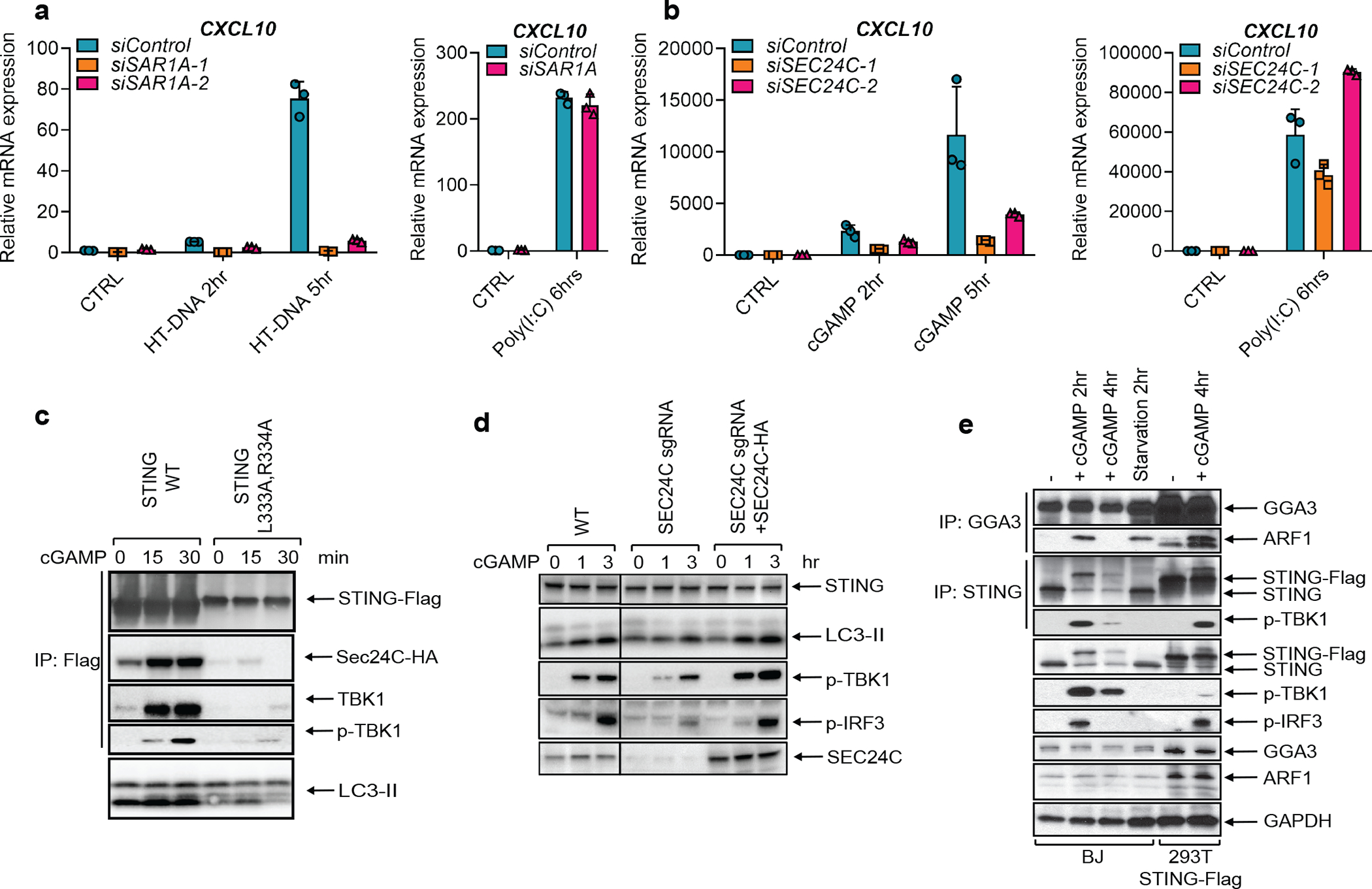
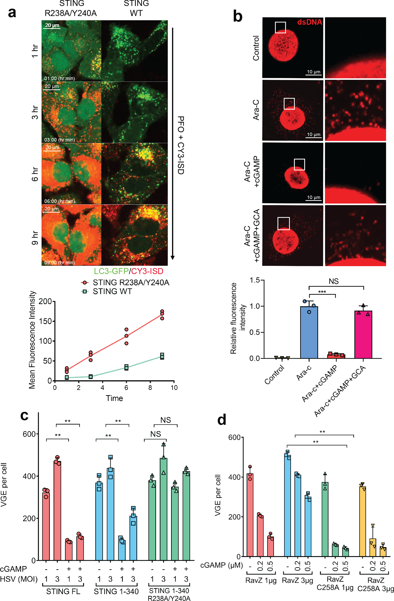
Cyclic GMP-AMP (cGAMP) synthase (cGAS) detects infections or tissue damage by binding to microbial or self DNA in the cytoplasm1. Upon binding DNA, cGAS produces cGAMP that binds to and activates the adaptor protein STING, which then activates the kinases IKK and TBK1 to induce interferons and other cytokines2-6. Here we report that STING also activates autophagy through a mechanism that is independent of TBK1 activation and interferon induction. Upon binding cGAMP, STING translocates to the endoplasmic reticulum-Golgi intermediate compartment (ERGIC) and the Golgi in a process that is dependent on the COP-II complex and ARF GTPases. STING-containing ERGIC serves as a membrane source for LC3 lipidation, which is a key step in autophagosome biogenesis. cGAMP induced LC3 lipidation through a pathway that is dependent on WIPI2 and ATG5 but independent of the ULK and VPS34-beclin kinase complexes. Furthermore, we show that cGAMP-induced autophagy is important for the clearance of DNA and viruses in the cytosol. Interestingly, STING from the sea anemone Nematostella vectensis induces autophagy but not interferons in response to stimulation by cGAMP, which suggests that induction of autophagy is a primordial function of the cGAS-STING pathway.


Conflict of interest statement

The authors declare no competing financial interests.

Figures

Extended Data Figure 1.
Extended Data Figure 1.. cGAMP-induced LC3 lipidation requires vesicle trafficking but not TBK1 or IKK.
a, DNA and cGAMP but not RNA trigger LC3 lipidation. BJ cells were stimulated with cGAMP by digitonin permeabilization or transfected with ISD or poly (I:C). Cell lysates were analyzed by immunoblotting with the indicated antibodies. b, DNA virus but not RNA virus induces LC3 conversion. BJ cells were infected with wild type (WT) HSV-1, HSV-1 ΔICP34.5, or Sendai virus (SeV) at the indicated multiplicity of infection (MOI) for 6 hr followed by immunoblotting. c, cGAMP induces STING degradation in the lysosome. HeLa cells stably expressing STING-Flag were treated with cGAMP or starved in the presence or absence of chloroquine, followed by immunoblotting. d, Inhibition of TBK1 or IKK does not impair LC3 lipidation. Inhibitors of TBK1 (BX-795 or MRT 67307) or IKK (TPCA1) were incubated with BJ cells before stimulation of the cells with cGAMP. Cell lysates were analyzed by immunoblotting. e, Control experiment showing that TPCA1 inhibits IκBα degradation (by inhibiting IKK). f, Quantification of double-membrane autophagosomes in BJ WT and BJ TBK1−/− cells. The cells were stimulated with cGAMP as indicated. Quantification was shown as the number of double-membrane autophagosomes per cell by counting on BJ cells (n=13, 12, 17, 11). Mean ±SEM are shown. **** p < 0.0001 (two-tailed Student’s t-test); NS, not significant (significance level, α = 0.01). g, STING S366 phosphorylation by TBK1 is essential for IRF3 phosphorylation but not LC3 conversion. HEK293T cells stably expressing WT or mutant STING (S366A or R238/Y240A) were transfected with a cGAS expression plasmid followed by immunoblotting. h, Hela cells stably expressing GFP-LC3 and different STING mutants (R238A Y240A or V155M) were stimulated with cGAMP followed by confocal immunofluorescence microscopy. i, Quantification of the cells with colocalization of LC3 and STING puncta. The percentage of cells with colocalized LC3 and STING was quantified from 100 cells (n = 2). N.D., not detectable. j&k, HeLa cells stably expressing GFP-LC3 and STING-Flag were treated with BafA1 or BFA followed by stimulation with cGAMP and confocal immunofluorescence microscopy. Quantification of the cells with colocalization of LC3 and STING puncta. The percentage of cells with colocalized LC3 and STING was quantified from 100 cells (n = 3, mean ±SD, two-tailed Student’s t-test). N.D., not detectable. l, BJ cells were treated with brefeldin A (BFA), lysosome inhibitors (bafilomycin A1 [BafA1] or chloroquine), or proteasome inhibitors (MG132 or Velcade) before stimulation with cGAMP. Cell lysates were analyzed by immunoblotting.
Extended Data Figure 2.
Extended Data Figure 2.. Delineation of the STING region (residues 330–334) required for LC3 lipidation.
a, Expression plasmids encoding truncated STING mutants were transiently transfected into HEK293T cells for 24 hr, followed by stimulation with cGAMP for 4 hr. Cell lysates were analyzed by immunoblotting. b, Expression plasmids of indicated STING truncation mutants were transfected into HeLa GFP-LC3 cells for 24 hr. The cells were stimulated with cGAMP followed by immunostaining and fluorescence microscopy. c&d, L333 and R334 of STING are important for LC3 lipidation and TBK1 activation. c, Expression plasmids encoding full-length STING harboring the indicated mutations were transiently transfected into HEK293T cells, followed by stimulation with cGAMP. Cell lysates were analyzed by immunoblotting. FL: full-length. d, Indicated STING mutants were transfected and stimulated as described in c followed by immunostaining and fluorescence microscopy. e, Schematic of functional domains and residues of human (huSTING) and sea anemone STING (nvSTING), highlighting the evolutionary conservation of the CDN binding domain but not the C-terminal activation domain.
Extended Data Figure 3.
Extended Data Figure 3.. STING-induced LC3 conversion is a primordial function of the cGAS-STING pathway.
a, Sea anemone STING (nvSTING) induces LC3 conversion but not TBK1 activation in response to 2’3’-cGAMP. HEK293T cells were transfected with expression plasmids encoding full-length (FL) human STING (huSTING), huSTING (1–340), or nvSTING for 24 hr and then treated with 2’3’-cGAMP for 3 hr. Cell lysates were analyzed by immunoblotting. b-d, Sea anemone cGAS (nvcGAS) produces 2’3’-cGAMP. b, Expression vectors encoding human cGAS (hucGAS), nvcGAS, or DncV were transfected into HEK293T cells for 24 hr. Small molecules were extracted from cells for analysis of cGAMP isomers by tandem mass spectrometry. Mass spectra of cGAMP from nvcGAS-expressing cells match those from hucGAS-expressing cells but not those from DncV-expressing cells. c, Similar to b except that the small molecule extracts were fractionated using a monoQ column, and each fraction was delivered into THP1-ISG luciferase reporter cells for measuring the CDN activity. d, CDNs produced by hucGAS and nvcGAS, but not that by DncV, is resistant to digestion by RNase T1. Small molecule extracts from HEK293T cells expressing the indicated CDN synthases were treated with RNase T1 or RNaseT2 or left untreated before delivery into THP1-ISG luciferase reporter cells to measure the CDN activity. Mean±SEM was shown (n=3) in the group treated with RNase T1. e, Domain organization of STING from human (hu), Danio rerio (dr), and Xenopus tropicalis (xt). f, Xenopus STING stimulates LC3 lipidation but not IRF3 phosphorylation. HEK293T cells were transfected with expression plasmids for STING-Flag from human, Danio, or Xenopus for 24 hr and then stimulated with cGAMP for 1 hr. A Flag antibody was used to immunoprecipitate STING from cell lysates followed by immunoblotting with the indicated antibodies. g, cGAMP stimulates formation of perinuclear puncta of STING from different species. Hela cells transiently expressing human, Danio, or Xenopus STING-Flag were stimulated with cGAMP for 3 hr. Cells were immunostained with a Flag antibody followed by fluorescence microscopy. h, Quantification of the percentage of cells with STING peri-nuclear foci formation. The percentage of cells with STING foci formation was quantified from 100 cells (n = 2). All results in this figure are representative of at least two independent experiments. N.D., not detectable.
Extended Data Figure 4.
Extended Data Figure 4.. cGAMP stimulates STING translocation to ERGIC vesicles that promote LC3 lipidation.
a, STING co-localizes with the ERGIC and autophagosomes in response to cGAMP stimulation. HeLa cells stably expressing GFP-LC3 and STING-Flag were stimulated with cGAMP for the indicated time followed by immunofluorescence microscopy. b, STING trafficking to the ERGIC is blocked by GCA. BJ cells were stimulated with cGAMP for 3 hr in the presence or absence of GCA. Cells were stained with DAPI or the indicated antibodies and examined by confocal microscopy. c, Procedures for in vitro reconstitution of cGAMP-induced LC3 lipidation and membrane fractionation. d, ATG5−/− 293T cells stably expressing STING-Flag were transfected with a cGAS expression plasmid or an empty vector for 24 hr. Membrane pelleted at 25,000 g (P25) from these cells was incubated with cytosolic extracts (S100) from starved or untreated 293T cells in the presence of GTP and an ATP regenerating (ATPR) system. After incubation at 30oC for 90 min, the reaction mixtures were analyzed by immunoblotting. e, Similar to c except that different organelle membranes enriched by differential centrifugation were prepared and incubated with cytosol (S100) from HEK293T cells to detect LC3 lipidation. f, Similar to c except that P25 membranes were further fractionated by sucrose ultracentrifugation to generate P25P (pellet) and P25L (light) and incubated with cytosol (S100) from HEK293T cells to detect LC3 lipidation.
Extended Data Figure 5.
Extended Data Figure 5.. cGAMP-bound STING traffics through the Golgi and endosomes or the ERGIC and autophagosomes before reaching lysosomes.
a, BJ cells were stimulated with cGAMP for the indicated time. Cells were immunostained with a STING antibody together with an antibody against GM130 (cis-Golgi), TGN38 (trans-Golgi), GGA3 (post-Golgi vesicles), CD63 (late endosomes), or LAMP1 (lysosomes) followed by immunofluorescence microscopy. b, Quantification of the percentage of cells in which STING colocalized with different organelle markers in a. N.D., not detectable. The percentage was quantified from 50 cells (n = 2). All results in this figure are representative of at least two independent experiments. c, cGAMP induces trafficking of STING to ERGIC, COP-I vesicles, and LC3 autophagosomes. HeLa cells stably expressing STING-Flag and GFP-LC3 were stimulated with cGAMP for the indicated time. Cells were immunostained with antibodies specific for Flag (to detect STING), ERGIC53 (ERGIC) or beta-COP (COP1 vesicles) followed by fluorescence microscopy. d, Quantification of the percentage of cells in which STING colocalized with different organelle markers in c. N.D., not detectable. The percentage was quantified from 50 cells (n = 2). e, BJ cells were stimulated with cGAMP in the presence or absence of Bafilomycin A1 (BafA1). Cells were immunostained with an antibody specific for STING or RAB7A followed by microscopy. f, Quantification of the percentage of cells in which STING colocalized with RAB7A in e. N.D., not detectable. The percentage was quantified from 50 cells (n = 3, mean ±SD, two-tailed Student’s t-test). g, BJ cells were transfected with an siRNA targeting RAB7A or a control siRNA for 3 days before stimulation with cGAMP for indicated time. Cell lysates were analyzed by immunoblotting.
Extended Data Figure 6.
Extended Data Figure 6.. ULK kinases are dispensable for cGAMP-induced LC3 conversion and autophagosome formation.
a, ATG5 is required for LC3 lipidation but dispensable for STING degradation induced by cGAMP. ATG5−/− or ATG5-reconstituted BJ cells were stimulated with cGAMP for the indicated time followed by immunoblotting of cell lysates. b, ATG9 is dispensable for LC3 lipidation and STING degradation. Atg9−/− or Atg9-reconstituted BJ cells were stimulated with cGAMP for the indicated time followed by immunoblotting of cell lysates. c, STING activation does not induce mTOR inhibition. BJ cells were treated with cGAMP, HT-DNA, Torin 1, or Rapamycin for the indicated time followed by immunoblotting of cell lysates. d, Loss of ULK1 and ULK2 impairs LC3 conversion and p62 degradation induced by mTOR inhibition.. Wild type and Ulk1−/−Ulk2−/− MEF cells were treated with torin1 at the indicated time followed by immunoblotting of cell lysates. e, STING-induced LC3 conversion is independent of ULK1 and ULK2. Wild type and Ulk1−/−Ulk2−/− MEF cells were treated with cGAMP, HT-DNA, or DMXAA for the indicated times followed by immunoblotting of cell lysates. f, Electron micrographs of Wild type and Ulk1−/−Ulk2−/− MEF cells stimulated with cGAMP or Torin1. Boxed areas are enlarged to show double-membrane organelles that represent autophagosomes. Red arrows highlight double-membrane characteristic of autophagosomes in stimulated cells. Scale bar is 1 μm for original picture and 200 nm for the zoomed in picture.
Extended Data Figure 7.
Extended Data Figure 7.. STING-induced LC3 conversion does not require Beclin-1 (BECN1) or VPS34.
a, BECN1 is dispensable for LC3 conversion triggered by cGAMP. Wild type and BECN1−/− BMDM were stimulated with cGAMP or HT-DNA at the indicated time followed by immunoblotting of cell lysates. b, BECN1 is not essential for LC3 conversion in conventional autophagy. Wild type and BECN1−/− BMDM were stimulated with cGAMP or Torin 1 or cultured in EBSS starvation media at the indicated time followed by immunoblotting of cell lysates. c, VPS34 depletion delayed cGAMP-induced STING degradation but not LC3 lipidation. VPS34 knock-out BJ cells were treated with cGAMP for the indicated time followed by immunoblotting of cell lysates. d, Electron micrographs of 293T STING-Flag and 293T STING-Flag BECN1−/− cells, stimulated with cGAMP or Torin1. Boxed areas are enlarged to show double-membrane organelles that represent autophagosomes. Red arrow highlights double-membrane characteristic of autophagosomes in stimulated cells. Scale bar is 1 μm for original picture and 200 nm for the zoomed pictures. e&f, ULK1 and VPS34 are essential for LC3 puncta formation induced by Torin 1 but not by cGAMP. ULK1−/−, VPS34−/−, or ATG5−/− Hela GFP-LC3 cells were treated with Torin 1 or cGAMP for the indicated time. GFP-LC3 puncta formation was visualized by fluorescence microscopy (e) and the percentage of cells with GFP-LC3 peri-nuclear foci formation was quantified (f). N.D., not detectable. The percentage of cells with LC3-GFP puncta was quantified from 100 cells (n = 2).
Extended Data Figure 8.
Extended Data Figure 8.. STING membrane trafficking and signaling requires SAR1A, SEC24C, and ARF GTPases.
a&b, Hela STING-GFP cells were transfected with siRNAs targeting SAR1A, SEC24C, or luciferase (control) for 3 days before stimulation with cGAMP (75 nM) for 1 hr. STING-GFP puncta were detected by confocal microscopy (a) and quantified (b). The percentage of cells with STING-GFP puncta was quantified from three random fields (n = 3, mean ±SD).. c, BJ cells were transfected with siRNAs targeting SAR1A for 3 days before transfection with HT-DNA or Poly(I:C) for the indicated time. Total RNA was isolated to measure the expression of indicated genes by RT-qPCR. d, Similar to c except that Hela cells were transfected with siRNAs targeting SEC24C, and cells were stimulated with cGAMP or poly(I:C). Mean ± SD was shown. Data represent 2 independent experiments with 3 replicates. e, Membrane trafficking is essential for cytosolic DNA but not RNA signaling. BJ cells were stimulated with BFA or GCA before transfection with HT-DNA or poly (I:C) or Lipofectamine (Lipo) alone. Cell lysates were analyzed by native gel (for IRF3 dimerization) or SDS-PAGE followed by immunoblotting with the indicated antibodies. f&g, BJ cells were transfected with two different siRNAs targeting ARF1 before transfection with HT-DNA or Poly (I:C) for the indicated time. Total RNA was isolated to measure the expression of the indicated genes by RT-qPCR. Mean ± SD was shown. Data represent 2 independent experiments with 3 replicates. h&i, Hela STING-GFP cells were transfected with siRNAs targeting different ARF family members for 3 days and then stimulated with cGAMP (75 nM) for 1 hr. STING-GFP foci were detected by confocal microscopy (h) and quantified (i). The percentage of cells with STING-GFP puncta was quantified from three random fields (n = 3, mean ±SD).
Extended Data Figure 9.
Extended Data Figure 9.. cGAMP induces anti-viral defense through autophagy.
a, Cytosolic DNA colocalizes with LC3 vesicles in STING-expressing cells. CY3-ISD DNA was delivered into HeLa-GFP-LC3 cells (lacking endogenous STING) or those stably expressing STING in the presence of PFO, followed by fluorescence microscopy. Single cell images are shown, representing > 90 % of the cells under examination. b&c, cGAMP-induced activation of STING 1–340 enhances clearance of HIV-1 and HSV-1. 293T cells reconstituted with WT or mutant STING were stimulated with cGAMP and then infected with the pseudotyped HIV1-GFP virus for 24 hr (b) or HSV1-GFP for 18 hr (c). GFP+ cells were analyzed by FACS. The results are representative of two independent experiments. d, STING 1–340 does not induce IFN-β or TNFα. HEK293T cells stably expressing full length STING, STING (S336A), STING (1–340) or STING (1–340, R238A) were stimulated with cGAMP (2 μM) for 8 hr and mRNA was extracted for RT-qPCR analysis of IFN-β or TNFα gene expression. Representative data was shown from two independent experiments. n=2. Data are presented as mean ± SD. e, RavZ catalyzes LC3 deconjugation. 293T-STING stable cells were transfected with RavZ expression plasmids (WT or C258A mutant) for 36 hr and then stimulated with cGAMP for the indicated time. Cell lysates were analyzed by immunoblotting with the indicated antibodies. f, ATG5 knockout partially reverses cGAMP-mediated repression of HSV-1. ATG5 or TBK1 were knocked out using CRISPR in STING-expressing HEK293T cells. The cells were then infected with HSV-GFP with or without cGAMP treatment. FACS was performed to quantify relative virus GFP intensity in each cell line. g, ATG5 deficiency partially abrogated cGAMP-mediated suppression of HSV-1. BECN1, ATG5, or TBK1 was knocked out using CRISPR in STING-expressing HEK293T cells. The cells were then infected with HSV-1 ΔICP34.5 with or without cGAMP stimulation. qPCR using HSV-1 primers was performed to quantify relative viral genome equivalent (VGE) in each cell line. n=3. Data are presented as mean ± SD. n.s., not significant (two-tailed Student’s t-test).
Extended Data Figure 10.
A model of DNA-induced autophagy and signaling through the cGAS-STING pathway. Step 1: DNA from pathogens or damaged cells activates cGAS to synthesize cGAMP. cGAMP binds to STING and triggers STING translocation from the ER to the ERGIC and Golgi in a process that depends on SAR1, SEC24C, and ARF family members. Step 2: The ERGIC, which contains cGAMP-bound STING, serves as a membrane source for LC3 recruitment and lipidation through a WIPI2-dependent mechanism. LC3-positive membranes target DNA and pathogens to autophagosomes, which are subsequently fused with lysosomes. Step 3: cGAMP-bound STING can also translocate through the trans-Golgi network (TGN) and endosomes to lysosomes for degradation via the multivesicular body (MVB) pathway. Both the MVB and autophagosome fuse with lysosomes in a process that requires RAB7 GTPase.
Figure 1.
Figure 1.. Autophagy induction by STING is evolutionarily conserved and separable from interferon induction.
a, cGAMP induces autophagosome formation. Electron micrographs of BJ cells stimulated with cGAMP. Boxed areas are enlarged to show double-membrane organelles representing autophagosomes, as indicated by red arrowheads. b, Quantification of the number of double-membrane autophagosomes per cell was obtained by counting BJ cells stimulated with (n=32) or without cGAMP (n=39). Mean ±SEM are shown. **** p < 0.0001 (two-tailed Student’s t-test). c, DNA-induced LC3 lipidation requires cGAS and STING. WT, cGas−/−, or Stinggt/gt primary MEF cells were stimulated with cGAMP or transfected with HT-DNA for the indicated time, followed by immunoblotting. d, TBK1 is dispensable for LC3 conversion. WT, STING−/−, or TBK1−/− BJ cells were stimulated with cGAMP before cell lysates were analyzed by immunoblotting. e, HEK293T cells stably expressing the indicated STING proteins were transfected with a cGAS expression plasmid before cell lysates were analyzed by immunoblotting. f, nvSTING stimulates LC3 conversion but not TBK1 activation. HEK293T cells expressing huSTING or nvSTING were treated with indicated concentrations of 2’3’-cGAMP or 3’3’-cGAMP followed by immunoblotting.
Figure 2.
Figure 2.. STING translocates to the ERGIC to trigger autophagosome formation.
a, LC3 lipidation in vitro requires membranes from cGAS-stimulated cells and cytosolic extracts containing ATG5. ATG5−/− HEK293T cells stably expressing STING were transfected with a cGAS expression plasmid for 18 hr before membrane pellets (P25) were prepared by differential centrifugation. The membranes were incubated with cytosolic extracts (S100) from HEK293T cells followed by immunoblotting analysis. b, Membrane trafficking of STING is important for LC3 lipidation in vitro. Similar to a except that BFA was added to 293T-STING ATG5−/− cells at indicated concentrations before cells were transfected with a cGAS expression plasmid. c, ERGIC fractions are enriched with LC3 lipidation activity. Similar to a except that P25 membranes were further fractionated by Opti-Prep gradient ultracentrifugation. The reaction mixtures and each fraction from the ultracentrifugation were analyzed by immunoblotting with the indicated antibodies. d, WIPI2 is required for STING-induced LC3 conversion. WIPI2 knock-out BJ cells were treated with cGAMP, HT-DNA, Torin 1, or rapamycin for the indicated time followed by immunoblotting of cell lysates. e&f, cGAMP induces autophagosome formation independently of ULK1 and BECN1. WT and Ulk1−/−Ulk2−/− MEF cells (e), and 293T STING-Flag and 293T STING-Flag BECN1−/− cells (f) were stimulated with cGAMP or Torin 1 as indicated. Quantification was shown as the number of double-membrane autophagosomes per cell by counting in MEF cells (n=17, 13, 13, 25, 20, 29; left to right) or 293T STING cells (n=14, 15, 16, 23, 20, 24; left to right). Mean±SEM are shown. **** p < 0.0001 (two-tailed Student’s t-test).
Figure 3.
Figure 3.. SAR1A and SEC24C are essential for STING trafficking and signaling.
a, BJ cells were transfected with siRNAs targeting SAR1A for 3 days before transfection with HT-DNA or Poly(I:C) for the indicated time. Total RNA was isolated to measure the expression of indicated genes by RT-qPCR. b, Similar to a except Hela cells were transfected with siRNAs targeting SEC24C, and cells were stimulated with cGAMP or poly(I:C). Mean ± SD was shown. Data represent 2 independent experiments with 3 replicates c, cGAMP induces STING binding to SEC24C. HEK293T cells stably expressing SEC24C-HA and WT STING-FLAG or the indicated STING mutant were stimulated with cGAMP before cell lysates were prepared for immunoprecipitation using Flag antibody. Precipitated proteins were analyzed by immunoblotting. d, SEC24C is required for LC3 lipidation and IRF3 phosphorylation. HEK293T cells stably expressing STING were infected with lentiviruses harboring SEC24C sgRNA to deplete endogenous SEC24C. To restore SEC24C expression, an aliquot of the cells was infected with lentiviruses expressing sgRNA-resistant SEC24C cDNA. The cells were stimulated with cGAMP followed by immunoblotting. e, cGAMP stimulates ARF1 GTPase activity. BJ cells were treated with cGAMP or starved for the indicated time before cell lysates were immunoprecipitated with an antibody against GGA3 or STING, followed by immunoblotting with the indicated antibodies.
Figure 4.
Figure 4.. cGAMP-induced autophagy mediates the clearance of cytosolic DNA and DNA viruses.
a, cGAMP binding by STING enhances the clearance of cytosolic DNA. Cy3-ISD was delivered into HeLa-GFP-LC3 cells stably expressing WT or mutant (R238A/Y240A) STING. Live cell imaging was carried out with still frames shown at the indicated times. Each mean fluorescence intensity of Cy3-ISD was calculated using Image J from three different areas, each of which contains three cells (bottom graph). n=3. b, cGAMP enhances degradation of cytosolic DNA generated by DNA damage. MEF cells were treated with Ara-C for 12 hr and then stimulated with cGAMP in the presence or absence of golgicide A (GCA) for another 12 hr. Cytosolic DNA was stained with a dsDNA-specific antibody. Intensity of cytosolic DNA staining was determined using Image J by deducting nuclear staining. Calculations were based on five cells from three different areas (bottom graph). Data was shown as mean± SEM. *** p < 0.001 (n=3, two-tailed Student’s t-test); NS, not significant (significance level, α = 0.01). c, Autophagy induction through STING 1–340 is sufficient to suppress HSV-1 replication. HEK293T cells stably expressing WT or mutant STING were stimulated with cGAMP and then infected with HSV-1 ΔICP34.5 for 12 hr at a MOI of 1 or 3. Viral DNA in the infected cells was quantified by qPCR using primers targeting the HSV-1 genome. VGE: virus genome equivalent. Mean ± SD was shown. Data represent 2 independent experiments with 3 replicates. n.s., not significant. (n=3, two-tailed Student’s t-test). d, LC3 deconjugation by RavZ abrogates cGAMP’s anti-viral effects. HEK293T-STING stable cells transiently expressing WT or C258A RavZ were stimulated with indicated concentrations of cGAMP before infection by HSV-1 △ICP34.5 for 8 hr. Viral DNA in infected cells was measured by qPCR to calculate VGE. Data are presented as mean ± SD. (n=3, two-tailed Student’s t-test).

Comment in

References

    1. Sun L, Wu J, Du F, Chen X & Chen ZJ Cyclic GMP-AMP synthase is a cytosolic DNA sensor that activates the type I interferon pathway. Science 339, 786–791, doi:10.1126/science.1232458 (2013). - DOI - PMC - PubMed
    1. Wu J et al. Cyclic GMP-AMP is an endogenous second messenger in innate immune signaling by cytosolic DNA. Science 339, 826–830, doi:10.1126/science.1229963 (2013). - DOI - PMC - PubMed
    1. Ishikawa H & Barber GN STING is an endoplasmic reticulum adaptor that facilitates innate immune signalling. Nature 455, 674–678, doi:10.1038/nature07317 (2008). - DOI - PMC - PubMed
    1. Wu J & Chen ZJ Innate immune sensing and signaling of cytosolic nucleic acids. Annu Rev Immunol 32, 461–488, doi:10.1146/annurev-immunol-032713-120156 (2014). - DOI - PubMed
    1. Zhong B et al. The adaptor protein MITA links virus-sensing receptors to IRF3 transcription factor activation. Immunity 29, 538–550, doi:10.1016/j.immuni.2008.09.003 (2008). - DOI - PubMed

Publication types

MeSH terms

Substances