. 2018 Dec 17;10(6):1607-1618.
doi: 10.1039/c8sc04397c. eCollection 2019 Feb 14.

Testosterone meets albumin - the molecular mechanism of sex hormone transport by serum albumins

Affiliations

Testosterone meets albumin - the molecular mechanism of sex hormone transport by serum albumins

Mateusz P Czub et al. Chem Sci. .

Abstract

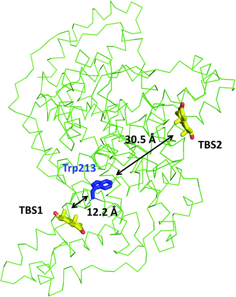
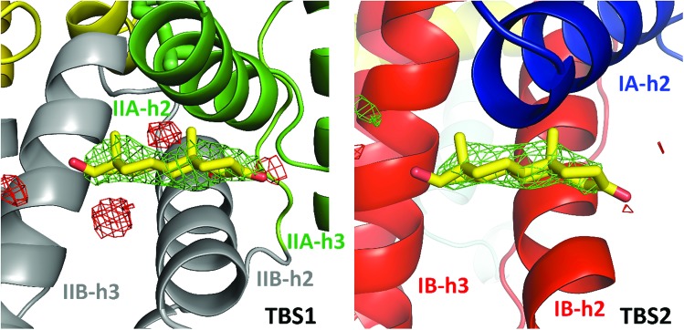
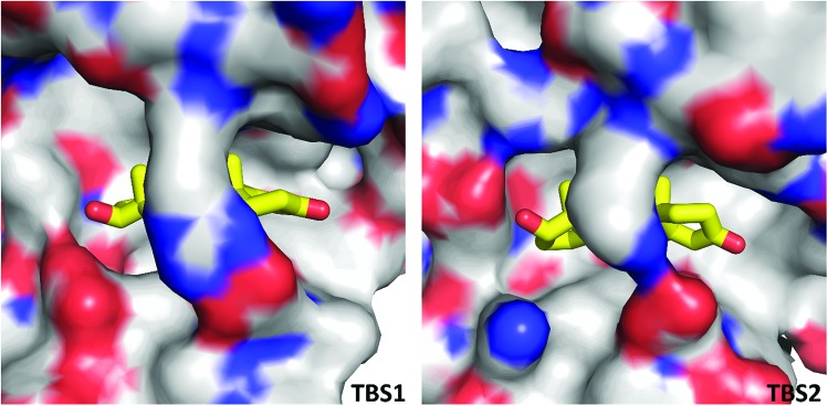
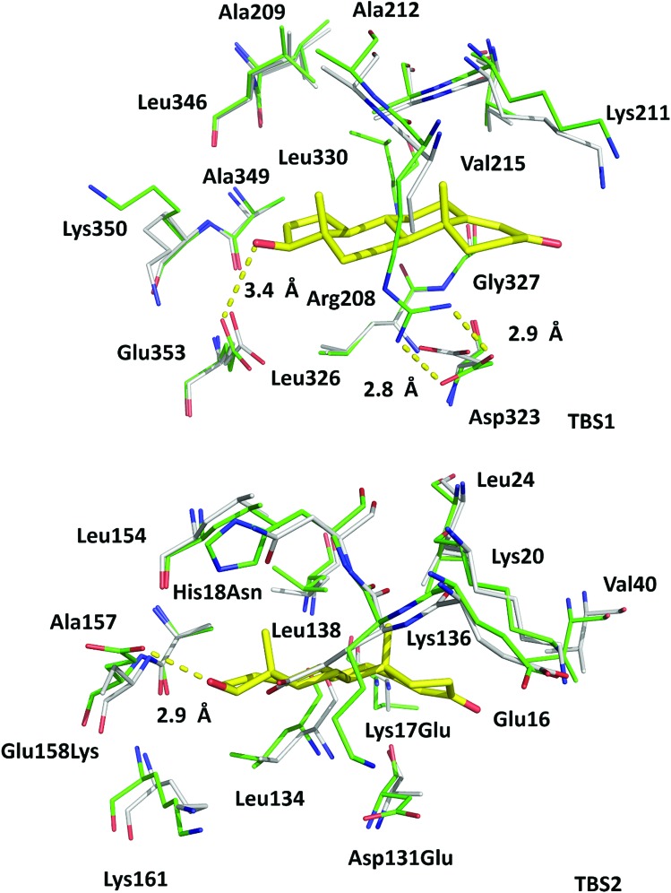
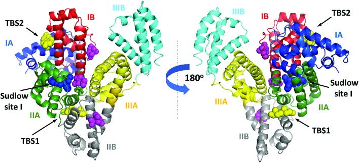
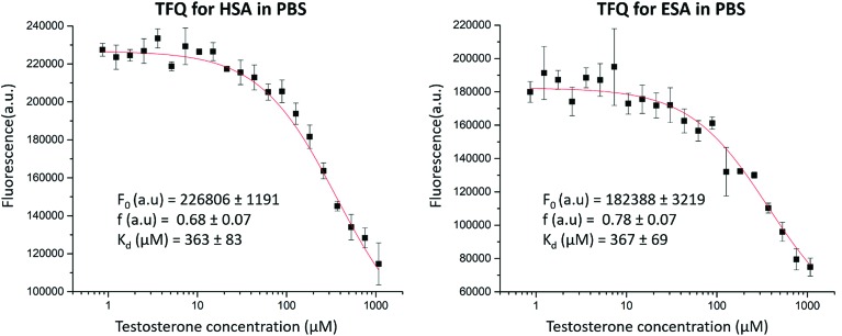
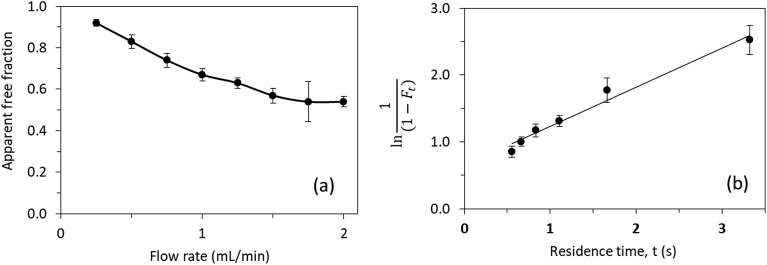
Serum albumin is the most abundant protein in mammalian blood plasma and is responsible for the transport of metals, drugs, and various metabolites, including hormones. We report the first albumin structure in complex with testosterone, the primary male sex hormone. Testosterone is bound in two sites, neither of which overlaps with the previously suggested Sudlow site I. We determined the binding constant of testosterone to equine and human albumins by two different methods: tryptophan fluorescence quenching and ultrafast affinity extraction. The binding studies and similarities between residues comprising the binding sites on serum albumins suggest that testosterone binds to the same sites on both proteins. Our comparative analysis of albumin complexes with hormones, drugs, and other biologically relevant compounds strongly suggests interference between a number of compounds present in blood and testosterone transport by serum albumin. We discuss a possible link between our findings and some phenomena observed in human patients, such as low testosterone levels in diabetic patients.


Figures

Fig. 1
Fig. 1. Testosterone binding sites and their distances from the tryptophan residue. Distances were calculated between the center of mass of tryptophan's indole ring center of mass and that of each testosterone molecule. The testosterone molecules (yellow) and the tryptophan's side chain (blue) are shown in stick representation and labeled.
Fig. 2
Fig. 2. Testosterone binding sites with omit electron density map (mFo–DFc map, calculated after 10 refinement cycles without testosterone, σ – 3.0) presented in green and red (positive and negative contours, respectively). TBS1 is located between subdomains IIA and IIB, while TBS2 is surrounded by subdomains IA and IB. Colors of helices correspond with the domains' colors in Fig. 5. The electron density can be inspected interactively at ; https://molstack.bioreproducibility.org/p/9CP6/.
Fig. 3
Fig. 3. Hydrophobic nature of testosterone binding sites. The color of the protein surface indicates the contributions of the particular atoms to the surface. The color scheme is as follows: gray for carbon atoms, red for oxygen atoms, and blue for nitrogen atoms. Testosterone's carbon atoms are shown in yellow.
Fig. 4
Fig. 4. Superposition of testosterone binding sites in ESA (PDB ID: 6MDQ) and analogous sites in HSA (PDB ID: ; 4K2C). All residues are shown in stick representation. Carbon atoms in ESA and HSA are shown in green and gray, respectively, oxygen atoms are shown in red, nitrogen in blue; testosterone molecules are shown with carbon atoms in yellow. Residue numbers correspond to positions in ESA; naming scheme is as follows: residue from ESA, residue number, residue from HSA (if different). A visualization of the ESA–testosterone complex superposed with ligand-free ESA (PDB ID: ; 3V08) and HSA (PDB ID: ; 4K2C) can be inspected interactively at ; https://molstack.bioreproducibility.org/p/6s7G/.
Fig. 5
Fig. 5. ESA domains and testosterone binding sites. The testosterone (yellow) and citrate molecules (magenta) are shown with atoms as spheres. Warfarin (from structure of HSA complexed with warfarin, PDB ID: 2BXD), which is bound at Sudlow site I, is shown with atoms as blue spheres. Testosterone was predicted to bind in Sudlow site I by Peters. The interactive collection of superpositions of the ESA–testosterone complex and other SA complexes with selected compounds that bind in TBS1 or TBS2 is available at ; https://molstack.bioreproducibility.org/c/hYYh/.
Fig. 6
Fig. 6. TFQ for HSA and ESA caused by testosterone. Standard deviation of fluorescence intensities is represented by error bars.
Fig. 7
Fig. 7. (a) Effect of flow rate on the measured free fraction of testosterone in the presence of ESA. (b) Analysis of the measured free fractions of testosterone/ESA mixtures based on eqn (5). The error bars represent a range of ± 1 S.D.

References

    1. Peters T. J., All About Albumin: Biochemistry, Genetics, and Medical Applications, Academic Press, San Diego, CA, USA, 1st edn, 1995.
    1. Handing K. B., Shabalin I. G., Kassaar O., Khazaipoul S., Blindauer C. A., Stewart A. J., Chruszcz M., Minor W. Chem. Sci. 2016;7:6635–6648. - PMC - PubMed
    1. Merlot A. M., Kalinowski D. S., Richardson D. R. Front. Physiol. 2014;5:299. - PMC - PubMed
    1. Sudlow G., Birkett D. J., Wade D. N. Mol. Pharmacol. 1976;12:1052–1061. - PubMed
    1. Sudlow G., Birkett D. J., Wade D. N. Mol. Pharmacol. 1975;11:824–832. - PubMed