Review
. 2019 May;39(5):788-801.
doi: 10.1111/liv.14091. Epub 2019 Apr 2.

Ischaemia reperfusion injury in liver transplantation: Cellular and molecular mechanisms

Affiliations
Review

Ischaemia reperfusion injury in liver transplantation: Cellular and molecular mechanisms

Wasim A Dar et al. Liver Int. 2019 May.

Abstract

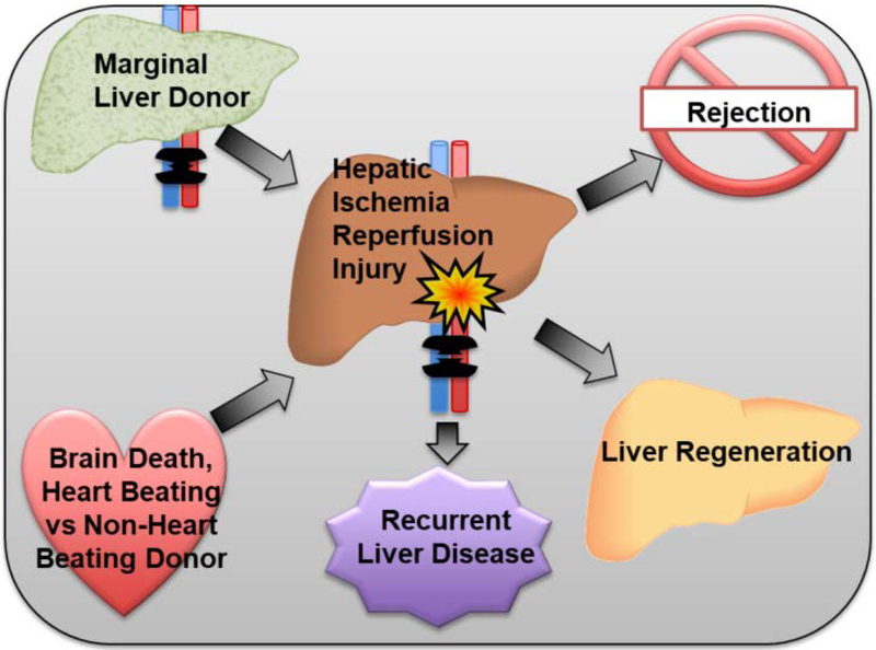
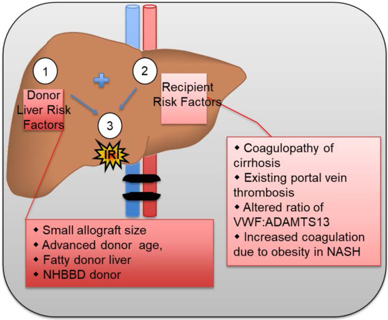
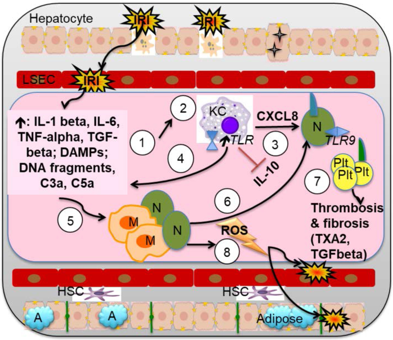
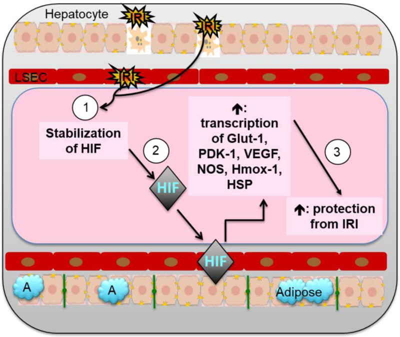
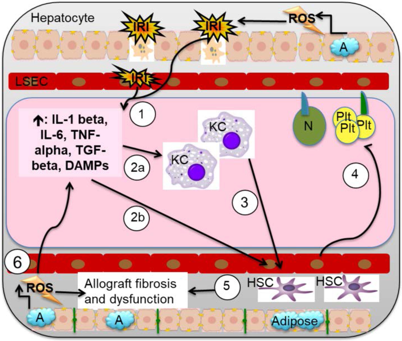
Liver disease causing end organ failure is a growing cause of mortality. In most cases, the only therapy is liver transplantation. However, liver transplantation is a complex undertaking and its success is dependent on a number of factors. In particular, liver transplantation is subject to the risks of ischaemia-reperfusion injury (IRI). Liver IRI has significant effects on the function of a liver after transplantation. The cellular and molecular mechanisms governing IRI in liver transplantation are numerous. They involve multiple cells types such as liver sinusoidal endothelial cells, hepatocytes, Kupffer cells, neutrophils and platelets acting via an interconnected network of molecular pathways such as activation of toll-like receptor signalling, alterations in micro-RNA expression, production of ROS, regulation of autophagy and activation of hypoxia-inducible factors. Interestingly, the cellular and molecular events in liver IRI can be correlated with clinical risk factors for IRI in liver transplantation such as donor organ steatosis, ischaemic times, donor age, and donor and recipient coagulopathy. Thus, understanding the relationship of the clinical risk factors for liver IRI to the cellular and molecular mechanisms that govern it is critical to higher levels of success after liver transplantation. This in turn will help in the discovery of therapeutics for IRI in liver transplantation - a process that will lead to improved outcomes for patients suffering from end-stage liver disease.

Keywords: hypoxia-inducible factors; ischaemia reperfusion; liver transplantation; therapeutics.


Conflict of interest statement

COI: Authors have no conflict of interests.

Figures

Figure 1:
Figure 1:. Risk factors and sequelae of hepatic IRI in liver transplant patients.
Factors such as use of marginal donor organs due to donor scarcity and critical donor hemodynamics, including NHBBD donors, can increase the risk of hepatic IRI. Higher levels of hepatic IRI consequently contribute to poor outcomes, including rejection, recurrence of liver disease, and liver regeneration.
Figure 2:
Figure 2:. Surgical, Donor, and Recipient factors that contribute to risk of IRI.
(1) Risks of IRI inherent to liver transplantation include: organwide ischemia during clamping and resection of donor organ, cold storage, re-anastomosis and circulation of widespread inflammatory factors, increased ischemic time. (2) Donor Liver risk factors can increase the severity of IRI: small allograft, advanced age, especially age >70, donor fatty liver, especially macrosteatosis > 30%, cause of donor death, NHBBD donor, use of marginal organs due to donor scarcity. (3) Recipient risk factors can further increase the severity of IRI: cirrhosis or hepatic fibrosis induced coagulopathy, nutritional coagulopathy, portal vein or hepatic artery thrombosis, altered VWF:ADAMTS13 ratio, history of NASH.
Figure 3:
Figure 3:. Cellular and molecular IRI pathway: A focus on the roles of Kupffer cells, macrophages, neutrophils, and platelets.
(1) IRI damage to LSECs and hepatocytes causes cell death and release of inflammatory cytokines IL-1 beta, IL-6, and TNF-alpha, TGF-beta, as well as DAMPS (HMGB1, FFA, and HSB), DNA fragments, and complement. (2) DAMPs cause KC to increase KC TLR activation, inhibit immunosuppressive IL-10 production, and increase KC cytokine production toward inflammatory phenotype. Inflammatory type KC release: (3) CXCL8, which amplifies recruitment and adhesion of neutrophils to LSEC; and (4) IL-1-beta, TNF-alpha, IFN-gamma, and IL-12. Together, the factors released from IRI damage to LSECs and hepatocytes, as well as those released by inflammatory KCs, (5) induce migration of neutrophils early in IRI and of macrophages late in IRI. (6) These factors also serve to promote further neutrophil adhesion and extravasation into the liver parenchyma via CD11b/CD18a on neutrophils and ICAM-1 on LSEC, while promoting platelet adhesion and activation via upregulation of P-selectin on LSEC. (7) Activated platelets release cytokines that affect vascular tone (TXA2 and serotonin), regulate local thrombosis (PAI-1), and induce fibrosis (TGF beta). (8) Simultaneously, neutrophils and macrophages cause further tissue injury and cellular destruction, through release of ROS and other destructive factors.
Figure 4:
Figure 4:. Cellular and molecular IRI pathway: A focus on the role of HIF.
(1) Hepatic IRI results in stabilization of HIF, which (2) enter the nucleus and induces transcription of genes in multiple pathways such as cellular metabolism (Glut-1, PDK-1), angiogenesis (VEGF, NOS), and cytoprotection (Hmox-1, HSP) to (3) ameliorate IRI induced hepatocyte injury and cell death.
Figure 5:
Figure 5:. Cellular and molecular IRI pathway: A focus on the roles of HSC, adiposity, and ROS.
(1) IRI damage to hepatocytes and LSECs causes cell death and release of inflammatory cytokines IL-1 beta, IL-6, and TNF-alpha, DAMPS, HMGB1, TGF-beta, FFA, and HSB, as well as DNA fragments, which cause (2a) KC to lose tolerogenic profile and increasing cytokine production in KC toward inflammatory phenotype and (2b) hepatic stellate cell (HSC) activation. (3) KC activation is associated with increased KC cytokine production, which further recruits HSCs. The activation and recruitment of HSC to areas of IRI results in (4) ingress of platelets and neutrophils, (5) MMP release, ECM destruction, and endothelial vasoconstriction through ET-1. Together, this results in collagen deposition and graft fibrosis, increasing the risk of graft failure, morbidity, and mortality. (6) After organ reperfusion, lipid peroxidation due to high levels of steatosis causes release of ROS and DAMPs. This results in increased release of IL6 and TNF-alpha, resulting in further acute liver injury, ineffective autophagy, early graft dysfunction or non-function, and increased long-term fibrosis.

References

    1. Marcellin P, Kutala BK. Liver diseases: A major, neglected global public health problem requiring urgent actions and large-scale screening. Liver Int 2018; 38 Suppl 1: 2–6. - PubMed
    1. JENNINGS RB, SOMMERS HM, SMYTH GA, FLACK HA, LINN H. Myocardial necrosis induced by temporary occlusion of a coronary artery in the dog. Arch Pathol 1960; 70: 68–78. - PubMed
    1. Eltzschig HK, Eckle T. Ischemia and reperfusion--from mechanism to translation. Nat Med 2011; 17: 1391–401. - PMC - PubMed
    1. Siniscalchi A, Gamberini L, Laici C, et al. Post reperfusion syndrome during liver transplantation: From pathophysiology to therapy and preventive strategies. World J Gastroenterol 2016; 22: 1551–69. - PMC - PubMed
    1. Stine JG, Northup PG. Coagulopathy Before and After Liver Transplantation: From the Hepatic to the Systemic Circulatory Systems. Clin Liver Dis 2017; 21: 253–74. - PubMed

Publication types

Substances