. 2019 Mar 7;15(3):e1006844.
doi: 10.1371/journal.pcbi.1006844. eCollection 2019 Mar.

A new clustering and nomenclature for beta turns derived from high-resolution protein structures

Affiliations

A new clustering and nomenclature for beta turns derived from high-resolution protein structures

Maxim Shapovalov et al. PLoS Comput Biol. .

Abstract

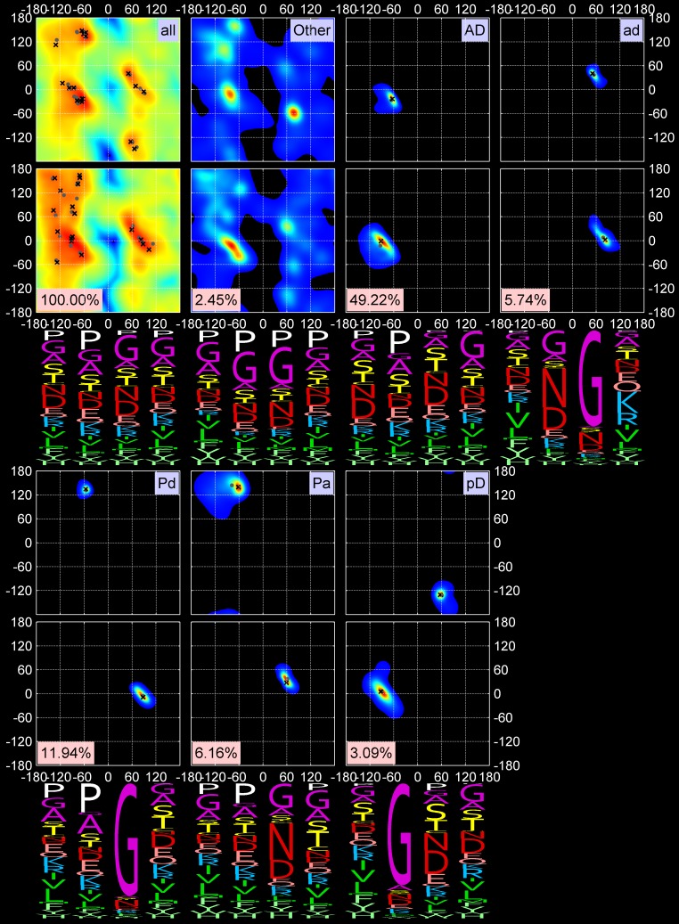
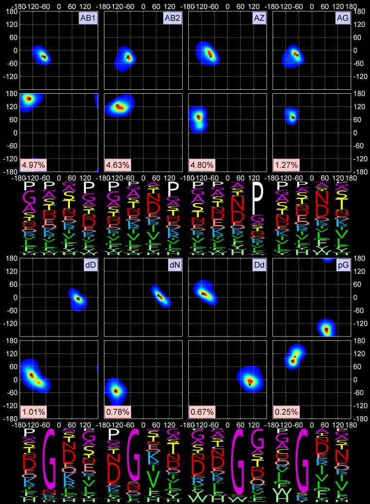
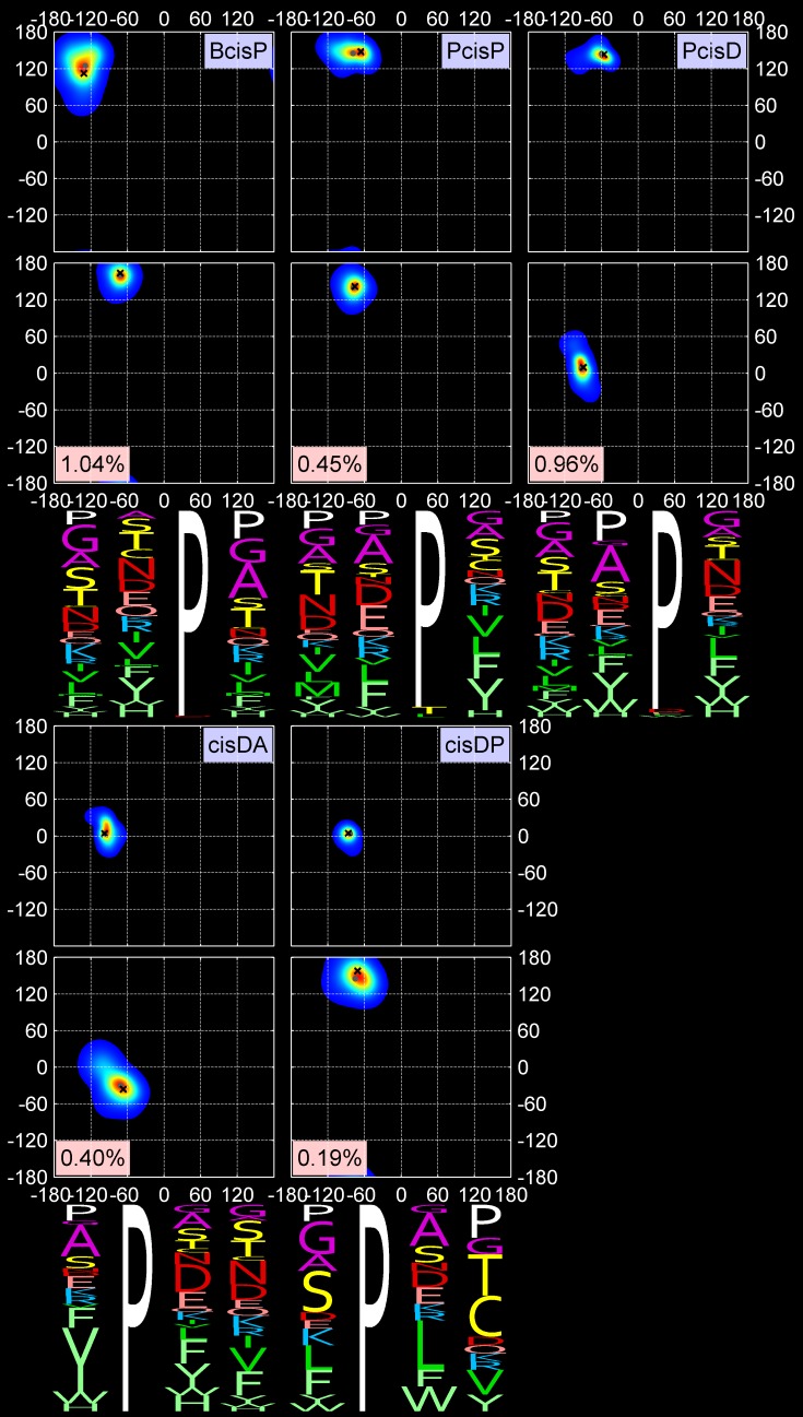
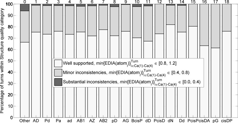
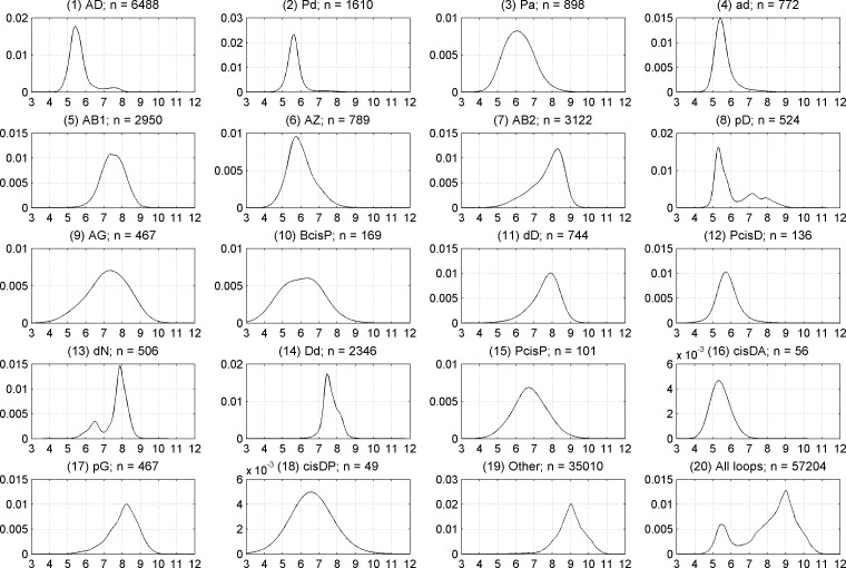
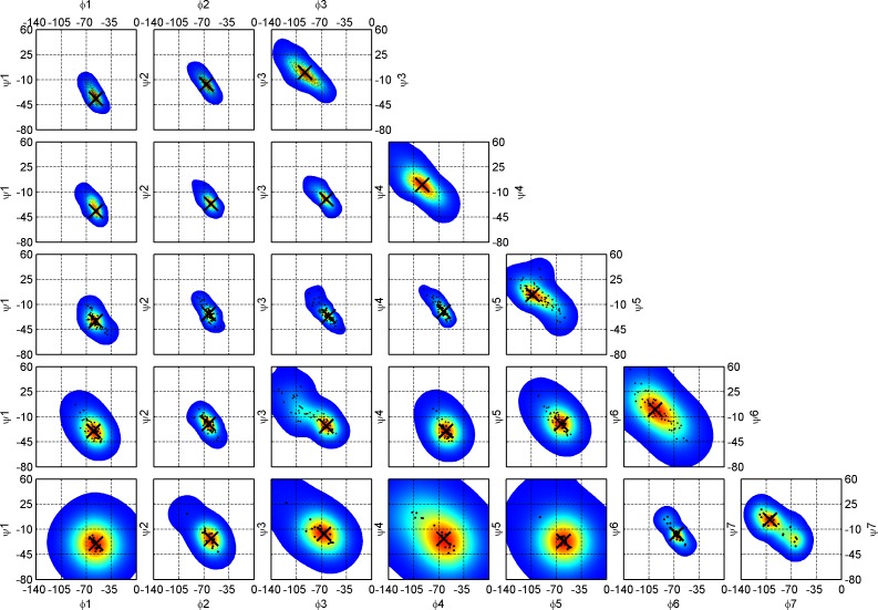
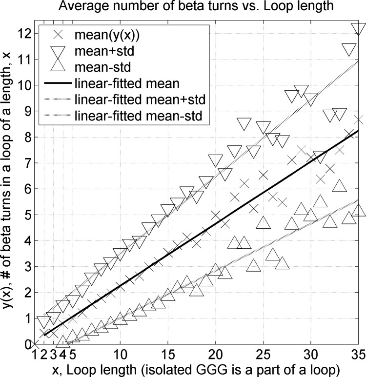
Protein loops connect regular secondary structures and contain 4-residue beta turns which represent 63% of the residues in loops. The commonly used classification of beta turns (Type I, I', II, II', VIa1, VIa2, VIb, and VIII) was developed in the 1970s and 1980s from analysis of a small number of proteins of average resolution, and represents only two thirds of beta turns observed in proteins (with a generic class Type IV representing the rest). We present a new clustering of beta-turn conformations from a set of 13,030 turns from 1074 ultra-high resolution protein structures (≤1.2 Å). Our clustering is derived from applying the DBSCAN and k-medoids algorithms to this data set with a metric commonly used in directional statistics applied to the set of dihedral angles from the second and third residues of each turn. We define 18 turn types compared to the 8 classical turn types in common use. We propose a new 2-letter nomenclature for all 18 beta-turn types using Ramachandran region names for the two central residues (e.g., 'A' and 'D' for alpha regions on the left side of the Ramachandran map and 'a' and 'd' for equivalent regions on the right-hand side; classical Type I turns are 'AD' turns and Type I' turns are 'ad'). We identify 11 new types of beta turn, 5 of which are sub-types of classical beta-turn types. Up-to-date statistics, probability densities of conformations, and sequence profiles of beta turns in loops were collected and analyzed. A library of turn types, BetaTurnLib18, and cross-platform software, BetaTurnTool18, which identifies turns in an input protein structure, are freely available and redistributable from dunbrack.fccc.edu/betaturn and github.com/sh-maxim/BetaTurn18. Given the ubiquitous nature of beta turns, this comprehensive study updates understanding of beta turns and should also provide useful tools for protein structure determination, refinement, and prediction programs.


Conflict of interest statement

The authors have declared that no competing interests exist.

Figures

Fig 1
Fig 1. Two-dimensional (φ, ψ) kernel density estimates (KDE) of turn residue 2 (top row) and 3 (bottom row) for four multi-modal clusters returned by DBSCAN (of 11 total).
These 4 clusters have multiple peaks in their densities and were further individually divided into subclusters with one or more rounds of k-medoids. Their final medoids and modes are marked with gray dots and black crosses respectively. The populations of the four clusters are shown in pink. (A) Cluster for classical Type I and VIII turns exhibit 5 peaks. Type I has the strongest peak and account for 76% of the data points in the cluster. (B) Classical Type II turns can be subdivided into two new clusters; (C) Classical VIb turns with a cis peptide bond at residue 3 can be divided into two types. (D) A DBSCAN cluster with residue 2 in the left-handed helical region and residue 3 adjacent to the right-handed helical region. The data have 3 obvious density peaks but the smallest peak (φ3, ψ3 ~ -90°,-20°) did not have a distinct amino acid profile and had a very small number of points. We divide this cluster into two beta-turn types.
Fig 2
Fig 2. Two-dimensional KDEs for (φ2, ψ2) and (φ3, ψ3) and amino-acid sequence profiles for 5 out 18 turn types, all turns, and Other turns.
First row: All data points, Other data points (unclassified), AD (classical Type I) and ad (Type I’) turns. “All” data points KDE is shown on a log scale. Second row: Pd and Pa turns (Type II) and pD turns (Type II’). The mode and medoid of each cluster are shown with a black cross and gray dot respectively. The four-letter profiles are shown for the four amino-acid residues composing a beta turn.
Fig 3
Fig 3. Two-dimensional KDEs for (φ2, ψ2) and (φ3, ψ3) and amino-acid sequence profiles for the next 8 out of 18 turn types.
First row: AB1, AB2, AZ, and AG turns (all formerly Type VIII). Second row: new turn types dD and dN and their approximate inverse, Dd, and new turn type pG. The formatting is the same as in Fig 2.
Fig 4
Fig 4. Two-dimensional KDEs for (φ2, ψ2) and (φ3, ψ3) and amino-acid sequence profiles for the last 5 out of 18 turn types.
First row: cis3 turn types, BcisP (Type VIb), PcisP (new), and PcisD (Type VIa1). Second row: new cis2 turns, cisDA and cisDP. The formatting is the same as in Fig 2.
Fig 5
Fig 5. Single-letter region codes for the Ramachandran map.
Greek letter designations were converted to Roman letter counterparts. The left side of the map with negative φ is encoded in upper-case letters, while the right side of the map with positive φ is in lower-case letters. Regions related by symmetry through the origin are denoted with the same letter (e.g., A and a; P and p). The original gamma and gamma prime regions are labeled g and G respectively. The new label N is used for the region below the A-D axis. All cluster medoids of residues 2 and 3 are marked with orange dots. The black dots represent data from all amino acids in the RefinedSet, including regular secondary structures and loops.
Fig 6
Fig 6. Structure quality of Other turns and 18 turn types based on agreement between atom positions and electron-density maps measured by EDIA software with the minimum of EDIA values for all 13 backbone atoms (N, Cα, C, O) in a turn connecting Cα1 to Cα4.
Fig 7
Fig 7. 2Fo-Fc electron density distributions for each of the 18 new beta-turn types, plus two examples from the Other category.
The PDB entries and other data for each turn are given in BetaTurnLib18 (S1 Table and S1 Text). Contours are shown at 2σ. The two Other examples (bottom row, 3rd and 4th images) are: (1) PDB entry 2EAB, chain A, residues 759–762, sequence YHAP, conformation APcis4, [φ22; φ33] = [-92°,-39°; -76°,165°]; (2) PDB entry 4RJZ, chain A, residues 366–369, sequence PRGP, conformation Bbcis4, [φ22; φ33] = [-78°,120°; 82°,-127°]. Residue 4 is a cis proline in both cases.
Fig 8
Fig 8. Frequency of beta-turn types at different resolutions.
Beta turns were identified in structure sets at different resolution ranges (1–1.2 Å, 1.1–1.3 Å, etc.) produced by the PISCES webserver. The y-axis on each plot covers 2 percentage points in total frequency of each beta-turn type.
Fig 9
Fig 9. Kernel density estimates of Cα1-Cα4 distances when no 1–4 distance cutoff is applied.
Plots are shown for each of 18 beta-turn clusters, the Other group, and all four-residue segments in loop regions of RefinedSet (bottom right).
Fig 10
Fig 10. Ramachandran distributions for residues 2 and 3 of AD turns with no Cα1-Cα4 distance cutoff.
In the left column, all points are plotted with those with Cα1-Cα4 distance ≤ 7.0 Å in blue and Cα1-Cα4 distance > 7.0 Å in red. Mean (φ,ψ) values are denoted with dark blue and red crosses for each group. The distance histogram is provided in the bottom row. In the middle column, only the points with Cα1-Cα4 distance ≤ 7.0 Å are plotted, and in the last column only those with Cα1-Cα4 distance > 7.0 Å are plotted. Figures for the other clusters are provided in S2 Supporting information.
Fig 11
Fig 11. Ramachandran maps for 310 helices of increasing length, n from 3 (DSSP code GGG, top row) to 7 (GGGGGGG, bottom row).
Optimized kernel density estimates overlaid with (φ,ψ) sample scatter plots are drawn for each 310 helix residue starting from the first left position, 1 (first column) to the last right position, 7 (last column). Modes (peaks) for each 2D distribution are shown with a large black cross. The first (1) and last (n) residue conformations of 310 helices are A (-64°, -23°) and D (-92°, -1°) conformations respectively. The intervening residues are in between, sometimes with conformations spanning both populations. The exact values for (φ,ψ) modes of 310 helices are reported in Table 4.
Fig 12
Fig 12. Frequency of loop length in CompleteSet of 17,176 loops.
Short 310 helices (DSSP of GGG) are considered part of loops. The frequency of a loop decreases exponentially after the length of 5 with approximately a 10-fold reduction every time a loop adds 10 more residues. Between loop lengths 2 and 5, there is a frequency fluctuation with two peaks at length 2 and 4 observed primarily due to beta turns between two beta-sheet strands. 95% of loops are 1 to 16 residues long. A similar figure for traditional (GGG excluded) loops is provided in Fig E in S1 Supporting information.
Fig 13
Fig 13. An average number of beta turns in a loop (isolated GGG included in the loops) as a function of loop length.
Beta turns are impossible (0 beta turns) in loops with a length of 1 residue because both 2nd and 3rd residues of a beta turn have to be in a loop region. For every 4.8 loop residues, there is on average a single beta turn. A similar figure for traditional loops (GGG excluded) is provided in Fig F in S1 Supporting information.

Similar articles

Cited by

References

    1. Venkatachalam C. Stereochemical criteria for polypeptides and proteins. V. Conformation of a system of three linked peptide units. Biopolymers 6:1425–1436 (1968). 10.1002/bip.1968.360061006 - DOI - PubMed
    1. Richardson J.S., The anatomy and taxonomy of protein structure, in Adv. Protein Chem. 1981, Elsevier; p. 167–339. - PubMed
    1. Wilmot C., Thornton J. Analysis and prediction of the different types of β-turn in proteins. J. Mol. Biol. 203:221–232 (1988). - PubMed
    1. Wilmot C.M., Thornton J.M. Beta-turns and their distortions: a proposed new nomenclature. Protein Eng. 3:479–93 (1990). - PubMed
    1. Koch O., Klebe G. Turns revisited: a uniform and comprehensive classification of normal, open, and reverse turn families minimizing unassigned random chain portions. Proteins 74:353–67 (2009). 10.1002/prot.22185 - DOI - PubMed

Publication types