. 2019 May 6;58(19):6300-6305.
doi: 10.1002/anie.201901141. Epub 2019 Apr 3.

Heterochromatin Protein HP1α Gelation Dynamics Revealed by Solid-State NMR Spectroscopy

Affiliations

Heterochromatin Protein HP1α Gelation Dynamics Revealed by Solid-State NMR Spectroscopy

Bryce E Ackermann et al. Angew Chem Int Ed Engl. .

Abstract

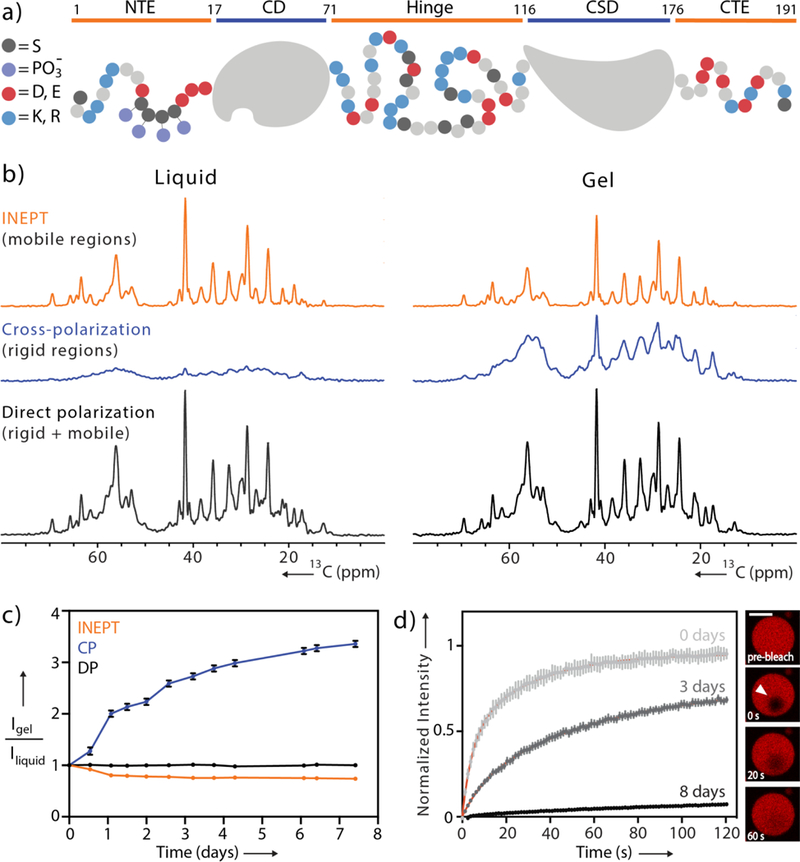
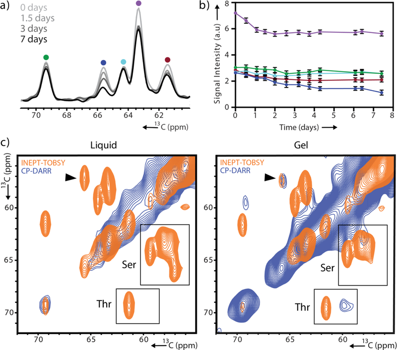
Heterochromatin protein 1α (HP1α) undergoes liquid-liquid phase separation (LLPS) and forms liquid droplets and gels in vitro, properties that also appear to be central to its biological function in heterochromatin compaction and regulation. Here we use solid-state NMR spectroscopy to track the conformational dynamics of phosphorylated HP1α during its transformation from the liquid to the gel state. Using experiments designed to probe distinct dynamic modes, we identify regions with varying mobilities within HP1α molecules and show that specific serine residues uniquely contribute to gel formation. The addition of chromatin disturbs the gelation process while preserving the conformational dynamics within individual bulk HP1α molecules. Our study provides a glimpse into the dynamic architecture of dense HP1α phases and showcases the potential of solid-state NMR to detect an elusive biophysical regime of phase separating biomolecules.

Keywords: biophysics; heterochromatin protein 1α (HP1α); liquid-liquid phase separation (LLPS); solid-state NMR spectroscopy; structural biology.


Figures

Figure 1.
Figure 1.
pHP1α dynamics monitored during gelation. (a) Domain map of monomeric pHP1α. Negatively charged residues are colored in red, positively charged residues are in blue, serine residues are in black and phosphorylation sites are in purple. (b) 1D 13C INEPT, CP and DP spectra acquired during the initial dense liquid state and the final gel state. (c) Integrated signal of the aliphatic regions of the spectra collected every 12 hours. Data are normalized to the starting point of each respective experiment. For error analysis, see SI. (d) FRAP recovery curves for pHP1α collected immediately upon droplet formation, 3 days and 8 days later. Red lines represent exponential fits. Inset: FRAP of a single pHP1α droplet at day 3, scale bar 20 μm.
Figure 2.
Figure 2.
Site-specific variation in the gelation dynamics of pHP1α. (a) 1D INEPT spectra collected 0, 1.5, 3 and 7 days after initiation of phase separation. (b) Signal intensity of the five peaks shown in (a) tracked over time with colors corresponding to the color scheme in (a). For error analysis, see SI. (c) Serine/threonine region of 2D 13C-13C CP-DARR and INEPT-TOBSY spectra recorded 3 hours after droplet formation and 7 days later. A black arrow denotes the serine cross-peak with greatest signal loss.
Figure 3.
Figure 3.
2D 13C-13C correlation spectra of pHP1α condensates. (a) Overlay of CP-DARR and INEPT-TOBSY spectra, probing the rigid and highly mobile sites of pHP1α 4 days after droplet formation. Amino acid types with characteristic and identifiable chemical shifts are marked in boxes (INEPT-TOBSY) and circles (CP-DARR). (b) Complete sequence of HP1α showing structured regions (gray), and disordered regions with identified amino acid types (orange).

References

    1. Mitrea DM, Kriwacki RW, Cell Comm. Signal. 2016, 14, DOI 10.1186/s12964-015-0125-7. - DOI - PMC - PubMed
    1. Kroschwald S, Maharana S, Mateju D, Malinovska L, Nüske E, Poser I, Richter D, Alberti S, eLife 2015, 4, DOI 10.7554/eLife.06807. - DOI - PMC - PubMed
    1. Strom AR, Emelyanov AV, Mir M, Fyodorov DV, Darzacq X, Karpen GH, Nature 2017, 547, 241–245. - PMC - PubMed
    1. Patel A, Lee HO, Jawerth L, Maharana S, Jahnel M, Hein MY, Stoynov S, Mahamid J, Saha S, Franzmann TM, et al. , Cell 2015, 162, 1066–1077. - PubMed
    1. Su X, Ditlev JA, Hui E, Xing W, Banjade S, Okrut J, King DS, Taunton J, Rosen MK, Vale RD, Science 2016, 352, 595–599. - PMC - PubMed

Publication types

MeSH terms

LinkOut - more resources