. 2019 Jun;18(3):e12939.
doi: 10.1111/acel.12939. Epub 2019 Mar 7.

Hormone replacement therapy attenuates hearing loss: Mechanisms involving estrogen and the IGF-1 pathway

Affiliations

Hormone replacement therapy attenuates hearing loss: Mechanisms involving estrogen and the IGF-1 pathway

Tanika T Williamson et al. Aging Cell. 2019 Jun.

Abstract

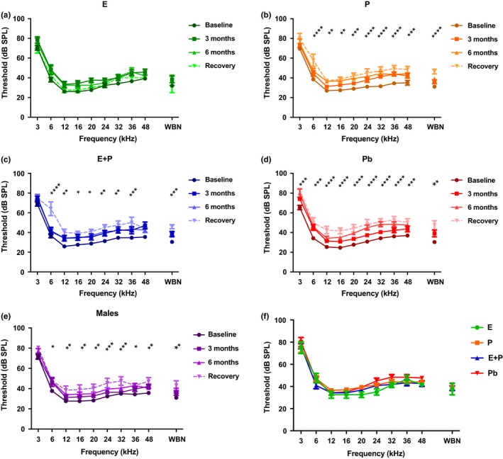
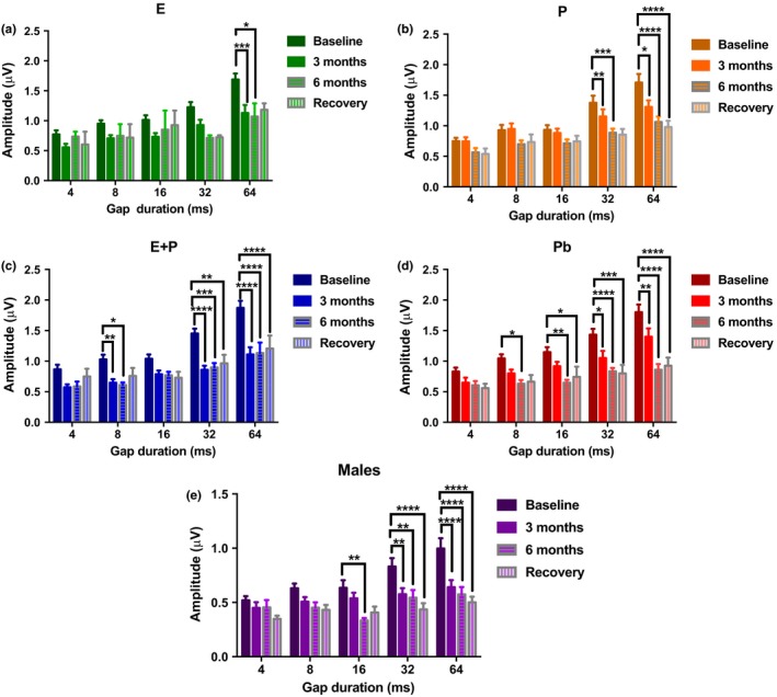
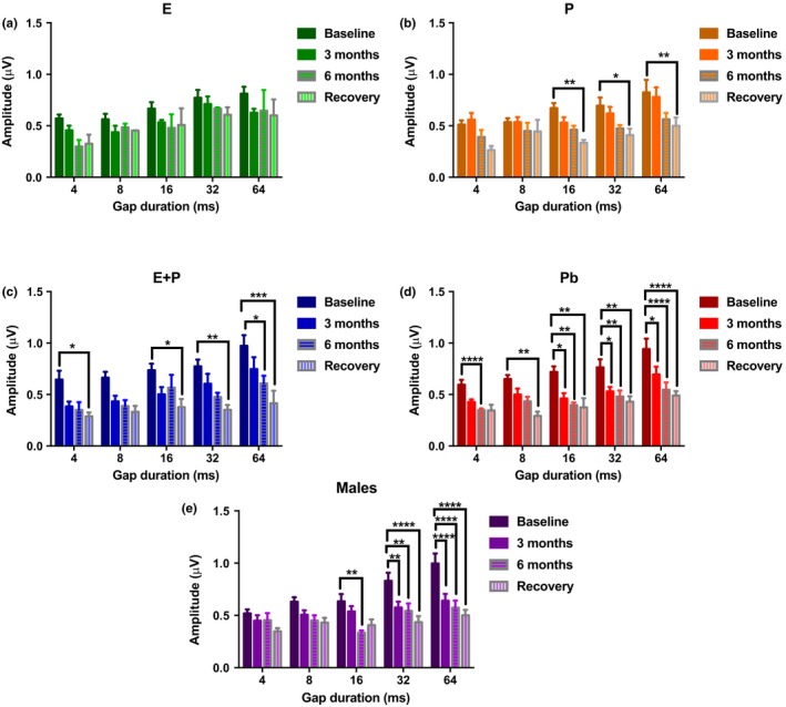
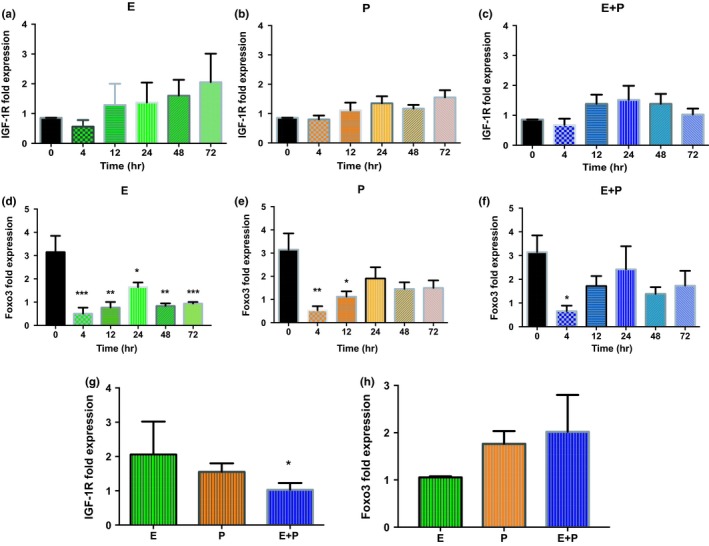
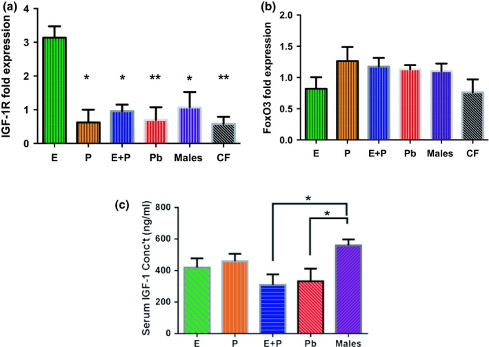
Estradiol (E) is a multitasking hormone that plays a prominent role in the reproductive system, and also contributes to physiological and growth mechanisms throughout the body. Frisina and colleagues have previously demonstrated the beneficial effects of this hormone, with E-treated subjects maintaining low auditory brainstem response (ABR) thresholds relative to control subjects (Proceedings of the National Academy of Sciences of the United States of America, 2006;103:14246; Hearing Research, 2009;252:29). In the present study, we evaluated the functionality of the peripheral and central auditory systems in female CBA/CaJ middle-aged mice during and after long-term hormone replacement therapy (HRT) via electrophysiological and molecular techniques. Surprisingly, there are very few investigations about the side effects of HRT in the auditory system after it has been discontinued. Our results show that the long-term effects of HRT are permanent on ABR thresholds and ABR gap-in-noise (GIN) amplitude levels. E-treated animals had lower thresholds and higher amplitude values compared to other hormone treatment subject groups. Interestingly, progesterone (P)-treated animals had ABR thresholds that increased but amplitude levels that remained relatively the same throughout treatment. These results were consistent with qPCR experiments that displayed high levels of IGF-1R in the stria vascularis (SV) of both E and P animal groups compared to combination treatment (E + P) animals. IGF-1R plays a vital role in mediating anti-apoptotic responses via the PI3K/AKT pathway. Overall, our findings gain insights into the neuro-protective properties of E hormone treatments as well as expand the scientific knowledge base to help women decide whether HRT is the right choice for them.

Keywords: HRT; age-related hearing loss; aging; auditory system; hormone replacement therapy; neurodegeneration.


Conflict of interest statement

None declared.

Figures

Figure 1
Figure 1
Auditory brainstem response (ABR) and ABR gap‐in‐noise techniques. (a) Five to seven waves are typically generated during Auditory Brainstem Response (ABR) testing for humans clinically, and animals such as mice. Starting at 90 decibels (dB), various frequencies (3–48 kHz) are presented at sound levels that decrease in intervals of 5 dB. The threshold is considered the lowest sound level at which a waveform can be detected. The figure shows the ABR results for a 15‐month‐old CBA/CaJ mouse at 90 decibels (dB). (b) The ABR gap‐in‐noise (GIN) technique measures auditory temporal processing. Subjects were presented with a 25‐millisecond (ms) noise burst (NB1) followed by a series of silent gap durations, ranging from 0 to 64 ms. A second 25‐ms noise bursts (NB2) was presented to measure the ability of the auditory system to recover from NB1 and efficiently respond to NB2. For the present study, the ABR GIN analysis focused on the amplitude levels (red arrow) for Peak 1 and Peak 4. The figure displays the baseline ABR GIN response at 8 ms for a 15‐month‐old mouse
Figure 2
Figure 2
Auditory brainstem response thresholds over the course of hormone treatments as well as during the recovery period. (a) E animals show no significant signs of ARHL over the course of hormone therapy, thus indicating that E possesses protective properties for auditory function. (b) P animals show significantly poorer hearing at almost all of the tested frequencies. (c) The E + P group displayed elevations in ABR thresholds as early as 3 months. Notable worsening of hearing could be seen in this group over time. (d) Pb control animals' thresholds changed drastically over the 6‐month time period. Significant ARHL changes were observed for all frequencies. (e) Changes observed in the male group, more specifically during the recovery period (8 months), could be attributed to ARHL. (f) Recovery period group comparison shows that E‐treated animals had lower thresholds at 12, 16, 20, 24, and 32 kHz compared to all the other HRT animals. Pb females had higher thresholds among the HRT groups at 24 and 32 kHz. These data, in conjunction with Figure 5, suggest that the results of long‐term HRT on ABR thresholds are permanent. No statistical differences were seen among the hormone groups during the recovery period. It should be noted that statistical differences for (a) through (e) are a comparison between the baseline and recovery. Statistical test: 2‐way ANOVA followed by Bonferroni; *p < 0.05, **p < 0.01, ***p < 0.001, ****p < 0.0001
Figure 3
Figure 3
Auditory brainstem response gap‐in‐noise P1 amplitude levels for NB2 for subject groups during HRT and for the recovery period. (a) Few changes were observed in E‐treated animals during the course of the longitudinal experiment. Significant changes were only seen at the largest gap interval, 64 ms. (b) The P group displayed greater declines while undergoing long‐term HRT. (c) Amplitude levels were sharply reduced once treatment began for E + P animals. Significant changes were seen as early as 3 months at 8, 32, and 64 ms. (d) ARHL was observed in Pb animals throughout the course of the experiment. These changes could have been exacerbated due to the removal of E from the circulatory system during middle age. (e) The males also displayed signs of presbycusis throughout the course of the experiment, as steep declines were detected for P1 amplitude levels. These findings suggest that females treated with E or P retain temporal processing abilities better than males. No signs of recovery were observed in any of the subject groups. Statistical test: 2‐way ANOVA followed by Bonferroni; *p < 0.05, **p < 0.01, ***p < 0.001, ****p < 0.0001
Figure 4
Figure 4
Auditory brainstem response gap‐in‐noise P4 amplitude levels for NB2 for subject groups during HRT and for the recovery period. (a) Amplitude levels for E‐treated animals decline marginally once treatment began. (b) The P group amplitude levels showed a steady decline throughout the advancement of treatment, with the largest differences seen during the recovery period. (c) E + P animals also displayed a significant decrease in P4 amplitude levels. Most striking differences were observed during the recovery period. (d) Amplitude values declined as early as 3 months for Pb animals. This group exhibited the worst reduction in amplitude levels among the female groups. (e) The males also showed signs of ARHL at longer gap intervals, starting at 16 ms. All of the groups, except E displayed changes during recovery, that is, for these groups P4 amplitudes worsened after HRT was discontinued. Statistical test: 2‐way ANOVA followed by Bonferroni; *p < 0.05, **p < 0.01, ***p < 0.001, ****p < 0.0001
Figure 5
Figure 5
In vitro quantitative IGF‐1R and FoxO3 gene expression in SVK‐1 cells after different time intervals for the duration of HRT. (a) IGF‐1R gene expression displayed an upward trend once E treatment began. Most notably, this expression more than doubled after 72 hr of E hormone therapy. Contrastingly, (b) P‐ and (c) E + P‐treated cells had IGF‐1R expression levels that were relatively the same over the course of the experiment, suggesting that these hormone treatments had little to no effect. (d) E‐treated cells exhibited FoxO3 levels that significantly declined. Statistical differences were observed throughout the course of HRT. (e) P, and (f) E + P displayed similar trends in which FoxO3 expression levels dramatically decreased after 4 hr of treatment; however, gene levels began to somewhat increase, especially by 24 hr. The statistical differences are relative to the untreated cells at 0 hr. Statistical test: 1‐way ANOVA followed by Bonferroni; *p < 0.05, **p < 0.01, ***p < 0.001. Comparison among the hormone‐treated cell groups after 72 hr of HRT for (g) IGF‐1R and (h) FoxO3 gene expression. (g) E maintained high IGF‐1R levels over time; meanwhile, E + P cells displayed gene expression levels that continued to decline at 72 hr, causing this cell group to have the lowest expression. A significant difference was observed for the E + P group relative to the E group. (h) Although E had FoxO3 expression levels that were lower than both P and E + P SVK‐1 cells, no significant differences were observed among the groups at 72 hr. Statistical test: Welch's t test followed by Bonferroni; *p < 0.033
Figure 6
Figure 6
In vivo 1‐month post‐treatment IGF‐1R and FoxO3 expression levels. (a) E animals had the highest IGF‐1R fold expression levels among the subject groups for SV tissue samples. Interestingly, Pb and control female (CF) animals had the most significant differences among the groups, relative to E. This implies that lack of HRT during the aging process could possibly decrease IGF‐1R levels. (b) FoxO3 gene expression was comparatively similar among the SV tissue sample groups. Congruous findings were observed for in vitro FoxO3 experiments. It can be noted that overall the CF group had the lowest expression levels for both genes. (c) Post‐treatment IGF‐1 concentration levels in the serum of HRT mice showed no significant differences among the female HRT groups. Only E + P and Pb groups displayed statistical variances in comparison to the control male animals. Statistical test: 1‐way ANOVA followed by Bonferroni; *p < 0.05, **p < 0.01 (E n = 3; P n = 3; E + P n = 3; Pb n = 3, Males n = 3; CF n = 3). Note: The CF group consists of age‐matched females with their ovaries intact that did not undergo any type of HRT

Similar articles

Cited by

References

    1. Barati, B. , Abtahi, S. H. , Hashemi, S. M. , Okhovat, S. A. , Poorqasemian, M. , & Tabrizi, A. G. (2013). The effect of topical estrogen on healing of chronic tympanic membrane perforations and hearing threshold. Journal of Research in Medical Sciences, 18(2), 99–102. - PMC - PubMed
    1. Berent‐Spillson, A. , Briceno, E. , Pinsky, A. , Simmen, A. , Persad, C. C. , Zubieta, J. K. , & Smith, Y. R. (2015). Distinct cognitive effects of estrogen and progesterone in menopausal women. Psychoneuroendocrinology, 59, 25–36. 10.1016/j.psyneuen.2015.04.020 - DOI - PMC - PubMed
    1. Bittar, R. S. M. , Cruz, O. L. M. , Lorenzi, M. C. , Marone, S. A. M. , & Miniti, A. (2001). Morphological and functional study of the cochlea after administration of estrogen and progesterone in guinea pig. International Tinnitus Journal, 7(1), 41–45. - PubMed
    1. Bonnard, A. , Sahlin, L. , Hultcrantz, M. , & Simonoska, R. (2013). No direct nuclear effect of progesterone in the inner ear: Other possible pathways. Acta Oto‐Laryngologica, 133(12), 1250–1257. 10.3109/00016489.2013.825377 - DOI - PubMed
    1. Chan, M. , Chow, C. , Hamson, D. K. , Lieblich, S. E. , & Galea, L. A. (2014). Effects of chronic oestradiol, progesterone and medroxyprogesterone acetate on hippocampal neurogenesis and adrenal mass in adult female rats. Journal of Neuroendocrinology, 26(6), 386–399. 10.1111/jne.12159 - DOI - PubMed

Publication types