Review
. 2019 Jul;92(1099):20180864.
doi: 10.1259/bjr.20180864. Epub 2019 May 13.

Complications from percutaneous microwave ablation of liver tumours: a pictorial review

Affiliations
Review

Complications from percutaneous microwave ablation of liver tumours: a pictorial review

Cheng Fang et al. Br J Radiol. 2019 Jul.

Abstract

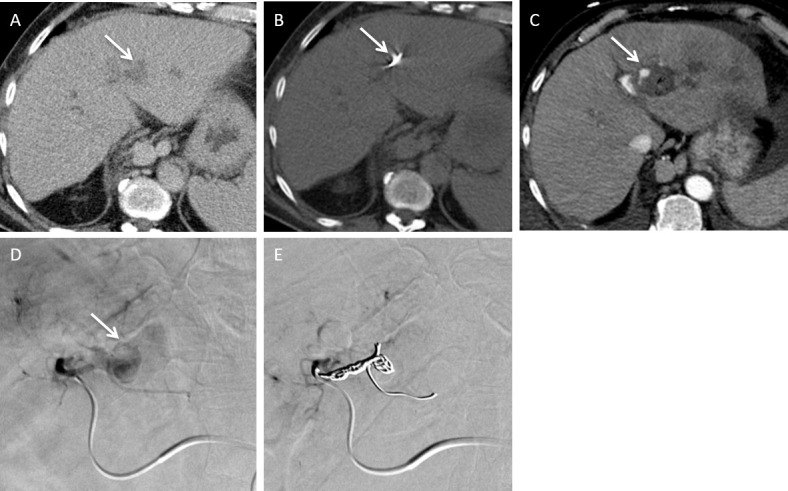
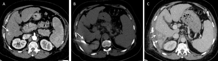
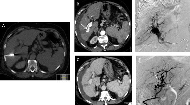
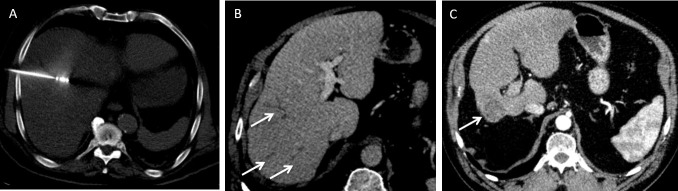
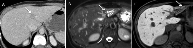
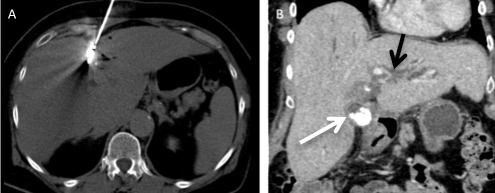
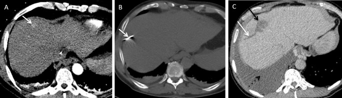
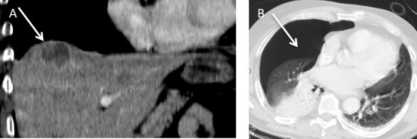
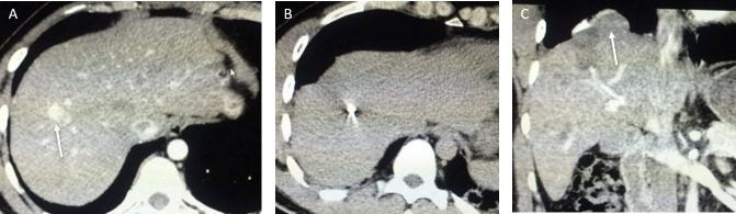
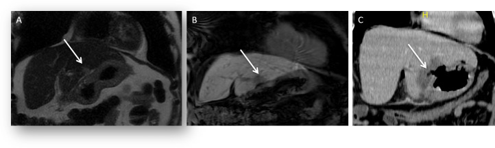
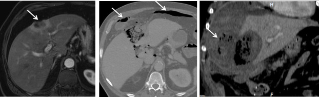
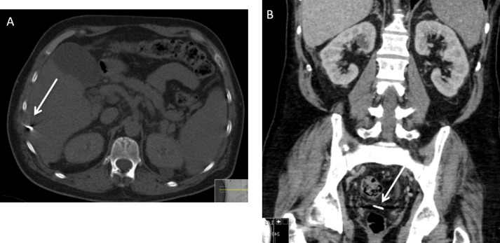
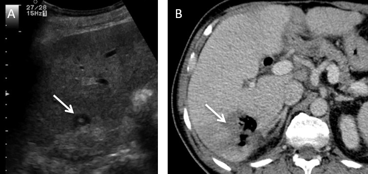
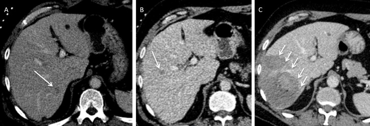
Percutaneous microwave ablation of liver tumours is a well-established technique that has been proven to be effective in the curative and palliative treatment of small volume primary and secondary liver tumours. Microwave ablation is designed to achieve larger areas of necrosis compared to radiofrequency ablation and has a good safety profile among liver tumour treatments. Mortality is unreported and major complications are rare. Knowledge of potential complications is essential for interventional radiologists performing liver ablation in order to reduce patient morbidity. The aim of this review is to illustrate major complications post microwave ablation in a pictorial format as well as a discussion on how best to avoid these complications.


Figures

Figure 1.
Figure 1.
A 66-year-old male with chronic liver disease who developed a 26 mm current hepatocellular carcinoma in the left lateral liver was treated with microwave ablation. There was no immediate post-procedural complications. Patient presented 4 weeks later with acute drop in haemoglobin. (A) Axial contrast-enhanced CT demonstrates the position of the lesion (arrow). (B) Axial contrast-enhanced CT demonstrates the position of the microwave needle. (C) Arterial phase CT shows pseudoaneurysm which was confirmed on angiography (D) and the pseudoaneurysm was coil embolised successfully (E).
Figure 2.
Figure 2.
A 51-year-old male with chronic viral hepatitis B and C infection developed a small hepatocellular carcinoma recurrence in Segment VI after previous ablation. 3 days later, he developed right upper quadrant pain. (A) Axial arterial phase CT prior to MWA shows a small arterialized nodule (arrow) representing a recurrent hepatocellular carcinoma. (B) Axial non-contrast-enhanced CT shows the position of the needle (arrow). (C) The repeat CT shows the presence of a small subcapsular haematoma (arrows). This was treated conservatively and the patient recovered with no further complications.
Figure 3.
Figure 3.
A surveillance CT of a 73-year-old female with non-alcoholic fatty liver disease related cirrhosis showed a 20 mm hepatocellular carcinoma in Segment V. This was treated with microwave ablation. (A) Axial non-contrast-enhanced CT demonstrates the position of the microwave needle. (B) Follow up axial arterial and (C) Axial portovenous post contrast-enhanced CT shows new onset of moderate ascites with blood results demonstrating worsening of hepatic function in keeping with decompensated portal hypertension. Note there is early opacification of the right portal vein on the arterial phase imaging (arrow). (D) Catheter hepatic angiography demonstrates an arterioportal shunt. (E) Angiography images demonstrate successful closure of arterioportal shunt with coil. The patient’s ascites and patient’s liver function improved.
Figure 4.
Figure 4.
A 76-year-old male with crytogenic cirrhosis was treated for a 18 mm hepatocellular carcinoma found in Segment VIII on surveillance scan. (A) Axial non-contrast-enhanced CT demonstrates the position of the microwave needle. (B) Axial contrast-enhanced CT demonstrates initial segmental peripheral biliary duct dilation on an immediate follow-up study (arrows). (C) Axial portovenous phase contrast-enhanced CT demonstrates the treated tumour (arrow) and marked atrophy of the right lobe of the liver as a result of right portal vein thrombosis 18 months after the initial procedure.
Figure 5.
Figure 5.
A 55-year-old male developed a solitary colorectal metastatic deposit. (A) Axial post contrast-enhanced CT shows the position of the liver metastasis (arrow) in the left liver lobe (Segment II/III) in close relation to the left portal vein. This was ablated under ultrasound guidance. (B) Follow-up imaging from axial T2 weighted fat suppressed image shows lobar intrahepatic biliary dilatation (arrow) post-ablation secondary to a thermal biliary stricture. (C) Axial 20 min delayed hepatobiliary phase post-administration of gadoxetic acid image shows end-stage atrophy of segments II–III and no excretion of hepatobiliary contrast medium (arrow).
Figure 6.
Figure 6.
A 73-year-old female with chronic viral hepatitis B infection had viable hepatocellular tumour in segment IV following trans-arterial chemo-emobolisation was treated with microwave ablation. (A) Axial non-contrast-enhanced CT shows the centre of the microwave needle which is within close proximity to the gallbladder. (B) Coronal contrast enhanced CT shows thickened gallbladder wall with calculi (white arrow) and intrahepatic biliary duct dilatation (black arrow). Patient was clinically symptomatic and a diagnosis of acute cholecystitis was made.
Figure 7.
Figure 7.
A 68-year-old male with viral hepatitis C related cirrhosis developed a 1.4 cm hepatocellular carcinoma within Segment VIII. (A) Axial contrast enhanced CT demonstrates a small hepatocellular carcinoma (arrow). (B) Axial CT (bone window) demonstrates the microwave needle position during ablation. (C) Axial post contrast enhanced CT demonstrates ablation zone (black arrow) with adjacent dilated biliary duct (white arrow) and associated large right pleural effusion (dotted black arrow). The appearance suggests an underlying biliary pleural fistula.
Figure 8.
Figure 8.
A 50-year-old male patient developed a colorectal liver metastasis in Segment VIII superiorly. (A) Coronal contrast enhanced CT demonstrates a hypoattenuate metastasis (arrow) which was subsequently treated with microwave ablation under ultrasound guidance. (B) Post procedure, the patient developed shortness of breath. Axial CT (lung window) revealed a moderate size pneumothorax (arrow).
Figure 9.
Figure 9.
A 32-year-old female with a background of chronic viral hepatitis B infection developed marginal recurrence of hepatocellular carcinoma at the previous ablation site in Segment VIII. (A) Axial contrast-enhanced CT shows recurrent hepatocellular carcinoma (arrow). (B) Axial non-contrast-enhanced CT shows the tip of the microwave needle within the centre of the lesion. (C) Coronal image from contrast enhanced CT at 6 week follow-up scan shows a small diaphragmatic hernia (arrow).
Figure 10.
Figure 10.
A 48-year-old male with a history of renal cell carcinoma presented with a solitary hepatic metastatic deposit located at the inferior free edge of the left liver lobe. This was ablated under ultrasound guidance. (A) Coronal T 2 and (B) T 1 coronal images shows location of the metastatic liver lesion (arrow). Patient developed epigastric pain 1 week after the procedure. (C) Coronal contrast enhanced CT shows a perforation at the lesser curvature of the stomach (arrow). Patient had a laparoscopic distal gastrectomy, with an uneventful post-operative recovery.
Figure 11.
Figure 11.
A 63-year-old male developed a peripheral cholangiocarcinoma and was treated with ultrasound-guided microwave ablation. (A) Axial contrast-enhanced CT demonstrates the lesion is in a subcapsular location in segment IVa (arrow). (B) Axial CT image (bone window) demonstrates free intraperitoneal gas (arrows) likely a result from thermal injury to the adjacent hepatic flexure (arrow). (C) Coronal contrast enhanced CT demonstrates gas containing perihepatic fluid collection (arrow) as a result of colonic perforation. The patient was treated conservatively with percutaneous drainage and recovered fully.
Figure 12.
Figure 12.
A 56-year-old male with cirrhosis due to alcohol liver disease developed an 18 mm hepatocellular carcinoma in Segment VI which was subsequently treated with ablation. (A) Axial CT on bone window demonstrates a dislodged ablation needle tip (arrow). The needle traverses the intercostal muscles and capsule of the liver in the region of Segment VI. After discussion with the surgical team, the needle was left in situ. (B) Subsequent follow-up post contrast enhanced coronal CT image shows that the dislodged needle has migrated into the pelvic peritoneal cavity (arrow).
Figure 13.
Figure 13.
A 62-year-old male with an operable pancreatic head ductal adenocarcinoma was resected via a Whipple’s procedure. (A) Intercostal view of greyscale ultrasound shows a solitary metastatic deposit located in the medial subcapsular portion of Segment VI (arrow). An ultrasound guided microwave ablation was performed. Patient presented with chills and rigors post procedure. (B) Axial contrast enhanced CT shows a pyogenic liver abscess (arrow).
Figure 14.
Figure 14.
A 64-year-old male with colorectal liver metastasis in segments VII and VIII were treated with microwave ablation. Post-procedure, he developed fever and the blood test showed an elevated level of aspartate aminotransferase. (A, B) demonstrates the low attenuation hepatic metastases (arrows) in segments VII and VIII respectively. (C) Subsequent CT demonstrates an unexpectedly large ablation zone (arrows).

References

    1. Seki T , Wakabayashi M , Nakagawa T , Itho T , Shiro T , Kunieda K , et al. . Ultrasonically guided percutaneous microwave coagulation therapy for small hepatocellular carcinoma . Cancer 1994. ; 74 : 817 – 25 . doi: 10.1002/1097-0142(19940801)74:3<817::AID-CNCR2820740306>3.0.CO;2-8 - DOI - PubMed
    1. Simon CJ , Dupuy DE , Mayo-Smith WW . Microwave ablation: principles and applications . Radiographics 2005. ; 25 Suppl 1 S69 – S83 . doi: 10.1148/rg.25si055501 - DOI - PubMed
    1. Huo YR , Eslick GD . Microwave ablation compared to radiofrequency ablation for hepatic lesions: a meta-analysis . J Vasc Interv Radiol 2015. ; 26 : 1139 – 46 . doi: 10.1016/j.jvir.2015.04.004 - DOI - PubMed
    1. Xu Y , Shen Q , Wang N , Wu P-P , Huang B , Kuang M , et al. . Microwave ablation is as effective as radiofrequency ablation for very-early-stage hepatocellular carcinoma . Chin J Cancer 2017. ; 36 : 14 . doi: 10.1186/s40880-017-0183-x - DOI - PMC - PubMed
    1. Ogata Y , Uchida S , Hisaka T , Horiuchi H , Mori S , Ishibashi N , et al. . Intraoperative thermal ablation therapy for small colorectal metastases to the liver . Hepatogastroenterology 2008. ; 55 ( 82–83 ): 550 – 6 . - PubMed

MeSH terms