The glycoprotein, non-virion protein, and polymerase of viral hemorrhagic septicemia virus are not determinants of host-specific virulence in rainbow trout
- PMID: 30845963
- PMCID: PMC6407216
- DOI: 10.1186/s12985-019-1139-3
The glycoprotein, non-virion protein, and polymerase of viral hemorrhagic septicemia virus are not determinants of host-specific virulence in rainbow trout
Abstract
Background: Viral hemorrhagic septicemia virus (VHSV), a fish rhabdovirus belonging to the Novirhabdovirus genus, causes severe disease and mortality in many marine and freshwater fish species worldwide. VHSV isolates are classified into four genotypes and each group is endemic to specific geographic regions in the north Atlantic and Pacific Oceans. Most viruses in the European VHSV genotype Ia are highly virulent for rainbow trout (Oncorhynchus mykiss), whereas, VHSV genotype IVb viruses from the Great Lakes region in the United States, which caused high mortality in wild freshwater fish species, are avirulent for trout. This study describes molecular characterization and construction of an infectious clone of the virulent VHSV-Ia strain DK-3592B from Denmark, and application of the clone in reverse genetics to investigate the role of selected VHSV protein(s) in host-specific virulence in rainbow trout (referred to as trout-virulence).
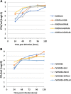
Methods: Overlapping cDNA fragments of the DK-3592B genome were cloned after RT-PCR amplification, and their DNA sequenced by the di-deoxy chain termination method. A full-length cDNA copy (pVHSVdk) of the DK-3592B strain genome was constructed by assembling six overlapping cDNA fragments by using natural or artificially created unique restriction sites in the overlapping regions of the clones. Using an existing clone of the trout-avirulent VHSV-IVb strain MI03 (pVHSVmi), eight chimeric VHSV clones were constructed in which the coding region(s) of the glycoprotein (G), non-virion protein (NV), G and NV, or G, NV and L (polymerase) genes together, were exchanged between the two clones. Ten recombinant VHSVs (rVHSVs) were generated, including two parental rVHSVs, by transfecting fish cells with ten individual full-length plasmid constructs along with supporting plasmids using the established protocol. Recovered rVHSVs were characterized for viability and growth in vitro and used to challenge groups of juvenile rainbow trout by intraperitoneal injection.
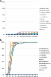
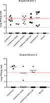
Results: Complete sequence of the VHSV DK-3592B genome was determined from the cloned cDNA and deposited in GenBank under the accession no. KC778774. The trout-virulent DK-3592B genome (genotype Ia) is 11,159 nt in length and differs from the trout-avirulent MI03 genome (pVHSVmi) by 13% at the nucleotide level. When the rVHSVs were assessed for the trout-virulence phenotype in vivo, the parental rVHSVdk and rVHSVmi were virulent and avirulent, respectively, as expected. Four chimeric rVHSVdk viruses with the substitutions of the G, NV, G and NV, or G, NV and L genes from the avirulent pVHSVmi constructs were still highly virulent (100% mortality), while the reciprocal four chimeric rVHSVmi viruses with genes from pVHSVdk remained avirulent (0-10% mortality).
Conclusions: When chimeric rVHSVs, containing all the G, NV, and L gene substitutions, were tested in vivo, they did not exhibit any change in trout-virulence relative to the background clones. These results demonstrate that the G, NV and L genes of VHSV are not, by themselves or in combination, major determinants of host-specific virulence in trout.
Keywords: Fish; Glycoprotein; Non-virion protein; Polymerase protein; Rhabdovirus; VHSV; Virulence determinant.
Conflict of interest statement
Ethics approval
Fish experiments were conducted in compliance with guidelines provided by the Guide for the Care and Use of Laboratory Animals and the United States Public Health Service Policy on the Humane Care and Use of Laboratory Animals. Fish challenge protocol was approved by the Institutional Animal Care and Use Committee of Western Fisheries Research Center and the studies were conducted in the Center’s Aquatic Biosafety Level 3 wetlab facility following strict containment procedures.
Consent for publication
Not applicable.
Competing interests
The authors declare that they have no competing interests.
Publisher’s Note
Springer Nature remains neutral with regard to jurisdictional claims in published maps and institutional affiliations.
Figures


References
-
- Smail DA, Snow M. Viral haemorrhagic septicaemia. 2011. In: Woo PTK, Bruno DW, eds. Fish diseases and disorders, Vol. 3, Viral, bacterial, and fungal infections. 2nd Edition. Wallingford, U.K.: CAB International; pp. 110–142.
-
- World Organisation for Animal Health. 2018. Manual of diagnostic tests for aquatic animals. Viral haemorrhagic septicaemia. World Organisation for Animal Health, Paris, France.
-
- ICTV Virus Taxonomy, 2018 release, available online at https://talk.ictvonline.org/taxonomy.
Publication types
MeSH terms
Substances
LinkOut - more resources
Full Text Sources
Research Materials

