Review
. 2019 Jun;27(6):497-507.
doi: 10.1016/j.tim.2019.01.008. Epub 2019 Mar 5.

Commensal Staphylococci Influence Staphylococcus aureus Skin Colonization and Disease

Affiliations
Review

Commensal Staphylococci Influence Staphylococcus aureus Skin Colonization and Disease

Corey P Parlet et al. Trends Microbiol. 2019 Jun.

Abstract

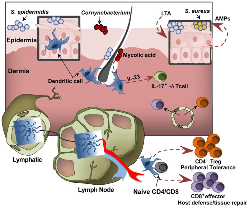
Commensal organisms that constitute the skin microbiota play a pivotal role in the orchestration of cutaneous homeostasis and immune competence. This balance can be promptly offset by the expansion of the opportunistic pathogen Staphylococcus aureus, which is responsible for the majority of bacterial skin infections. S. aureus carriage is also known to be a precondition for its transmission and pathogenesis. Recent reports suggest that skin-dwelling coagulase-negative staphylococci (CoNS) can prime the skin immune system to limit the colonization potential of invaders, and they can directly compete through production of antimicrobial molecules or through signaling antagonism. We review recent advances in these CoNS colonization resistance mechanisms, which may serve to aid development of pharmacologic and probiotic intervention strategies to limit S. aureus skin colonization and disease.

Keywords: MRSA; Staphylococcus aureus; colonization; host protection; quorum sensing; skin microbiota.


Figures

Figure 1.
Figure 1.. Skin interactions with commensal bacteria.
Enriched in the skin’s epidermal and dermal compartments, resident dendritic cells support cutaneous immune competence by shaping the functional repertoire of the skin’s T cell network. In response to encounters with commensal antigens, these skin dendritic cells migrate to the draining lymph nodes and orchestrate the priming of CD4 and CD8 T cells. The T cells fortify the cutaneous immune system through their respective roles in maintaining peripheral tolerance and mediating host immunity and tissue repair. In addition, commensal derived factors, such as mycolic acid from Corynebacterium sp., have been shown to augment specific modes of inflammation via expanding the cellular mediators of the IL-23/IL-17 inflammatory axis. Apart from altering the function of cutaneous T cells, commensal signals such as lipoteichoic acid (LTA) trigger antimicrobial peptide (AMP) production from the skin’s predominant cellular constituent, keratinocytes. Overall, the sensitivity of both innate and adaptive elements of the cutaneous immune system to specific commensal cues/signals appears to finely tune both effector and regulatory immune responses in a manner that maintains barrier integrity, encourages commensal occupation and opposes pathogen invasion.
Figure 2.
Figure 2.. The commensal skin microbiota employ diverse strategies to compete with S. aureus.
Healthy skin is populated by a diverse array of coagulase negative staphylococci (CoNS) as well as other commensal flora. CoNS compete with S. aureus on the skin by producing a wide array of small molecule products like antimicrobial peptides (AMPs) and lantibiotics. Another potential mediator of staphylococcal competition is the agr quorumsensing system. While relatively less studied than other small molecules, there is evidence to suggest that interspecies competition exists as shown by the ability of S. epidermidis to make an AIP that inhibits S. aureus quorum sensing. We recently published that another rare skin commensal, S. caprae, also makes an AIP that inhibits all classes of S. aureus quorum sensing with nanomolar potency (further described in Figure 4). Thus, non-cognate AIPs represent a potential source of competition that merits further investigation in more representative models of skin colonization.
Figure 3.
Figure 3.. Schematic of the Staphylococcal agr quorum-sensing system and signaling cross-talk.
The agr locus is conserved in all species of Staphylococci, and is composed of the agrBDCA 4-gene operon divergently transcribed from the RNAIII effector. The AgrB and AgrD proteins build the autoinducing peptide (AIP) signal, and the AgrC histidine kinase is the AIP receptor. When AIP binds to AgrC, phosphoryl transfer to AgrA occurs, inducing the P2 and P3 promoters, resulting in agrBCDA and RNAIII expression. RNAIII transcript is the primary effector of the system and upregulates toxins (in S. aureus) and exo-enzymes (in all staphylococci). The ‘cognate AIP’ shown is AIP-I from S. aureus agr type I strains (e.g. USA300), and the ‘competing AIP’ represents any signal with receptor antagonist activity. These competing AIPs can come from commensal staphylococci. This figure is adapted from [51].
Figure 4.
Figure 4.. Commensal competition with MRSA during skin infection.
This figure is adapted from our recent report demonstrating S. caprae can compete with MRSA in a skin model of infection [51]. S. caprae produces a competing AIP signal (Fig. 2) with nanomolar potency for the MRSA AgrC receptor, completely inhibiting agr function. In contrast, a similar skin commensal, S. capitis, has no ability to produce a competing AIP signal and could not prevent MRSA infection. Briefly: A. Representative images of in vivo bioluminescence induction 3.5 hours after challenge with MRSA agr P3-Lux +/− 10 μg of S. caprae AIP, or equal CFU of the CoNS S. caprae or S. capitis. B. Representative images show dermonecrosis 5 days after the bacterial challenge. C. Time course comparison of in vivo bioluminescence after intradermal challenge with the indicated conditions. Error bars represent SEM. Post test p value (*)=<0.05, (**)=<0.01, (***)=<0.005. D. Time course of dermonecrotic lesion size in the indicated challenge conditions. Error bars represent SEM. Post test p value (*)=<0.05, (**)=<0.01.

References

    1. Gallo RL (2017) Human Skin Is the Largest Epithelial Surface for Interaction with Microbes. J Invest Dermatol 137 (6), 1213–1214. - PMC - PubMed
    1. Christensen GJ and Bruggemann H (2014) Bacterial skin commensals and their role as host guardians. Benef Microbes 5 (2), 201–15. - PubMed
    1. Grice EA and Segre JA (2011) The skin microbiome. Nat Rev Microbiol 9 (4), 244–53. - PMC - PubMed
    1. Byrd AL et al. (2018) The human skin microbiome. Nat Rev Microbiol 16 (3), 143–155. - PubMed
    1. Oh J et al. (2016) Temporal Stability of the Human Skin Microbiome. Cell 165 (4), 854–66. - PMC - PubMed

Publication types

MeSH terms