

Interhemispheric plasticity is mediated by maximal potentiation of callosal inputs
- PMID: 30846552
- PMCID: PMC6442599
- DOI: 10.1073/pnas.1810132116
Interhemispheric plasticity is mediated by maximal potentiation of callosal inputs
Abstract
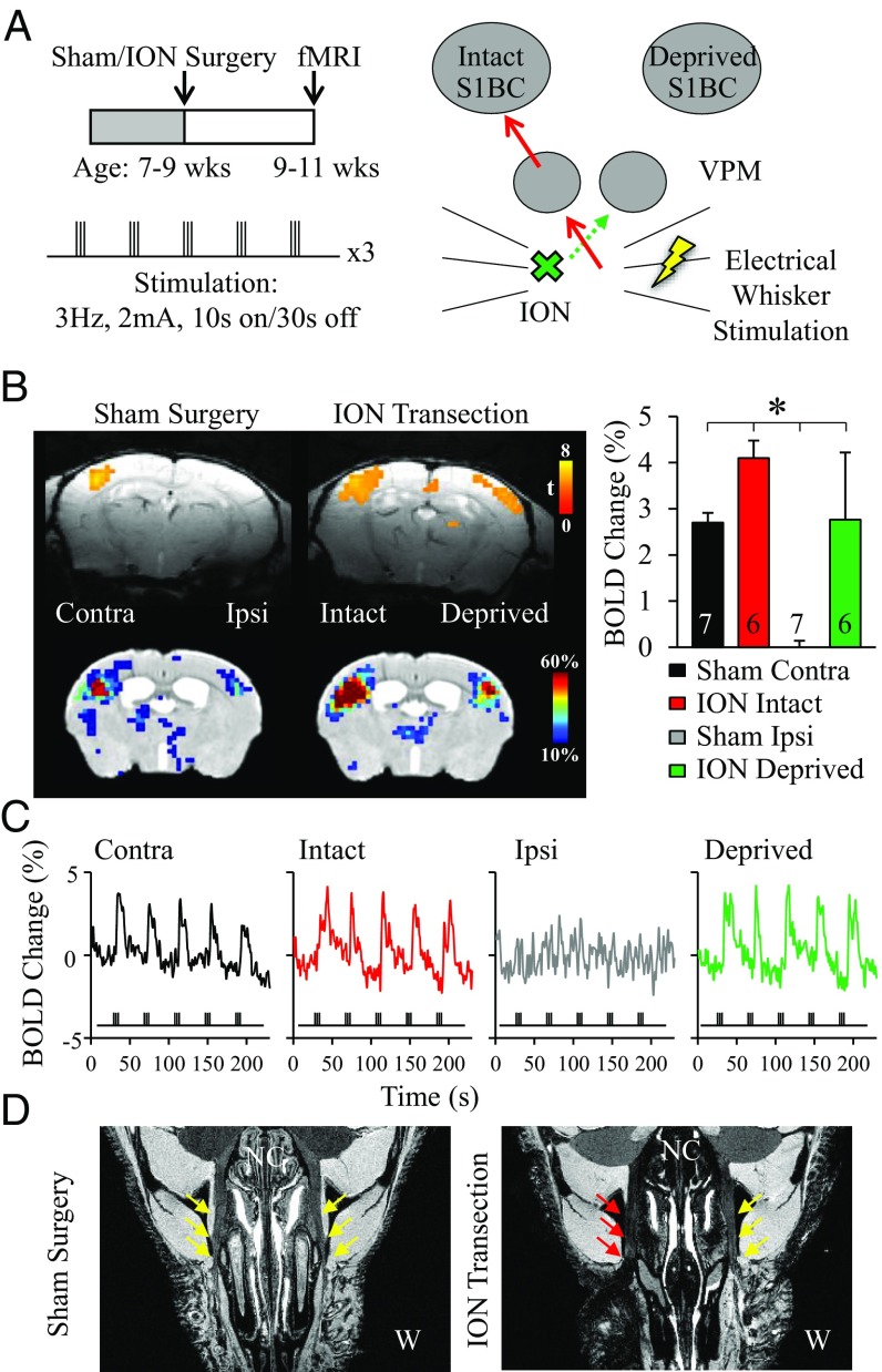
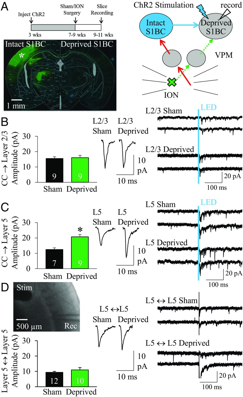
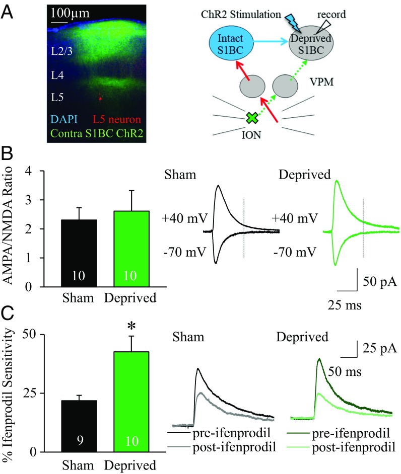
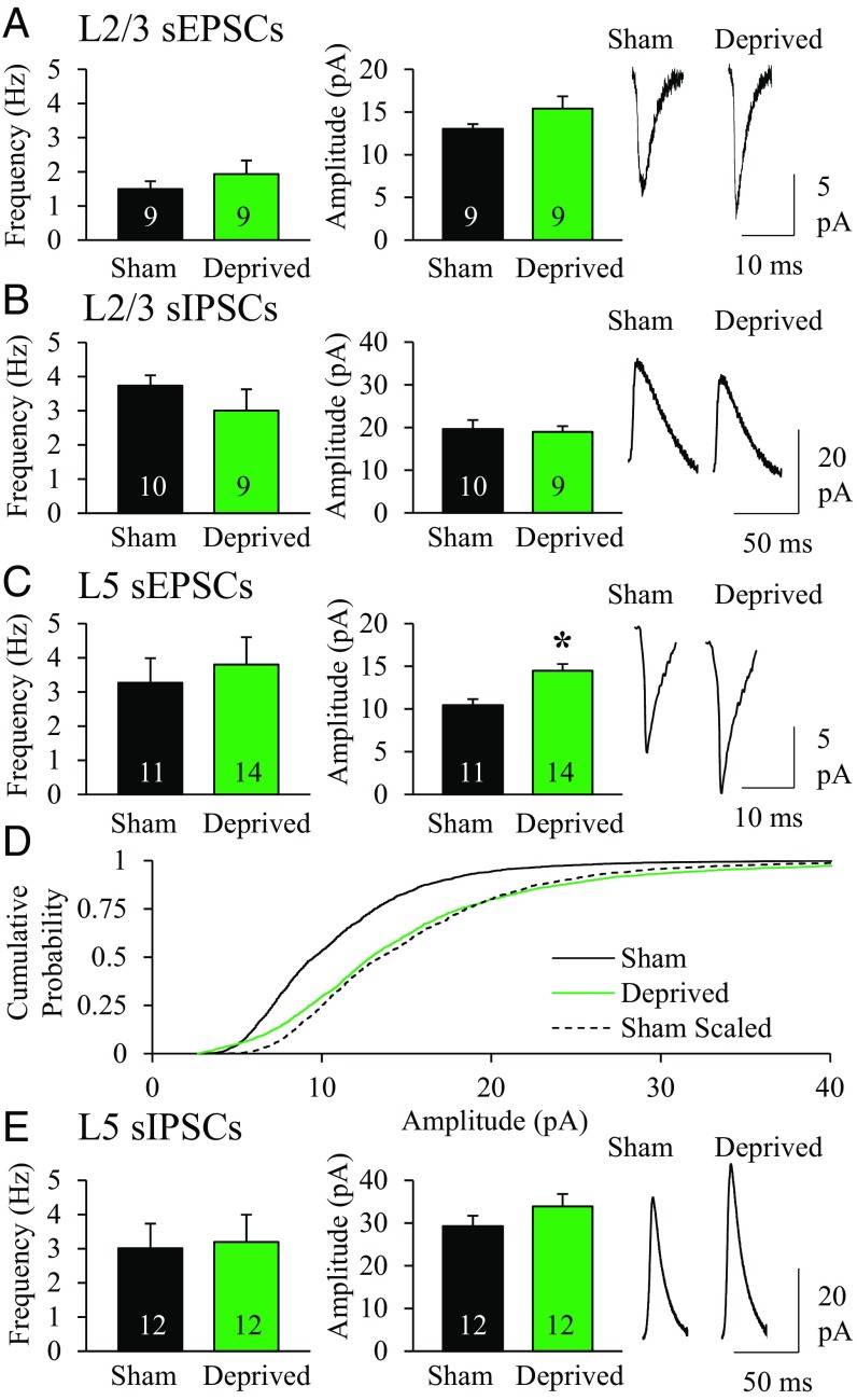
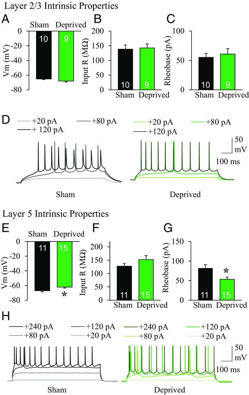
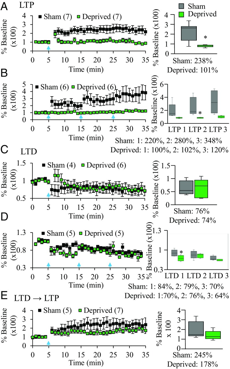
Central or peripheral injury causes reorganization of the brain's connections and functions. A striking change observed after unilateral stroke or amputation is a recruitment of bilateral cortical responses to sensation or movement of the unaffected peripheral area. The mechanisms underlying this phenomenon are described in a mouse model of unilateral whisker deprivation. Stimulation of intact whiskers yields a bilateral blood-oxygen-level-dependent fMRI response in somatosensory barrel cortex. Whole-cell electrophysiology demonstrated that the intact barrel cortex selectively strengthens callosal synapses to layer 5 neurons in the deprived cortex. These synapses have larger AMPA receptor- and NMDA receptor-mediated events. These factors contribute to a maximally potentiated callosal synapse. This potentiation occludes long-term potentiation, which could be rescued, to some extent, with prior long-term depression induction. Excitability and excitation/inhibition balance were altered in a manner consistent with cell-specific callosal changes and support a shift in the overall state of the cortex. This is a demonstration of a cell-specific, synaptic mechanism underlying interhemispheric cortical reorganization.
Keywords: corpus callosum; cortical circuit; interhemispheric plasticity.
Copyright © 2019 the Author(s). Published by PNAS.
Conflict of interest statement
The authors declare no conflict of interest.
Figures
Similar articles
-
Circuit-Specific Plasticity of Callosal Inputs Underlies Cortical Takeover.J Neurosci. 2020 Sep 30;40(40):7714-7723. doi: 10.1523/JNEUROSCI.1056-20.2020. Epub 2020 Sep 10. J Neurosci. 2020. PMID: 32913109 Free PMC article.
-
Potentiation of convergent synaptic inputs onto pyramidal neurons in somatosensory cortex: dependence on brain wave frequencies and NMDA receptor subunit composition.Neuroscience. 2014 Jul 11;272:271-85. doi: 10.1016/j.neuroscience.2014.04.062. Epub 2014 May 9. Neuroscience. 2014. PMID: 24814019
-
Time course of experience-dependent synaptic potentiation and depression in barrel cortex of adolescent rats.J Neurophysiol. 1996 Apr;75(4):1714-29. doi: 10.1152/jn.1996.75.4.1714. J Neurophysiol. 1996. PMID: 8727408
-
Integrated technology for evaluation of brain function and neural plasticity.Phys Med Rehabil Clin N Am. 2004 Feb;15(1):263-306. doi: 10.1016/s1047-9651(03)00124-4. Phys Med Rehabil Clin N Am. 2004. PMID: 15029909 Review.
-
Intracortical processes regulating the integration of sensory information.Prog Brain Res. 1990;86:129-41. doi: 10.1016/s0079-6123(08)63172-6. Prog Brain Res. 1990. PMID: 1982365 Review.
Cited by
-
Characterization of brain-wide somatosensory BOLD fMRI in mice under dexmedetomidine/isoflurane and ketamine/xylazine.Sci Rep. 2021 Jun 23;11(1):13110. doi: 10.1038/s41598-021-92582-5. Sci Rep. 2021. PMID: 34162952 Free PMC article.
-
Circuit-Specific Plasticity of Callosal Inputs Underlies Cortical Takeover.J Neurosci. 2020 Sep 30;40(40):7714-7723. doi: 10.1523/JNEUROSCI.1056-20.2020. Epub 2020 Sep 10. J Neurosci. 2020. PMID: 32913109 Free PMC article.
-
Cortical HFS-Induced Neo-Hebbian Local Plasticity Enhances Efferent Output Signal and Strengthens Afferent Input Connectivity.eNeuro. 2025 Feb 10;12(2):ENEURO.0045-24.2024. doi: 10.1523/ENEURO.0045-24.2024. Print 2025 Feb. eNeuro. 2025. PMID: 39809536 Free PMC article.
-
Targeting microglial NAAA-regulated PEA signaling counters inflammatory damage and symptom progression of post-stroke anxiety.Cell Commun Signal. 2025 May 1;23(1):211. doi: 10.1186/s12964-025-02202-2. Cell Commun Signal. 2025. PMID: 40312408 Free PMC article.
-
Symmetry in frontal but not motor and somatosensory cortical projections.bioRxiv [Preprint]. 2024 May 29:2023.06.02.543431. doi: 10.1101/2023.06.02.543431. bioRxiv. 2024. Update in: J Neurosci. 2024 Aug 14;44(33):e1195232024. doi: 10.1523/JNEUROSCI.1195-23.2024. PMID: 37398221 Free PMC article. Updated. Preprint.
References
-
- Cao Y, Vikingstad EM, George KP, Johnson AF, Welch KMA. Cortical language activation in stroke patients recovering from aphasia with functional MRI. Stroke. 1999;30:2331–2340. - PubMed
-
- Kinsbourne M. The minor cerebral hemisphere as a source of aphasic speech. Arch Neurol. 1971;25:302–306. - PubMed
-
- Grefkes C, et al. Cortical connectivity after subcortical stroke assessed with functional magnetic resonance imaging. Ann Neurol. 2008;63:236–246. - PubMed
-
- Rehme AK, Fink GR, von Cramon DY, Grefkes C. The role of the contralesional motor cortex for motor recovery in the early days after stroke assessed with longitudinal FMRI. Cereb Cortex. 2011;21:756–768. - PubMed
-
- Shuler MG, Krupa DJ, Nicolelis MAL. Integration of bilateral whisker stimuli in rats: Role of the whisker barrel cortices. Cereb Cortex. 2002;12:86–97. - PubMed
Publication types
MeSH terms
Substances
LinkOut - more resources
Full Text Sources
