. 2019 Mar 8;363(6431):eaat4042.
doi: 10.1126/science.aat4042.

Endocytosis of commensal antigens by intestinal epithelial cells regulates mucosal T cell homeostasis

Affiliations

Endocytosis of commensal antigens by intestinal epithelial cells regulates mucosal T cell homeostasis

Mark S Ladinsky et al. Science. .

Abstract

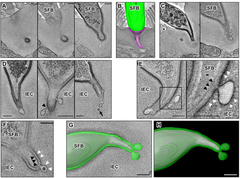
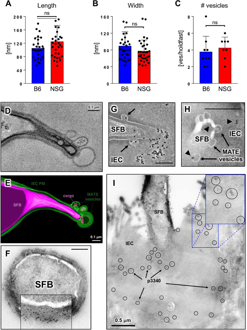
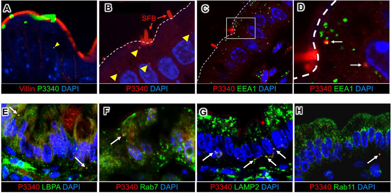
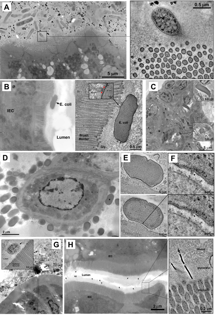
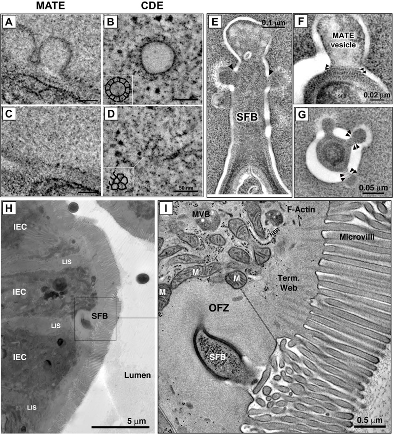
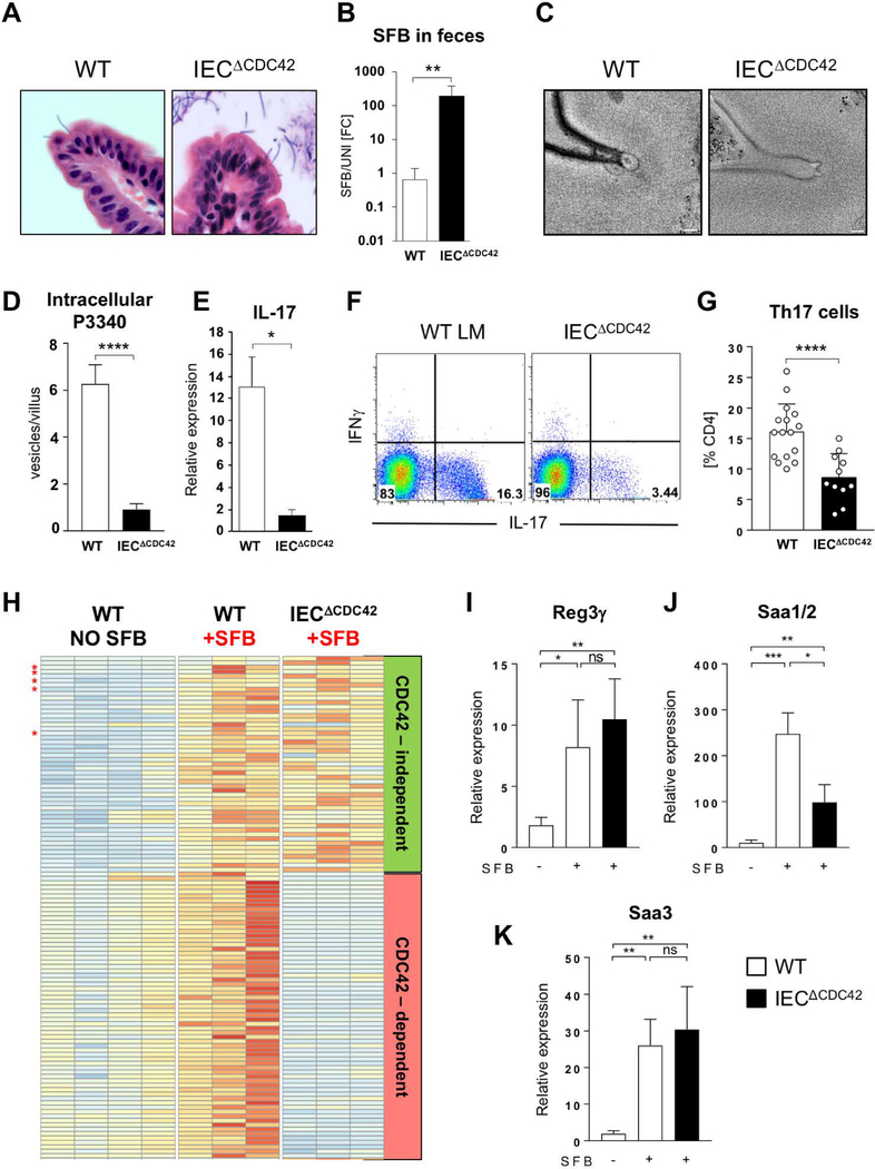
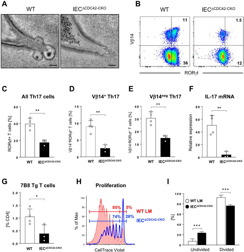
Commensal bacteria influence host physiology, without invading host tissues. We show that proteins from segmented filamentous bacteria (SFB) are transferred into intestinal epithelial cells (IECs) through adhesion-directed endocytosis that is distinct from the clathrin-dependent endocytosis of invasive pathogens. This process transfers microbial cell wall-associated proteins, including an antigen that stimulates mucosal T helper 17 (TH17) cell differentiation, into the cytosol of IECs in a cell division control protein 42 homolog (CDC42)-dependent manner. Removal of CDC42 activity in vivo led to disruption of endocytosis induced by SFB and decreased epithelial antigen acquisition, with consequent loss of mucosal TH17 cells. Our findings demonstrate direct communication between a resident gut microbe and the host and show that under physiological conditions, IECs acquire antigens from commensal bacteria for generation of T cell responses to the resident microbiota.


Conflict of interest statement

Competing interests: K.H. is a scientific advisory board member at Vedanta Biosciences.

Figures

Fig. 1.
Fig. 1.. Microbial Adhesion-Triggered Endocytosis (MATE) is induced following attachment of commensal segmented filamentous bacteria (SFB) to intestinal epithelial cells (IECs).
(A) Consecutive sections of an electron tomogram of an SFB-IEC synapse showing that double membrane phagosome-like vesicles (left panel) represent invaginations of the IEC plasma membrane. (B) A 3D reconstruction of an SFB-IEC holdfast, demonstrating separation of the IEC plasma membrane (PM) in purple and the SFB PM in green/gray. SFB do not penetrate the IEC PM. (C) Membrane vesicles at the tip of SFB holdfasts. (D) Holdfast vesicles form necks (arrowhead) and bud off (arrow) of the IEC PM into the IEC cytosol. (E, F) SFB PM (black arrowheads) remains uninterrupted and holdfast vesicles form exclusively from the host IEC PM (white arrowheads) and contain electron dense cargo (F). (G, H) Reconstruction of an SFB holdfast. IEC PM in green, SFB PM in gray. All scale bars are 200 nm.
Fig. 2.
Fig. 2.. MATE vesicles transfer an immunodominant SFB antigen inside IECs.
(A-C) Quantification of MATE vesicle size (A, B) and numbers per holdfast (C) in the terminal ileum of C57BL/6 (B6) and NOD.Scid.Il2rgnull (NSG) mice. Error bars, standard deviation. Statistics, unpaired two-tailed t test. (D, E) Single section and 3D reconstruction of an SFB-IEC synapse featuring MATE vesicles that contain electron dense cargo. Green, IEC PM; magenta, SFB PM. (F) Immuno-EM for P3340 on SFB in mouse intestine. Cross-section of an SFB cell. P3340 is present exclusively on the cell-wall of the bacterium. (G, H, I) Immuno-EM for P3340 on intestinal sections from terminal ileum. (G) An SFB cell interacting with a single IEC transfers P3340 into the IEC cytosol via MATE. Arrows, MATE vesicles containing P3340 labeling. (H) A close-up of the distal end of an SFB holdfast showing P3340 presence (black arrowheads) on the microbe, in a MATE vesicle, and inside the IEC cytosol. (I) Immuno-EM of P3340 in terminal ileum of C57BL/6 mice colonized with SFB. An IEC with an attached SFB is shown. P3340 immunogold labeling is present on the cell wall of the bacteria, as well as inside the IEC cytosol in the vicinity of the SFB-IEC synapse (black circles and arrows). All scale bars are 200 nm unless otherwise noted.
Fig 3.
Fig 3.. SFB antigens are shuttled through the IEC endosomal-lysosomal vesicular network.
(A, B) Immunofluorescence for P3340 on intestinal sections from terminal ileum showing SFB P3340 inside IECs (yellow arrowheads). (C, D) P3340 is present in EEA1+ early endosomes. White dashed line outlines the apical IEC surface. (E-H) Co-localization of intracellular SFB protein P3400 (red) with endosomal and lysosomal markers (green). P3340 co-localizes with late endosomes (LBPA, Rab7) (E, F), as well as basolateral lysosomes (LAMP2) (G). (H) Some P3340 also co-localizes with recycling endosomes (Rab11).
Fig 4.
Fig 4.. Electron tomography of host-microbe interactions in the intestine.
(A) Microbiota in the terminal ileum of SFB-negative Jackson C57BL/6 mice posess limited interactions with IECs and lack MATE. Inset: rare interaction between a bacterial cell and IEC microvilli. (B,C) Interactions of adherent-invasive Escherichia coli (AIEC) strains LF82 (B) and NRG857c (C) with IECs in terminal ileum. (A, right) Tomogram showing interaction with microvilli. (C) Internalized AIEC cells. (D) Citrobacter rodentium, overview showing numerous bacterial cells associating with IEC surface in the colon. (E) Tomograms of a Citrobacter pedestal. (F) Tomographic details of a presumptive T3SS needle connecting the microbe to IEC (arrowheads). (G) Mixture of 20 human Th17 cell-inducing strains. Inset; tomogram showing bacterial cells in the lumen, close to microvilli, but not making contact with IECs. (H) Bifidobacterium adolescentis (arrowheads) in terminal ileum. Inset: tomogram of B. adolescentis showing penetration of the glycocalyx and interaction with the IEC microvilli.
Fig 5.
Fig 5.. MATE is a type of clathrin-independent endocytosis.
(A-D) MATE vesicles do not contain clathrin coat. Tomographic reconstruction of cytoplasmic MATE vesicles (A, C) and clathrin-coated vesicles in cultured HeLa cells (B, D). (A, B) Tomographic slices taken near the equator of the vesicle. Clathrin spikes (arrows) are present on the surface in (B), but absent in (A). (C, D) Tomographic slices taken at the surface of the vesicles, showing clathrin cage composed of triskelions in (D), and lack of coating in (C). Insets; manual segmentation of the clathrin cage to highlight its features. (E-G) MATE vesicles contain dynamin-like rings. (E) Tomographic slice of an SFB holdfast interacting with an intestinal epithelial cell, showing thin bands (arrowheads) circumscribing the necks of MATE vesicles. (F, G) High-magnification details from negative-stain tomograms showing MATE vesicles in longitudinal (F) and cross (G) section. Dynamin-like bands are clearly visible circumscribing the necks of the vesicles (arrowheads). (H, I) SFB induce reorganization of the IEC actin cytoskeleton. An overview (H) and magnification (I) showing an interaction between SFB and an intestinal epithelial cell (IEC) in the terminal ileum. Reorganization of actin filaments in the IEC terminal web is evident immediately around the SFB holdfast. This reorganization seems to result in an organelle free zone (OFZ). The demarcation between normal terminal web and the OFZ is noted by a thin doubled dashed black line in the right panel. The OFZ contains vesicles and actin filaments organized differently than the terminal web in the neighboring area. LIS, lateral intercellular space; MVB, multivesicular body; M, mitochondrion.
Fig 6.
Fig 6.. MATE is CDC42-dependent and dynamin-dependent.
(A, B) Quantification of active CDC42-GTP and CDC42EP1 expression in IECs isolated from terminal ileum of C57BL/6J mice 10 days after colonization with SFB. CDC42-GTP levels in (A) were normalized to total CDC42 and CDC42EP1 levels in (B) were normalized to β-actin. Data points represent individual animals. (C-F) MATE is CDC42-dependent and dynamin-dependent. NSG mice were colonized with SFB for at least one month. After establishment of persistent colonization, chemical inhibitors were introduced into externalized terminal ileum intestinal loops of live animals by perfusion in vivo as described in Methods. (C) Representative electron tomograms of SFB-IEC synapses. All scale bars are 200 nm. (D-F) Quantification of MATE vesicle morphology. Data points represent individual MATE vesicles. All error bars, standard deviation. Statistics, (A, D-F) unpaired two-tailed t test, (B) Mann-Whitney-Wilcoxon test.
Fig 7.
Fig 7.. Epithelial CDC42 is required for MATE, SFB antigen acquisition and Th17 cell induction by SFB.
(A) H&E staining of sections from terminal ileum of WT and IECΔCDC42 mice. SFB attachment on the surface of the villi is evident in both groups. (B) SFB levels in feces from WT and IECΔCDC42 mice. (C) MATE vesicles in SFB-IEC synapses in terminal ileum of WT and IECΔCDC42 mice. (D) Decrease in acquisition of P3340 by IEC in terminal ileum of IECΔCDC42 mice. (E) Decrease in IL-17 mRNA in terminal ileum of SFB-colonized IECΔCDC42 mice. (F, G) Decrease in small intestinal lamina propria (LP) Th17 cells in IECΔCDC42 mice. Plots in (F) gated on TCRβ+CD4+ lymphocytes. (G) Combined data from several independent experiments. (H) Relative expression levels of SFB-induced genes in IECs from WT and IECΔCDC42 mice. Only genes induced by SFB are depicted. Red stars indicate Saa1, Saa2, Saa3, Nos2, and Reg3g. Complete list of gene names is included in Fig. S9. (I-K) RT-PCR for SFB-controlled genes on RNA isolated from IECs from WT and IECΔCDC42 mice as described in Materials and Methods. Statistics, unpaired t test. * p < 0.05, ** p < 0.01, *** p < 0.005, **** p < 0.001. Scale bars in (C) are 100 nm.
Fig. 8.
Fig. 8.. Epithelial CDC42 is required for activation of SFB-specific CD4 T cells and induction of SFB-specific Th17 cells.
WT and IECΔCDC42-CKO mice were treated with tamoxifen prior to SFB colonization and transfer of naïve P3340-specific 7B8 Tg CD4 T cells (for details see Materials and Methods). (A) MATE vesicles in SFB-IEC synapses in terminal ileum of tamoxifen-treated WT and IECΔCDC42-CKO mice. (B-E) Decrease in endogenous SI LP Th17 cells in tamoxifen-treated IECΔCDC42-CKO mice six days after SFB colonization. (B) Representative FACS plots of LP lymphocytes gated on TCRβ+CD4+ cells. (C-E) Statistic based on the gating in (B). (F) Decrease in IL-17 mRNA in terminal ileum of tamoxifen-treated IECΔCDC42-CKO mice. (G) Decreased expansion of adoptively transferred P3340-specific 7B8 Tg CD4 T cells in mesenteric lymph nodes (MLN) of tamoxifen-treated IECΔCDC42-CKO mice four days after transfer. (H, I) Proliferation of 7B8 Tg CD4 T cells in MLN of tamoxifen-treated WT littermate (WT LM) or IECΔCDC42-CKO recipient mice on Day 4 after adoptive transfer. One of three independent experiments with similar results. Error bars, standard deviation. Statistics, unpaired two-tailed t test. * p < 0.05, ** p < 0.01, *** p < 0.005, **** p < 0.001. Scale bars in (A) are 100 nm.

References

    1. Hooper LV, Macpherson AJ, Immune adaptations that maintain homeostasis with the intestinal microbiota. Nat Rev Immunol 10, 159–169 (2010). - PubMed
    1. Furusawa Y et al. , Commensal microbe-derived butyrate induces the differentiation of colonic regulatory T cells. Nature 504, 446–450 (2013). - PubMed
    1. Smith PM et al. , The microbial metabolites, short-chain fatty acids, regulate colonic Treg cell homeostasis. Science 341, 569–573 (2013). - PMC - PubMed
    1. Arpaia N et al. , Metabolites produced by commensal bacteria promote peripheral regulatory T-cell generation. Nature 504, 451–455 (2013). - PMC - PubMed
    1. Shen Y et al. , Outer membrane vesicles of a human commensal mediate immune regulation and disease protection. Cell host & microbe 12, 509–520 (2012). - PMC - PubMed

Publication types