. 2019 Mar 7;10(1):973.
doi: 10.1038/s41467-019-08858-y.

Brain tyrosinase overexpression implicates age-dependent neuromelanin production in Parkinson's disease pathogenesis

Affiliations

Brain tyrosinase overexpression implicates age-dependent neuromelanin production in Parkinson's disease pathogenesis

Iria Carballo-Carbajal et al. Nat Commun. .

Abstract

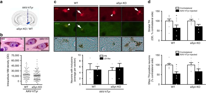
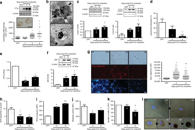
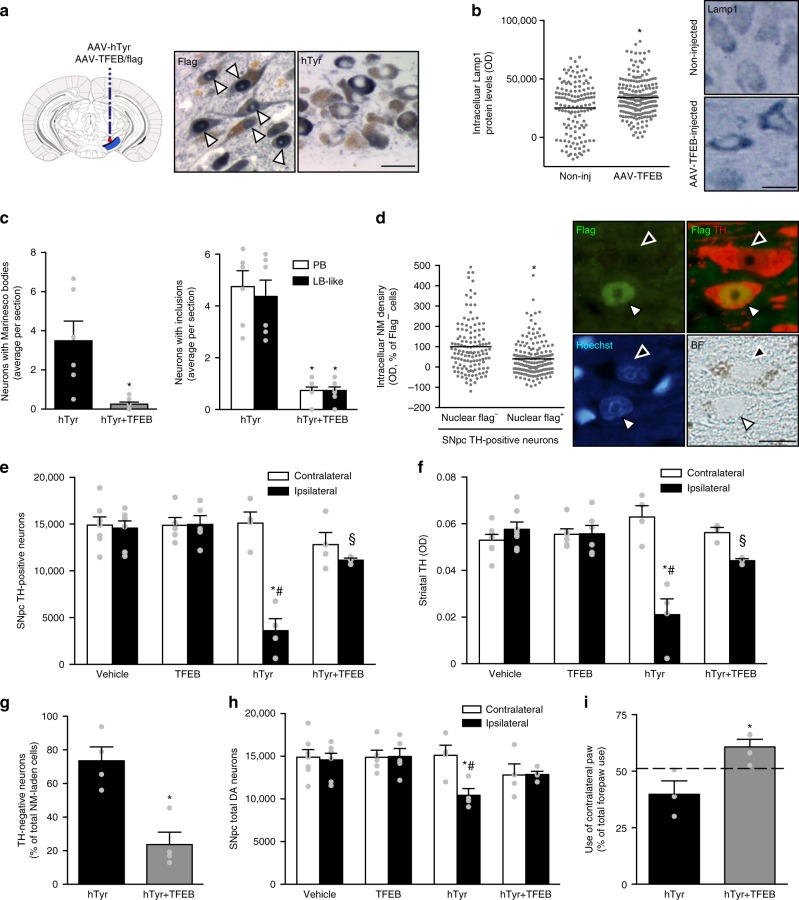
In Parkinson's disease (PD) there is a selective degeneration of neuromelanin-containing neurons, especially substantia nigra dopaminergic neurons. In humans, neuromelanin accumulates with age, the latter being the main risk factor for PD. The contribution of neuromelanin to PD pathogenesis remains unknown because, unlike humans, common laboratory animals lack neuromelanin. Synthesis of peripheral melanins is mediated by tyrosinase, an enzyme also present at low levels in the brain. Here we report that overexpression of human tyrosinase in rat substantia nigra results in age-dependent production of human-like neuromelanin within nigral dopaminergic neurons, up to levels reached in elderly humans. In these animals, intracellular neuromelanin accumulation above a specific threshold is associated to an age-dependent PD phenotype, including hypokinesia, Lewy body-like formation and nigrostriatal neurodegeneration. Enhancing lysosomal proteostasis reduces intracellular neuromelanin and prevents neurodegeneration in tyrosinase-overexpressing animals. Our results suggest that intracellular neuromelanin levels may set the threshold for the initiation of PD.


Conflict of interest statement

VHIR has applied for a use patent for the modulation of neuromelanin levels in the treatment of Parkinson's disease and/or brain aging.

Figures

Fig. 1
Fig. 1
Human-like NM production in AAV-hTyr-injected rats. a Schematic representation of the site of AAV-hTyr unilateral stereotaxic injection above the SNpc of the rat brain. b Representative photomicrographs of a 30-μm-thick ipsilateral SNpc section from an AAV-hTyr-injected rat (1 m post-AAV injection) immunostained for TH (red) and hTyr (green). Scale bar, 500 μm. c Left, representative unstained AAV-hTyr-injected rat brain (2 m post-AAV injection) mounted in a cryostat in which ipsilateral SNpc can be detected macroscopically as a brown, darkened area (dashed outline). A hole was made in the contralateral hemisphere as anatomical reference. Right, representative unstained midbrain from a 62-year-old human control subject (Hu) in which the SNpc can be detected macroscopically (bilateral dashed outlines). d Representative NM-sensitive high-resolution T1-weighted magnetic resonance imaging of an AAV-hTyr-injected rat brain at 2 m post-AAV injection (left, ex-vivo) and of a 59-year-old human control brain (Hu, right, in vivo). SNpc can be detected as a unilateral (AAV-hTyr-injected rodent) or bilateral (human) hyperintense area (dashed outlines). e Representative photomicrograph of an unstained 30-μm-thick ipsilateral SNpc section from an AAV-hTyr-injected rat (2 m post-AAV injection) in which NM is shown in brown. Inset, high magnification of a melanized neuron. Scale bars, 100 μm and 12.5 μm (inset). f Representative photomicrographs of Masson-Fontana melanin staining (NM in dark brown) in 5-μm-thick SNpc sections from an AAV-hTyr-injected rat at 2 m post-AAV injection (left) and an 80-year-old human control subject (Hu, right). Scale bars, 25 μm (left) and 12.5 μm (right). g Representative electron micrograph of NM granules in the ipsilateral SNpc of an AAV-hTyr-injected rat at 4 m post-AAV injection (left) and a 75-year-old human control subject (Hu, right). NM pigment is detected as an electron dense matrix. Characteristic associated lipid droplets are indicated with asterisks. Scale bars, 500 nm
Fig. 2
Fig. 2
Age-dependent NM accumulation in AAV-hTyr-injected rats. a Hematoxylin-eosin (H&E)-stained brain sections showing progressive intracellular NM accumulation (brown) within ipsilateral SNpc DA neurons from AAV-hTyr-injected rats. Scale bar, 12.5 μm. b Quantification of intracellular NM optical density in ipsilateral SNpc DA neurons of AAV-hTyr-injected rats. *p < 0.05, compared to 0.5 m; #p < 0.05, compared to 1 m; §p < 0.05, compared to 2&4 m. c Left, quantification of intracellular NM optical density in post-mortem SNpc sections from elderly human (average 80 years old) control subjects, age-matched ILBD subjects and age-matched idiopathic PD patients. *p < 0.05, compared to control subjects. Right, H&E-stained human brain sections. Scale bar, 12.5 μm. In b, c values are mean ± SEM. In b, n = 163 neurons from n = 7 rats (0.5 m), n = 136 neurons from n = 5 rats (1 m), n = 174 neurons from n = 5 rats (2 m), n = 162 neurons from n = 7 rats (4 m), n = 172 neurons from n = 6 rats (12 m), n = 179 neurons from n = 6 rats (24 m). In c, n = 1436 neurons from n = 6 control subjects, n = 640 neurons from n = 3 ILBD subjects, n = 644 neurons from n = 10 PD subjects. See Supplementary Table 1 for additional information on the human subjects used for the analyses in c. Statistical analyses: ANOVA on ranks; Dunn’s post-hoc test. Photomicrographs correspond to 5-μm-thick sections
Fig. 3
Fig. 3
Progressive nigrostriatal degeneration in AAV-hTyr-injected rats. a Left, ipsilateral SNpc and striatum (inset) sections immunostained for TH (blue). Right, ipsilateral SNpc sections stained with Masson-Fontana (NM in dark brown; inset, higher magnification of melanized neurons). Scale bars, 300 μm (left), 2 mm (left inset), 150 μm (right), and 12.5 μm (right inset). b Stereological cell counts of SNpc TH-positive neurons in AAV-hTyr-injected rats. *p < 0.05, compared to respective contralateral (non-injected) side; #p ≤ 0.05, compared to ipsilateral naive, 0.5, 1, and 2 m; §p < 0.05, compared to ipsilateral 4 and 12 m (two-way ANOVA; Student–Newman–Keuls post-hoc test). c Optical densitometry of striatal TH-positive fibers in AAV-hTyr-injected rats. *p < 0.05, compared to 0.5 m; #p < 0.05, compared to naive (ANOVA on ranks; Dunn’s post-hoc test). d Stereological cell counts of SNpc NM + /TH + (white arrowhead) and NM + /TH− (black arrowhead) neurons vs total NM + neurons in AAV-hTyr-injected rats. *p < 0.05, compared to 0.5 m; #p < 0.05, compared to 1 and 2 m; §p < 0.05, compared to 12 m (one-way ANOVA; Student–Newman–Keuls post-hoc test). e Stereological cell counts of total SNpc DA neurons (including TH-immunopositive and TH-immunonegative melanized neurons) in AAV-hTyr-injected rats. *p < 0.05, compared to respective contralateral side; #p < 0.05, compared to ipsilateral naive, 0.5 and 1 m; §p < 0.05, compared to ipsilateral 2, 4, and 12 m (two-way ANOVA; Student–Newman–Keuls post-hoc test). In all panels, values are mean ± SEM. In ae, n = 6 (Naïve), n = 8 (0.5 m), n = 7 (1 m), n = 5 (2 m), n = 8 (4 m), n = 7 (12 m), and n = 6 (24 m). Photomicrographs correspond to 5-μm-thick sections
Fig. 4
Fig. 4
Early dopaminergic dysfunction in AAV-hTyr-injected rats. a Contralateral forepaw use in AAV-hTyr- and AAV-EV-injected rats, as assessed with the cylinder test. *p ≤ 0.05, compared to AAV-EV-injected animals at the same time-point; #p < 0.05, compared to 0.5 m AAV-hTyr animals (two-way ANOVA; Student–Newman–Keuls post-hoc test). b Striatal DA release in AAV-hTyr- and vehicle-injected rats measured by microdialysis in the ipsilateral striatum following local amphetamine (left) or veratridine (right) administration by reverse-dialysis. *p < 0.05, compared to vehicle-injected animals (ANOVA for repeated measures of the DA values during the specified time periods; Tukey’s post-hoc test). Baseline DA concentration (fmol/fraction-20 min): amphetamine experiments, 82.83 ± 19.16 (vehicle) vs 94.38 ± 21.82 (AAV-hTyr); veratridine experiments, 25.52 ± 4.33 (vehicle) vs 29.93 ± 6.46 (AAV-hTyr). c Striatal DA release in AAV-hTyr- and vehicle-injected rats measured by microdialysis in the ipsilateral striatum following electrical stimulation of the MFB for 10-min periods under S1 and S2 conditions (S1, 2.0 Hz, 0.1 mA, 0.2 ms; S2, 10 Hz, 0.1 mA, 1 ms). *p < 0.05, compared to stimulated AAV-hTyr-injected rats (ANOVA for repeated measures; Tukey’s post-hoc test). Baseline DA concentration (fmol/fraction-10 min): 16.8 ± 2.38 (vehicle) vs 23.02 ± 6.37 (AAV-hTyr). In all panels, values are mean ± SEM. In a, AAV-EV-injected rats n = 20 (0.5 m), n = 20 (1 m), n = 9 (2 m), n = 9 (3 m), n = 8 (6 m), n = 12 (12 m), n = 4 (24 m); and AAV-hTyr-injected rats n = 29 (0.5 m), n = 8 (1 m), n = 21 (2 m), n = 14 (3 m), n = 7 (6 m), n = 14 (12 m), n = 6 (24 m). In b, c, experiments were performed at 1-2 m post-AAV injection. In b, n = 6 vehicle-injected, n = 6 AAV-hTyr-injected rats + Amphetamine and n = 8 AAV-hTyr-injected rats + Veratridine. In c, n = 4 animals per group
Fig. 5
Fig. 5
Extracellular NM and neuronophagia in AAV-hTyr-injected rats. a Left, extracellular NM aggregates in ipsilateral SNpc from AAV-hTyr-injected rats. *p < 0.05, compared to 0.5&1 m; #p < 0.05, compared to 2 m (one-way ANOVA; Student–Newman–Keuls post-hoc test). Middle, hematoxylin-eosin (H&E)-stained ipsilateral SNpc sections from AAV-hTyr-injected rats (4 m). Scale bar, 25 μm. Right, Pearson correlation analysis between total SNpc DA neurons and extracellular NM. b Left, neuronophagia in ipsilateral SNpc from AAV-hTyr-injected rats. *p < 0.05, compared to 0.5 m (ANOVA on ranks; Dunn’s post-hoc test). Middle, H&E-stained ipsilateral SNpc sections from AAV-hTyr-injected rats (4 and 12 m). Scale bar, 62.5 μm. Right, Pearson correlation analysis between extracellular NM and neuronophagia. c H&E-stained midbrain sections from PD post-mortem brains and AAV-hTyr-injected rats. Scale bars, 12.5 μm (top & bottom) and 25 μm (middle). d Ipsilateral SNpc sections from AAV-hTyr-injected rats (2–12 m) immunostained for the microglial marker Iba1 (blue, top) or the macrophage marker CD68 (blue, bottom). Scale bars, 375 μm (Iba1, low magnification), 12.5 μm (Iba1, high magnification), 150 μm (CD68, low magnification), and 25 μm (CD68, high magnification). e Ipsilateral SNpc sections from AAV-hTyr-injected rats (4 m) immunostained for TH (red) and Iba1 (green) and co-stained with the nuclear marker Hoechst (blue). Arrowheads, intact TH-positive NM-laden neurons; single arrow, neuronophagia directed at extracellular NM debris; double arrows, neuronophagia directed at a TH-immunonegative NM-laden neuron. Scale bar, 25 μm. f Top, Ipsilateral SNpc section from an AAV-hTyr-injected rat (4 m) immunostained with Iba1 (green) and astrocytic marker GFAP (purple), co-stained with blood vessel marker tomato lectin (red) and Hoechst (blue). Arrowheads, microglial cells carrying NM towards blood vessels. Arrow, perivascular NM. Scale bar, 25 μm. Bottom, Pearson correlation analysis between neuronophagia and perivascular NM. In a, b histograms, values are mean ± SEM. In a, n = 8 (0.5 m), n = 7 (1 m), n = 5 (2 m), n = 8 (4 m), n = 7 (12 m), n = 6 (24 m). In b, n = 6 (0.5 m), n = 5 (1 m), n = 5 (2 m), n = 7 (4 m), n = 6 (12 m), n = 6 (24 m). For Pearson correlation analyses each point represents the average value for the corresponding parameter at any given time post-AAV-hTyr injection (0.5, 1, 2, 4, 12, and 24 m). BF bright-field. In all panels, unstained NM appears as brown. Photomicrographs correspond to 5-μm-thick sections
Fig. 6
Fig. 6
PD-type inclusion formation in AAV-hTyr-injected rats. a Quantification of NM-laden neurons with p62-positive intranuclear Marinesco bodies (MB) in ipsilateral SNpc from AAV-hTyr-injected rats. *p < 0.05, compared to 0.5 m (ANOVA on ranks; Dunn’s post-hoc test). b Quantification of NM-laden neurons with p62-positive total intracytoplasmic inclusion bodies (left) and pale bodies (PB) and/or Lewy body (LB)-like p62-positive inclusions (right) in ipsilateral SNpc from AAV-hTyr-injected rats. Left, *p < 0.05, compared to 0.5, 1, 4, 12, and 24 m (one-way ANOVA; Student–Newman–Keuls post-hoc test). Right, *p < 0.05, compared to PB at 0.5, 4, 12, and 24 m; #p < 0.05, compared to LB-like inclusions at 0.5, 1, 4, and 24 m (two-way ANOVA; Holm-Sidak post-hoc test). ce Midbrain sections from PD post-mortem brains (top) and AAV-hTyr-injected rats (bottom) exhibiting MB (c, white arrowheads), PB (d, blue arrowheads) and LB-type aSyn-positive inclusions (e, arrows). In c, d, Hematoxylin-eosin (H&E) staining. In e, aSyn immunostaining (in blue). Scale bars, 12.5 μm. f Ipsilateral SNpc sections from AAV-hTyr-injected rats exhibiting NM-laden neurons with MB (white arrowhead) detected by immunofluorescence with p62 (red) and ubiquitin (Ub, green). Nuclei are stained with Hoechst (blue). Scale bar, 12.5 μm. g Ipsilateral SNpc sections from AAV-hTyr-injected rats exhibiting NM-laden neurons with p62-positive multipunctate cytoplasmic inclusions (yellow arrowhead), PB (blue arrowhead) or LB-like inclusions (arrows). Scale bar, 12.5 μm. h Ipsilateral SNpc section from an AAV-hTyr-injected rat exhibiting a NM-laden neuron with an intracytoplasmic LB-like inclusion immunopositive for p62 (red), aSyn (purple) and ubiquitin (Ub, green). Scale bar, 12.5 μm. i Ipsilateral SNpc section from an AAV-hTyr-injected rat immunostained for p62 (red) and TH (purple) in which a NM-laden neuron containing both a PB (blue arrowhead) and a MB (white arrowhead) exhibits decreased TH immunostaining (asterisk). Nuclei are stained with Hoechst (blue). Scale bar, 25 μm. In a, b, values are mean ± SEM. n = 8 (0.5 m), n = 5 (1 m), n = 6 (2 m), n = 5 (4 m), n = 6 (12 m), n = 5 (24 m). Ub ubiquitin, BF bright-field. In all panels, unstained NM appears as brown. Photomicrographs correspond to 5-μm-thick sections
Fig. 7
Fig. 7
Dispensability of aSyn for PD-like inclusion formation and neurodegeneration linked to NM accumulation. a Schematic representation of the site of AAV-hTyr unilateral stereotaxic injection above the SNpc of aSyn knockout (KO) and wild-type (WT) mice. b Top, representative photomicrographs of NM-laden neurons in hematoxylin-eosin (H&E)-stained ipsilateral SNpc brain sections from AAV-hTyr-injected aSyn KO and WT mice at 6 m post-AAV injection. Scale bar, 12.5 μm. Bottom, quantification of intracellular NM optical density in ipsilateral SNpc DA neurons of AAV-hTyr-injected aSyn KO and WT mice. p = 0.879 (two-tailed t-test). c Top, ipsilateral SNpc sections from AAV-hTyr-injected aSyn KO and WT mice exhibiting NM-laden neurons with p62-positive (red) PB (arrowhead) and LB-like inclusions (arrow) at 2 m post-AAV injection. aSyn immunofluorescence is shown in green. Scale bar, 12.5 μm. Bottom, quantification of NM-laden neurons with p62-positive PB or LB-like inclusions in AAV-hTyr-injected aSyn KO and WT mice at 2 m post-AAV injection. p = 0.866 (two-way ANOVA). d Top, optical densitometry of striatal TH-positive fibers in AAV-hTyr-injected aSyn KO and WT mice at 6 m post-AAV injection. *p < 0.05, compared to respective contralateral (non-injected) side (two-way ANOVA; Student–Newman–Keuls post-hoc test). Bottom, stereological cell counts of SNpc TH-positive neurons in AAV-hTyr-injected aSyn KO and WT mice at 6 m post-AAV injection. *p < 0.05, compared to respective contralateral (non-injected) side (two-way ANOVA; Student–Newman–Keuls post-hoc test). In all panels, values are mean ± SEM. In b, n = 76 neurons from n = 4 mice (WT), n = 112 neurons from n = 4 mice (aSyn KO). In c, n = 3 (WT), n = 4 (aSyn KO) mice. In d (top), n = 9 (WT), n = 5 (aSyn KO) mice. In d (bottom), n = 8 (WT), n = 6 (aSyn KO) mice. BF bright-field. In all panels, unstained NM appears as brown. Photomicrographs correspond to 5-μm-thick sections
Fig. 8
Fig. 8
General proteostasis failure in NM-producing cells. a Top, Induction of hTyr expression in differentiated neuroblastoma SH-SY5Y cells before (OFF) and after (ON) treatment with doxycycline. Bottom, quantification of intracellular NM optical density post-hTyr induction. *p < 0.05, compared to OFF and ON 1d (ANOVA on ranks; Dunn’s post-hoc test). Scale bar, 12.5 μm. b Electron micrographs of NM granules in hTyr-expressing cells (6d post-hTyr induction). Arrowhead, double-membrane; asterisk, lipid droplet. Scale bar, 0.5 μm. c Immunoblot levels of Lamp1 (left) and LC3 (right) post-hTyr induction. d Lysosomal proteolysis post-hTyr induction. e Ubiquitin-proteasome (UPS) activity post-hTyr induction. f Immunoblot levels of p62 post-hTyr induction. g Representative images (left) and quantification (right) of aSyn oligomers (red) in hTyr-expressing cells as detected by aSyn-proximity ligation assay. Hoechst, nuclei (blue). BF bright-field. Scale bar, 25 μm. h Mitochondrial oxygen consumption rate (OCR) post-hTyr induction. i ROS production post-hTyr induction. MFI mean fluorescence intensity. j Cellular metabolic activity post-hTyr induction. k Number of surviving cells post-hTyr induction. l Representative bright-field photomicrographs with superimposed nuclear Hoechst fluorescent staining (blue) of hTyr-expressing cells before (OFF) and after (ON, 1–8d) hTyr induction. For clarity purposes, cell contour in weakly or no melanized cells are highlighted in white. Scale bar, 12.5 μm. In all panels, values are mean ± SEM. In a, d, e and i, n = 3 independent experiments. In c, h, j and k, n = 4 independent experiments. In a (bottom), n = 499 (OFF), n = 155 (1d), n = 111 (3d), and n = 48 (6d) cells. In g, n = 153 (OFF), n = 201 (3d), and n = 135 (6d) cells. Immunoblot densitometry was normalized to β-actin expression levels. BF bright-field, KDa kilodaltons. Unstained NM appears as brown. In cf, h, j, *p < 0.05, compared to OFF (one-way ANOVA; Student–Newman–Keuls post-hoc test). In g, *p < 0.05, compared to OFF; #p < 0.05, compared to ON 3d (ANOVA on ranks; Dunn’s post-hoc test). In i, *p < 0.05, compared to OFF; #p < 0.05, compared to ON 3d (one-way ANOVA; Student–Newman–Keuls post-hoc test). In k, *p < 0.05, compared to OFF (ANOVA on ranks; Dunn’s post-hoc test)
Fig. 9
Fig. 9
Therapeutic enhancement of lysosomal proteostasis in NM-producing rats. a Ipsilateral SNpc sections from rats co-injected with AAV-hTyr/flagged-AAV-TFEB immunostained with Flag (blue, left) or hTyr (blue, right) at 5 m post-AAV. NM, brown. Arrowheads, nuclear Flag/TFEB. Scale bar, 25 μm. b Lamp1 immunolabeling (blue) in ipsilateral SNpc of AAV-TFEB-injected rats (12 m post-AAV). *p < 0.05, compared to contralateral (non-injected) SNpc (Mann–Whitney rank sum test). Scale bar, 12.5 μm. c NM-laden neurons with p62-positive MB (left) and PB/LB-like inclusions (right) in ipsilateral SNpc from AAV-hTyr- and AAV-hTyr/TFEB-injected rats (2 m post-AAV). *p < 0.05, compared to AAV-hTyr-injected animals (left, Mann–Whitney rank sum test; right, two-way ANOVA, Student–Newman–Keuls post-hoc test). d Intracellular NM density in ipsilateral SNpc TH-positive neurons from rats co-injected with AAV-TFEB/AAV-hTyr (12 m post-AAV). White arrowhead, Flag/TFEB-positive nucleus; black arrowhead, Flag/TFEB-negative nucleus. Scale bar, 12.5 μm. *p < 0.05, compared to Flag/TFEB-negative neurons (Mann–Whitney rank sum test). e SNpc TH-positive neurons at 12 m post-AAV/vehicle injections. f Striatal TH-positive fibers at 12 m post-AAV/vehicle injections. In e, f, *p < 0.05, compared to respective contralateral side; #p < 0.05, compared to ipsilateral vehicle- and AAV-TFEB-injected animals; §p < 0.05, compared to ipsilateral AAV-hTyr-injected animals (two-way ANOVA; Student–Newman–Keuls post-hoc test). g TH downregulation within NM-laden neurons (12 m post-AAV). *p < 0.05, compared to AAV-hTyr-injected animals (two-tailed t-test). h Total SNpc DA neurons at 12 m post-AAV/vehicle injections. *p < 0.05, compared to respective contralateral side; #p < 0.05, compared to ipsilateral vehicle- and AAV-TFEB-injected animals (two-way ANOVA; Student–Newman–Keuls post-hoc test). i Contralateral forepaw use in AAV-hTyr- and AAV-hTyr/TFEB-injected rats (12 m post-AAV). *p < 0.05, compared to AAV-hTyr (only)-injected animals (two-tailed t-test). Dashed line indicates average contralateral forepaw use in control-injected rats. In all panels, values are mean ± SEM. In b, n = 138 neurons from n = 4 non-injected rats and n = 203 neurons from n = 5 AAV-TFEB-injected rats. In c, n = 6 (hTyr), n = 8 (hTyr + TFEB) rats. In d, n = 132 nuclear flag- neurons and n = 173 nuclear flag + neurons from n = 4 AAV-hTyr + TFEB-injected rats. In e and h, n = 7 (vehicle), n = 4 (TFEB), n = 5 (hTyr), n = 4 (hTyr + TFEB) rats. In f, n = 7 (vehicle), n = 6 (TFEB), n = 4 (hTyr), n = 3 (hTyr + TFEB) rats. In g, n = 4 animals per group. In i, n = 12 (control), n = 3 (hTyr), n = 4 (hTyr + TFEB) rats. BF bright-field. Photomicrographs correspond to 5-μm-thick sections

Comment in

Similar articles

Cited by

References

    1. Hirsch E, Graybiel AM, Agid YA. Melanized dopaminergic neurons are differentially susceptible to degeneration in Parkinson’s disease. Nature. 1988;334:345–348. doi: 10.1038/334345a0. - DOI - PubMed
    1. Dickson DW, et al. Neuropathological assessment of Parkinson’s disease: refining the diagnostic criteria. Lancet Neurol. 2009;8:1150–1157. doi: 10.1016/S1474-4422(09)70238-8. - DOI - PubMed
    1. Sulzer D, Surmeier DJ. Neuronal vulnerability, pathogenesis, and Parkinson’s disease. Mov. Disord. 2013;28:715–724. doi: 10.1002/mds.25187. - DOI - PubMed
    1. Bensaid M, Michel PP, Clark SD, Hirsch EC, Francois C. Role of pedunculopontine cholinergic neurons in the vulnerability of nigral dopaminergic neurons in Parkinson’s disease. Exp. Neurol. 2016;275(Pt 1):209–219. doi: 10.1016/j.expneurol.2015.11.004. - DOI - PubMed
    1. Szego EM, Gerhardt E, Outeiro TF, Kermer P. Dopamine-depletion and increased alpha-synuclein load induce degeneration of cortical cholinergic fibers in mice. J. Neurol. Sci. 2011;310:90–95. doi: 10.1016/j.jns.2011.06.048. - DOI - PubMed

Publication types

MeSH terms