

Subacute to chronic Alzheimer-like alterations after controlled cortical impact in human tau transgenic mice
- PMID: 30846870
- PMCID: PMC6405988
- DOI: 10.1038/s41598-019-40678-4
Subacute to chronic Alzheimer-like alterations after controlled cortical impact in human tau transgenic mice
Abstract
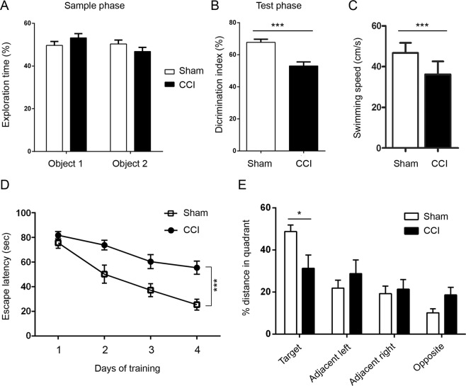
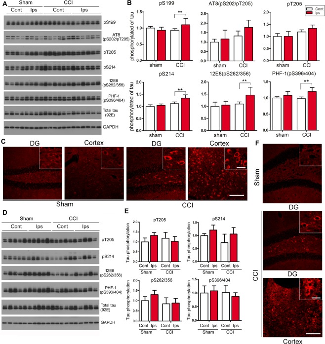
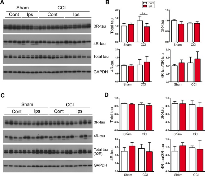
Repetitive traumatic brain injury (TBI) has been linked to late life development of chronic traumatic encephalopathy (CTE), a neurodegenerative disorder histopathologically characterized by perivascular tangles of hyperphosphorylated tau at the depth of sulci to later widespread neurofibrillary pathology. Although tau hyperphosphorylation and neurofibrillary-like pathology have been observed in the brain of transgenic mice overexpressing human tau with aggregation-prone mutation after TBI, they have not been consistently recapitulated in rodents expressing wild-type tau only. Here, we characterized Alzheimer-like alterations behaviorally, biochemically and immunohistochemically 6 weeks and 7 months after unilateral mild-to-moderate controlled cortical impact (CCI) in 5-7-month-old Tg/htau mice, which express all six isoforms of non-mutated human tau in a mouse tau null background. We detected hyperphosphorylation of tau at multiple sites in ipsilateral hippocampus 6 weeks but not 7 months after CCI. However, neuronal accumulation of AT8 positive phospho-tau was sustained in the chronic phase, in parallel to prolonged astrogliosis, and decreased neural and synaptic markers. The mice with CCI also exhibited cognitive and locomotor impairment. These results indicate subacute to chronic Alzheimer-like alterations after CCI in Tg/htau mice. This is the first known study providing insight into the role of CCI in Alzheimer-like brain alterations in young adult mice expressing only non-mutated human tau.
Conflict of interest statement
The authors declare no competing interests.
Figures


Similar articles
-
Involvement of Activation of Asparaginyl Endopeptidase in Tau Hyperphosphorylation in Repetitive Mild Traumatic Brain Injury.J Alzheimers Dis. 2018;64(3):709-722. doi: 10.3233/JAD-180177. J Alzheimers Dis. 2018. PMID: 29889065 Free PMC article.
-
Alzheimer's disease-like tau neuropathology leads to memory deficits and loss of functional synapses in a novel mutated tau transgenic mouse without any motor deficits.Am J Pathol. 2006 Aug;169(2):599-616. doi: 10.2353/ajpath.2006.060002. Am J Pathol. 2006. PMID: 16877359 Free PMC article.
-
Chronic Repetitive Mild Traumatic Brain Injury Results in Reduced Cerebral Blood Flow, Axonal Injury, Gliosis, and Increased T-Tau and Tau Oligomers.J Neuropathol Exp Neurol. 2016 Jul;75(7):636-55. doi: 10.1093/jnen/nlw035. Epub 2016 May 31. J Neuropathol Exp Neurol. 2016. PMID: 27251042 Free PMC article.
-
Traumatic brain injury (TBI) in collision sports: Possible mechanisms of transformation into chronic traumatic encephalopathy (CTE).Metabolism. 2019 Nov;100S:153943. doi: 10.1016/j.metabol.2019.07.007. Metabolism. 2019. PMID: 31610856 Review.
-
Does neuroinflammation drive the relationship between tau hyperphosphorylation and dementia development following traumatic brain injury?Brain Behav Immun. 2017 Feb;60:369-382. doi: 10.1016/j.bbi.2016.09.027. Epub 2016 Sep 28. Brain Behav Immun. 2017. PMID: 27686843 Review.
Cited by
-
An update on the association between traumatic brain injury and Alzheimer's disease: Focus on Tau pathology and synaptic dysfunction.Neurosci Biobehav Rev. 2021 Jan;120:372-386. doi: 10.1016/j.neubiorev.2020.10.020. Epub 2020 Nov 7. Neurosci Biobehav Rev. 2021. PMID: 33171143 Free PMC article. Review.
-
The Citron homology domain of MAP4Ks improves outcomes of traumatic brain injury.Neural Regen Res. 2025 Nov 1;20(11):3233-3244. doi: 10.4103/NRR.NRR-D-24-00113. Epub 2024 Sep 24. Neural Regen Res. 2025. PMID: 39314140 Free PMC article.
-
Diffusion Tensor Imaging Detects Acute Pathology-Specific Changes in the P301L Tauopathy Mouse Model Following Traumatic Brain Injury.Front Neurosci. 2021 Feb 24;15:611451. doi: 10.3389/fnins.2021.611451. eCollection 2021. Front Neurosci. 2021. PMID: 33716645 Free PMC article.
-
Heightened Tameness and Accelerated Handling-Habituation in 3×Tg-AD Mice on a B6;129 Genetic Background.J Alzheimers Dis Rep. 2022 May 23;6(1):245-255. doi: 10.3233/ADR-220007. eCollection 2022. J Alzheimers Dis Rep. 2022. PMID: 35719714 Free PMC article.
-
Intra-hippocampal cis-P tau microinjection induces long-term changes in behavior and synaptic plasticity in mice.Behav Brain Funct. 2023 May 25;19(1):9. doi: 10.1186/s12993-023-00211-0. Behav Brain Funct. 2023. PMID: 37231523 Free PMC article.
References
-
- Oliveira CO, Ikuta N, Regner A. Outcome biomarkers following severe traumatic brain injury. Revista Brasileira de terapia intensiva. 2008;20:411–421. - PubMed
-
- Guskiewicz, K. M. et al. Association between recurrent concussion and late-life cognitive impairment in retired professional football players. Neurosurgery57, 719–726; discussion 719–726 (2005). - PubMed
Publication types
MeSH terms
Substances
LinkOut - more resources
Full Text Sources
Medical
Molecular Biology Databases
Miscellaneous
