Review
. 2019 Jul;16(7):389-406.
doi: 10.1038/s41569-019-0169-2.

Inflammation and its resolution in atherosclerosis: mediators and therapeutic opportunities

Affiliations
Review

Inflammation and its resolution in atherosclerosis: mediators and therapeutic opportunities

Magnus Bäck et al. Nat Rev Cardiol. 2019 Jul.

Abstract

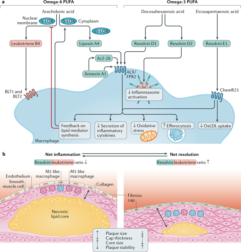
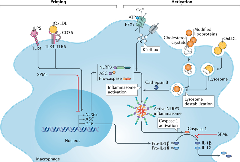
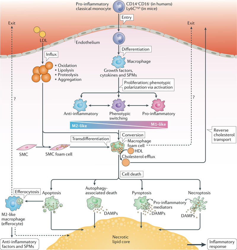
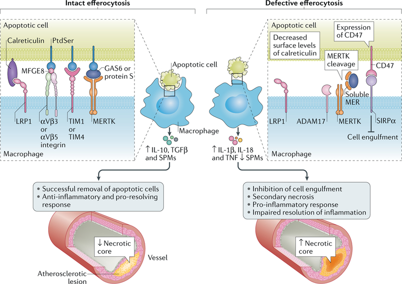
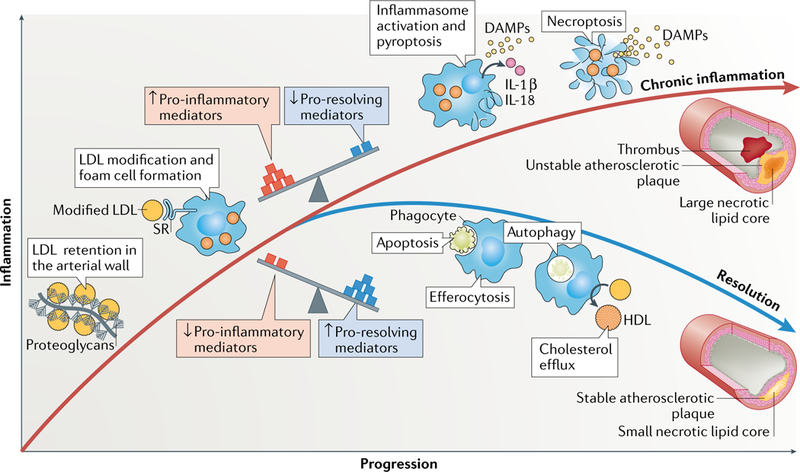
Atherosclerosis is a lipid-driven inflammatory disease of the arterial intima in which the balance of pro-inflammatory and inflammation-resolving mechanisms dictates the final clinical outcome. Intimal infiltration and modification of plasma-derived lipoproteins and their uptake mainly by macrophages, with ensuing formation of lipid-filled foam cells, initiate atherosclerotic lesion formation, and deficient efferocytotic removal of apoptotic cells and foam cells sustains lesion progression. Defective efferocytosis, as a sign of inadequate inflammation resolution, leads to accumulation of secondarily necrotic macrophages and foam cells and the formation of an advanced lesion with a necrotic lipid core, indicative of plaque vulnerability. Resolution of inflammation is mediated by specialized pro-resolving lipid mediators derived from omega-3 fatty acids or arachidonic acid and by relevant proteins and signalling gaseous molecules. One of the major effects of inflammation resolution mediators is phenotypic conversion of pro-inflammatory macrophages into macrophages that suppress inflammation and promote healing. In advanced atherosclerotic lesions, the ratio between specialized pro-resolving mediators and pro-inflammatory lipids (in particular leukotrienes) is strikingly low, providing a molecular explanation for the defective inflammation resolution features of these lesions. In this Review, we discuss the mechanisms of the formation of clinically dangerous atherosclerotic lesions and the potential of pro-resolving mediator therapy to inhibit this process.


Figures

Fig. 1 |
Fig. 1 |. Ligands and receptors transducing pro–resolving signalling in macrophages.
a |Specialized pro-resolving lipid mediators (in blue) include lipoxin A4, derived from the metabolism of the omega-6 polyunsaturated fatty acid (PUFA) arachidonic acid, and D-series and E-series resolvins formed from the metabolism of the omega-3 PUFAs docosahexaenoic acid and eicosapentaenoic acid, respectively. The protein pro-resolving mediator annexin A1 and the derivative peptide Ac2–26, as well as lipoxin A4 and resolvin D1, signal through N-formyl peptide receptor 2 (ALX/FPR2), whereas resolvin E1 transduces its effects through the ChemR23 receptor. These ligand-receptor interactions result in various effects, such as decreased secretion of inflammatory cytokines, reduced oxidative stress and improved efferocytosis. Resolvin E1 signalling through ChemR23 inhibits uptake of oxidized LDL (oxLDL), and resolvin D2 inhibits inflammasome activation. The ratio between pro-resolving and pro-inflammatory mediators (exemplified in the Figure by leukotriene B4 and its receptors BLT1 and BLT2) determines the macrophage phenotype. A disturbed balance in favour of pro-inflammatory pathways might serve as a marker of non-resolving inflammation in atherosclerosis. A pro-resolving amplification loop inhibits nuclear membrane localization of 5-lipoxygenase (5-LOX) and thereby further favours the formation of pro-resolving over pro-inflammatory mediators. b | Atherosclerotic plaques with net inflammation (left) and with net resolution of inflammation (right) are shown. Both atherosclerotic plaques are characterized by ongoing inflammation in which both pro-inflammatory and pro-resolving processes are concurrently active. However, the levels of these opposing processes are different in the two plaques: the resolvin:leukotriene ratio is low in the plaque with net inflammation and high in the plaque with net resolution. The plaque with net inflammation is larger and has a thin fibrous cap (with mainly pro-inflammatory M1-like macrophages, few smooth muscle cells and cleaved collagen) and a large necrotic lipid core. The plaque with net resolution is smaller and has a thicker fibrous cap (with mainly anti-inflammatory M2-like macrophages, abundant smooth muscle cells and intact collagen) and a smaller necrotic lipid core. The plaque with net inflammation is unstable and susceptible to rupture, whereas the plaque with net resolution is stable and not susceptible to rupture. During the long-lasting period of plaque development, the relative magnitude of pro-inflammatory and pro-resolving processes can vary over time. Therefore, during periods of net inflammation, plaque size tends to increase, and during periods of net resolution, plaque size tends to decrease — that is, plaque regression takes place.
Fig. 2 |
Fig. 2 |. Modified lipoproteins and cholesterol crystals induce inflammasome activation.
Activation of the NLRP3 inflammasome requires two steps: priming and activation. The priming step induces the expression of NLRP3 components and pro-IL-1β and can be triggered by lipopolysaccharide (LPS) and oxidized LDL (oxLDL). The activation step induces the activation of the NLRP3 inflammasome. Extracellular ATP activates the P2X7 receptor, which leads to K+ efflux with ensuing decreased intracellular K+ concentration and NLRP3 inflammasome activation. The activation step can also be triggered by cellular uptake of oxLDL. OxLDL uptake can lead to formation of cholesterol crystals in the lysosomes, with ensuing lysosomal destabilization and release of cathepsin B from disrupted lysosomes. The release of unesterified cholesterol from the disrupted lysosomes causes an increase in the content of unesterified cholesterol in intracellular membranes and can thereby cause NLRP3 activation. Moreover, uptake of cholesterol crystals and other modified lipoproteins (such as electronegative LDL, lipolysed LDL and VLDL and apolipoprotein B-containing lipid particles from atherosclerotic lesions) or immune complexes of oxLDL can induce activation of the NLRP3 inflammasome. Regardless of the activation mechanism, formation of the NLRP3 complex induces autocleavage and activation of caspase 1. Active caspase 1 can then cleave pro-IL-1β and the constitutively expressed pro-IL-18 to their mature, secretable forms. Specialized pro-resolving mediators (SPMs) can inhibit both the priming and the activation of the NLRP3 inflammasome. TLR, Toll-like receptor.
Fig. 3 |
Fig. 3 |. Macrophage life cycle and cholesterol round trip in atherosclerosis.
The pro-inflammatory subclass of circulating monocytes, the ‘classical monocytes’, are highly responsive to inflammatory signals and invade inflamed sites, such as atherosclerosis-susceptible areas of the arterial intima in which the cholesterol of locally modified LDL particles accumulates. In the atherosclerotic intima, monocytes differentiate into macrophages, which are then exposed to growth factors, cytokines and specialized pro-resolving mediators (SPMs). Different combinations of these factors determine the macrophage proliferation rate and their polarization into anti-inflammatory M2-like or pro-inflammatory M1-like phenotypes. Macrophages have high phenotypic plasticity, enabling phenotypic switching to provide the continuum of intermediate phenotypes that are often seen in advanced atherosclerotic lesions. A primary function of macrophages is to ingest modified LDL particles, which leads to accumulation of cytoplasmic cholesteryl ester droplets in the macrophages (that is, to foam cell formation). Smooth muscle cells (SMCs) can also form foam cells and then transdifferentiate into macrophage-like cells. HDL particles can induce cholesterol efflux from the foam cells, thereby initiating the process of reverse cholesterol transport for removal of cholesterol from the lesion. Some studies have provided evidence of egress of lesional macrophages during plaque regression (dotted lines). Thus, migration of foam cells to the blood might be an additional mechanism for cholesterol removal. Ultimately, old foam cells die via apoptotic or non-apoptotic forms of regulated cell death. Apoptosis is non-inflammatory and can even induce an anti-inflammatory response when the apoptotic cell is phagocytosed by an M2-like macrophage (efferocytosis) with ensuing release of anti-inflammatory factors and SPMs. The autophagic process itself is anti-inflammatory, but stressed cells undergoing extensive autophagy might die and can release pro-inflammatory mediators. Pyroptosis and necroptosis are pro-inflammatory via release of pro-inflammatory mediators, such as damage-associated molecular patterns (DAMPs). In advanced atherosclerotic lesions, influx of LDL particles and monocytes and formation of foam cells occur continually and, inevitably, some apoptotic foam cells escape efferocytosis and thereby contribute to the formation of a necrotic lipid core. Cholesterol-containing efferocytes can also die and contribute to the formation and growth of the necrotic lipid core. Therefore, unless foam cells (either of macrophage or SMC origin) and the macrophages that have ingested them return to the blood (dotted lines), all cholesteryl-ester-loaded cells, irrespective of their origin or mode of death, contribute to the generation and growth of a pro-inflammatory necrotic lipid core.
Fig. 4 |
Fig. 4 |. Defective efferocytosis drives necrotic core formation in atherosclerosis.
Intact efferocytosis by macrophages prevents necrotic core development in atherosclerotic lesions. However, when efferocytosis becomes defective, such as through cleavage of the tyrosine-protein kinase receptor MERTK by the protease ADAM17 on the macrophage, through inappropriate expression of CD47 on apoptotic cells resulting in CD47-SIRPα interaction or through reduced surface levels of calreticulin on apoptotic cells, atherosclerosis advances and necrotic core formation occurs. LRP1, LDL receptor-related protein 1; MFGE8, lactadherin; PtdSer, phosphatidylserine; SPM, specialized pro-resolving mediator; TIM, T cell immunoglobulin mucin receptor.
Fig. 5 |
Fig. 5 |. Resolution versus chronic inflammation in atherosclerosis.
The balance of pro-inflammatory and antiinflammatory processes controls the resolution of the lipid-driven inflammation in atherosclerotic lesions. Retention of LDL particles by arterial wall proteoglycans and subsequent modification of the retained LDL induce inflammation in the arterial wall. Macrophages ingest the modified LDL particles via scavenger receptor (SR)-mediated endocytosis and become foam cells. If the balance between pro-inflammatory and pro-resolving mediators is tilted towards inflammation, the resolving mechanisms fail. Under these conditions, pyroptosis (mediated by inflammasome activation) or necroptosis can ensue. These pro-inflammatory forms of cell death further promote inflammation and generation of a large necrotic lipid core. These unstable atherosclerotic plaques might ultimately lead to plaque rupture and a local occluding arterial thrombus. Conversely, if the balance between the mediators is tilted towards pro-resolving mediators, apoptosis and autophagy-associated cell death and cholesterol efflux from the lesions are favoured, and efferocytosis of the dead cells can lead to resolution of inflammation. These processes promote the formation of a stable plaque with a small necrotic lipid core. DAMPs, damage-associated molecular patterns.

Similar articles

Cited by

References

    1. Williams KJ & Tabas I The response-to-retention hypothesis of early atherogenesis. Arteriosder. Thromb. Vasc. Biol. 15, 551–561 (1995). - PMC - PubMed
    1. Williams KJ & Tabas I The response-to-retention hypothesis of atherogenesis reinforced. Curr. Opin. Lipidol. 9, 471–474 (1998). - PubMed
    1. Tabas I, Williams KJ & Boren J Subendothelial lipoprotein retention as the initiating process in atherosclerosis: update and therapeutic implications. Circulation 116, 1832–1844 (2007). - PubMed
    1. Galkina E & Ley K Immune and inflammatory mechanisms of atherosclerosis*. Annu. Rev. Immunol. 27, 165–197 (2009). - PMC - PubMed
    1. Hansson GK Inflammation, atherosclerosis, and coronary artery disease. N. Engl. J. Med. 352, 1685–1695 (2005). - PubMed

Publication types