. 2019 Jan 31:18:101-112.
doi: 10.1016/j.jare.2019.01.013. eCollection 2019 Jul.

Therapeutic potential of endothelial progenitor cells in a rat model of epilepsy: Role of autophagy

Affiliations

Therapeutic potential of endothelial progenitor cells in a rat model of epilepsy: Role of autophagy

Shimaa O Ali et al. J Adv Res. .

Abstract

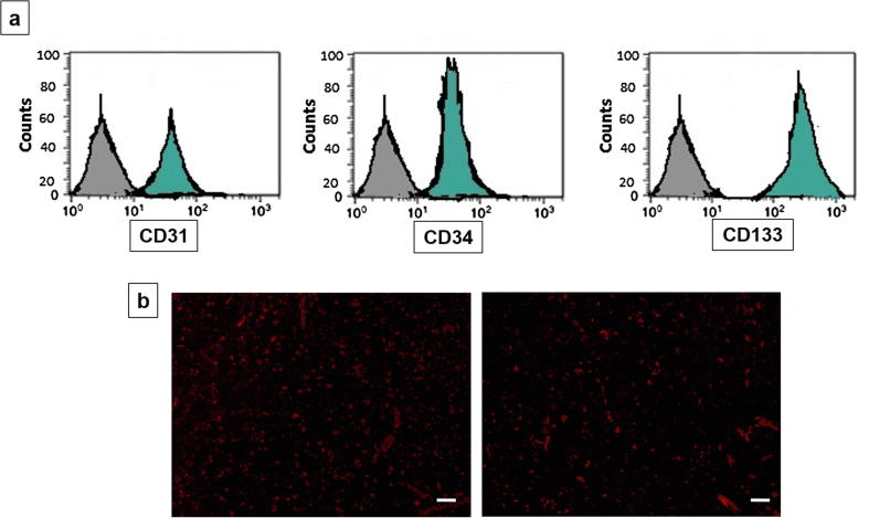
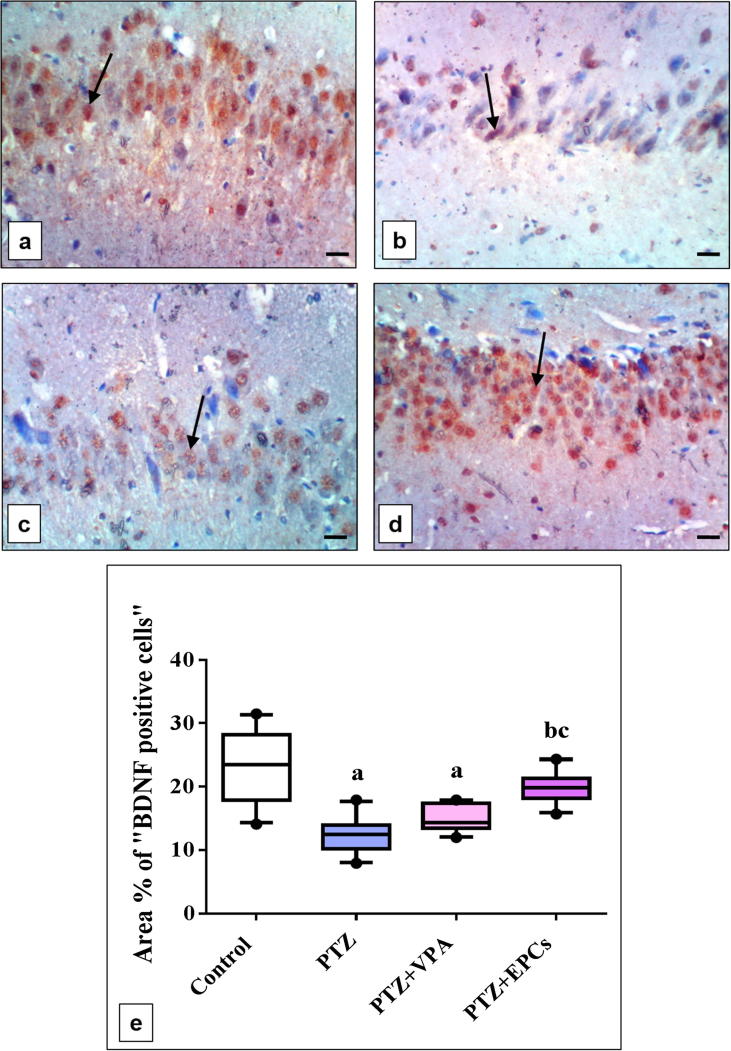
Epilepsy is one of the most well-known neurological conditions worldwide. One-third of adult epileptic patients do not respond to antiepileptic drugs or surgical treatment and therefore suffer from the resistant type of epilepsy. Stem cells have been given substantial consideration in the field of epilepsy therapeutics. The implication of pathologic vascular response in sustained seizures and the eminent role of endothelial progenitor cells (EPCs) in maintaining vascular integrity tempted us to investigate the potential therapeutic effects of EPCs in a pentylenetetrazole (PTZ)-induced rat model of epilepsy. Modulation of autophagy, a process that enables neurons to maintain an equilibrium of synthesis, degradation and subsequent reprocessing of cellular components, has been targeted. Intravenously administered EPCs homed into the hippocampus and amended the deficits in memory and locomotor activity. The cells mitigated neurological damage and the associated histopathological alterations and boosted the expression of brain-derived neurotrophic factor. EPCs corrected the perturbations in neurotransmitter activity and enhanced the expression of the downregulated autophagy proteins light chain protein-3 (LC-3), beclin-1, and autophagy-related gene-7 (ATG-7). Generally, these effects were comparable to those achieved by the reference antiepileptic drug, valproic acid. In conclusion, EPCs may confer therapeutic effects against epilepsy and its associated behavioural and biochemical abnormalities at least in part via the upregulation of autophagy. The study warrants further research in experimental and clinical settings to verify the prospect of using EPCs as a valid therapeutic strategy in patients with epilepsy.

Keywords: Autophagy; Endothelial progenitor cells; Epilepsy; Neuronal damage; Pentylenetetrazole.


Figures

None
Graphical abstract
Fig. 1
Fig. 1
Characterization and recognition of fluorescent signals from EPCs in the rat hippocampus. a) A flow cytometric assay showed the presence of CD31, 34, and 133 surface markers on EPCs engrafted to the hippocampus after their administration. b) Homing of EPCs to the hippocampus of rats was verified through recognition of red fluorescent signals from PKH26-marked cells (scale bar 100 µm). EPCs, endothelial progenitor cells.
Fig. 2
Fig. 2
Effect of EPCs on PTZ-induced changes in neurobehavioural function: Y-maze test (a–c) and OFT (d–g). Each horizontal line inside the box plots of SAP (a), SAR scores (b), total arm entries (c), grooming (e), rearing, and (f) ambulation frequency, and (g) represents the median; the boxes mark the interval between the 25th and 75th percentiles. The whiskers denote the intervals between the 10th and 90th percentiles. Filled circles indicate data points outside the 10th and 90th percentiles. These parameters were analysed using the Kruskal–Wallis test followed by Dunn’s post-test for multiple comparisons. Latency time (d), for which each column with a vertical line represents the mean ± SEM, was analysed using one-way ANOVA followed by Tukey-Kramer test. a significantly different from the control group, b significantly different from the PTZ group, c significantly different from the VPA group at P < 0.05. PTZ, pentylenetetrazole; VPA, valproic acid; EPCs, endothelial progenitor cells; SAP, spontaneous alternation percentage; SAR, same-arm returns.
Fig. 3
Fig. 3
Effect of EPCs on PTZ-induced changes in hippocampal neurotransmitters. GABA (a), glutamate (b), serotonin (c), dopamine (d), and ACH (e) concentrations. Each column with a vertical line represents the mean ± SEM. a significantly different from the control group, b significantly different from the PTZ group, and c significantly different from the VPA group at P < 0.05 using one-way ANOVA followed by a Tukey-Kramer test. PTZ, pentylenetetrazole; VPA, valproic acid; EPCs, endothelial progenitor cells; GABA, γ-aminobutyric acid; ACH, acetylcholine.
Fig. 4
Fig. 4
Effect of EPCs on PTZ-induced changes in hippocampal protein expression of the autophagy markers LC-3, beclin-1 and ATG-7. Band densities obtained by Western blotting analysis were quantified and normalized to those of β-actin (a). Each column with a vertical line of LC-3 (b), beclin-1 (c), and ATG-7 (d) represents the mean ± SEM. a significantly different from the control group, b significantly different from the PTZ group, and c significantly different from the VPA group at P < 0.05 using one-way ANOVA followed by a Tukey-Kramer test. PTZ, pentylenetetrazole; VPA, valproic acid; EPCs, endothelial progenitor cells; LC-3, light chain protein-3; ATG-7, autophagy-related gene-7.
Fig. 5
Fig. 5
Effect of EPCs on histopathological alterations. a) Control group displaying normal histological structure of the hippocampus. b and c) PTZ-treated rats showing severe neuronal necrosis with vacuolation in the hippocampus (arrow). d and e) PTZ + VPA group displaying mild neuronal necrosis and vacuolation (arrow) of the hippocampus. f: PTZ + EPCs-treated group demonstrating mitigated histopathological alterations with almost normal hippocampal structure. PTZ, pentylenetetrazole; VPA, valproic acid; EPCs, endothelial progenitor cells (scale bar 100 µm). (0) no histopathological changes, (1) mild changes, (2) moderate changes, (3) severe changes.
Fig. 6
Fig. 6
Effect of EPCs on PTZ-induced changes in immunohistochemical staining of hippocampal BDNF. a) Control group. b) PTZ-treated group. c) PTZ + VPA group. d) PTZ + EPCs group. e) Quantification of BDNF (calculated as the area of BDNF-immunopositive cells as a percentage of the total area of the microscopic field across ten fields). Horizontal lines inside the box plots represent the median; the boxes mark the interval between the 25th and 75th percentiles. The whiskers denote the intervals between the 10th and 90th percentiles. Filled circles indicate data points outside the 10th and 90th percentiles. a significantly different from the control group, b significantly different from the PTZ group, and c significantly different from the VPA group at P < 0.05 using the Kruskal–Wallis test followed by Dunn’s post-test for multiple comparisons. PTZ, pentylenetetrazole; VPA, valproic acid; EPCs, endothelial progenitor cells; BDNF, brain-derived neurotrophic factor (scale bar 100 µm).
Suppl. Fig. 1
Suppl. Fig. 1
Suppl. Fig. 2
Suppl. Fig. 2

References

    1. WHO. WHO | Epilepsy Fact sheet fs999; 2017.
    1. Verma A., Kumar A. Sudden unexpected death in epilepsy: some approaches for its prevention and medico-legal consideration. Acta Neurol Belg. 2015;115(3):207–212. - PubMed
    1. Quintas R., Raggi A., Giovannetti A.M., Pagani M., Sabariego C., Cieza A. Psychosocial difficulties in people with epilepsy: a systematic review of literature from 2005 until 2010. Epilepsy Behav. 2012;25(1):60–67. - PubMed
    1. Guiard B.P., Di Giovanni G. Central serotonin-2A (5-HT2A) receptor dysfunction in depression and epilepsy: the missing link? Front Pharmacol. 2015;6:46. - PMC - PubMed
    1. Gan J., Qu Y., Li J., Zhao F., Mu D. An evaluation of the links between microRNA, autophagy, and epilepsy. Rev Neurosci. 2015;26(2):225–237. - PubMed

LinkOut - more resources