. 2018 Oct;2(10):732-740.
doi: 10.1038/s41551-018-0282-2. Epub 2018 Sep 3.

Personalized virtual-heart technology for guiding the ablation of infarct-related ventricular tachycardia

Affiliations

Personalized virtual-heart technology for guiding the ablation of infarct-related ventricular tachycardia

Adityo Prakosa et al. Nat Biomed Eng. 2018 Oct.

Abstract

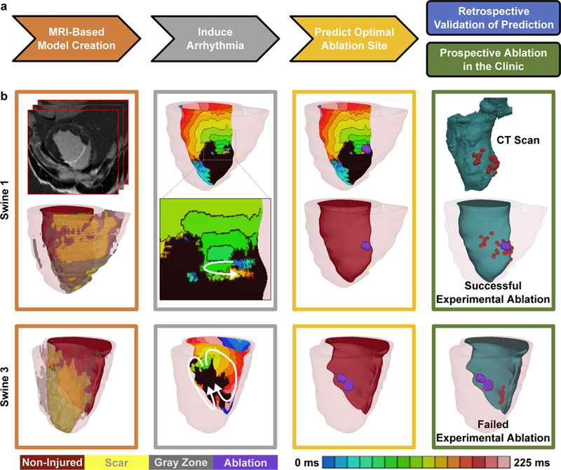
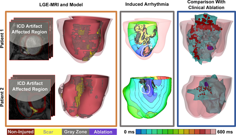
Ventricular tachycardia (VT), which can lead to sudden cardiac death, occurs frequently in patients with myocardial infarction. Catheter-based radiofrequency ablation of cardiac tissue has achieved only modest efficacy, owing to the inaccurate identification of ablation targets by current electrical mapping techniques, which can lead to extensive lesions and to a prolonged, poorly tolerated procedure. Here we show that personalized virtual-heart technology based on cardiac imaging and computational modelling can identify optimal infarct-related VT ablation targets in retrospective animal (5 swine) and human studies (21 patients) and in a prospective feasibility study (5 patients). We first assessed in retrospective studies (one of which included a proportion of clinical images with artifacts) the capability of the technology to determine the minimum-size ablation targets for eradicating all VTs. In the prospective study, VT sites predicted by the technology were targeted directly, without relying on prior electrical mapping. The approach could improve infarct-related VT ablation guidance, where accurate identification of patient-specific optimal targets could be achieved on a personalized virtual heart prior to the clinical procedure.


Conflict of interest statement

Competing Interests. NAT holds partial ownership of CardioSolv Ablation Technologies LLC. SN is a scientific advisor to CardioSolv Ablation Technologies LLC. The other authors declare no competing financial and non-financial interests.

Figures

Figure 1:
Figure 1:. Virtual-heart ablation-target (VAAT) prediction: protocol and results from the animal study.
(a) Flow-chart summarizing the protocol (arrowed steps) and retrospective and prospective studies. (b) In silico models and predictions for one case of successful mapping-guided ablation in Swine 1 (top), and one case of failed mapping-guided ablation in Swine 3 (bottom). Panels, from left to right: reconstructed ventricular model with different remodeled regions and the upper, top panel showing the LGE-MRI stack for Swine 1; endocardial electrical activation maps of the infarct-related VTs with white arrows showing the direction of propagation of the excitation wave, inset shows the zoomed-in propagation waves through channels in the scar in Swine 1; purple circles correspond to in silico predicted ablation targets on the ventricular endocardial surface; CARTO XP ventricular geometry from post-ablation CT scans co-registered with the MRI-based model for comparison of the predicted ablation targets with experimental mapping-based endocardial ablation locations, where red dots correspond to location of the tip of the catheter during ablation. Panel frame colors correspond to the protocol steps outline in Fig. 1a. Non-injured, scar, gray zone tissues and VAAT ablation targets are shown in red, yellow, gray, and purple respectively. Color scale indicates activation times, from earliest to latest, for images in column 2; black indicates tissue regions that did not activate.
Figure 2:
Figure 2:. Results from retrospective human study.
Representative examples of in silico models and predictions from 4 patients are shown (top to bottom). Left to right: reconstructed ventricular computational model with different structurally-remodeled regions; electrical activation maps of the infarct-related VTs after induced arrhythmia on the epi-or endocardial surfaces and white arrows showing the direction of propagation of the excitation wave; purple regions correspond to VAAT predicted ablation targets on the endocardial surface, insets show the zoomed-in VAAT predictions; and co-registration of VAAT targets with the CARTO 3 endocardial surface (green) showing clinical ablation where red dots correspond to locations of the tip of the catheter during ablation. Non-injured, scar, gray zone tissues and VAAT ablation targets are shown in red, yellow, gray, and purple respectively. Color scale indicates activation times, from earliest to latest, for images in column 2; black indicates tissue regions that did not activate.
Figure 3:
Figure 3:. Results from the prospective human study.
VAAT-guided ablation (a-d) for one patient at the University of Utah; (e-h) and at the University of Pennsylvania. (a,e) Reconstructed ventricular models with different remodeled regions, (b,f) Activation maps corresponding to the two VT morphologies induced in the Utah and Pennsylvania patient model respectively with white arrows depicting the direction of propagation of the excitation wave, (c,g) VAAT-predicted ablation targets for the two VT morphologies, (d,h) Co-registration of the VAAT-predicted targets (purple) with the CARTO 3 endocardial surface (green). The red dots correspond to locations of the tip of the catheter during ablation. The left ventricular endocardial surface is shown in green and the total infarct region is shown in gray (gray zone). Non-injured and scar tissues are shown in red and yellow respectively. Color scale indicates activation times, from earliest to latest, for images in column 2; black indicates tissue regions that did not activate.
Figure 4:
Figure 4:. Results from the retrospective human study in patients with ICDs.
Representative examples of in silico models and predictions from 2 patients are shown (top and bottom). The myocardial wall artifact burden was 59% and 46% for patient 1 and 2 respectively. Left panel: LGE MRI scans with ICD artifact burden and reconstructed ventricular models with different remodeled regions; middle panel: electrical activation maps of the infarct-related VTs on the epi-or endocardial surfaces (chosen for best visualization) and white arrows showing direction of VT propagation; right panel: and co-registration of ventricular model surfaces and the VAAT ablation targets (purple) with the CARTO endocardial surfaces (green) showing clinical ablation locations corresponding to red dots representing locations of the tip of the catheter during ablation. The CARTO endocardial surfaces (green surfaces, right panel) show the left ventricle for patient 1 and the right ventricle for patient 2 co-registered with the corresponding MRI shells obtained in the corresponding individuals at the time of their clinical procedure. In Patient 1, the predicted ablation target overlapped with the clinical at the same location. The clinical ablation also identified extensive lesions at the periphery of the infarct (the gray zone). In patient 2, the VAAT lesion was within the area ablated clinically. Non-injured, scar, gray zone tissues and VAAT ablation targets are shown in red, yellow, gray, and purple respectively. Color scale indicates activation times, from earliest to latest, for images in column 3; black indicates tissue regions that did not activate.

Comment in

References

    1. Stevenson WG et al. Radiofrequency catheter ablation of ventricular tachycardia after myocardial infarction. Circulation 98, 308–314 (1998). - PubMed
    1. Aliot EM et al. EHRA/HRS Expert Consensus on Catheter Ablation of Ventricular Arrhythmias. Heart Rhythm 6, 886–933, 10.1016/j.hrthm.2009.04.030 (2009). - DOI - PubMed
    1. Zhong H, Lacomis JM & Schwartzman D On the accuracy of CartoMerge for guiding posterior left atrial ablation in man. Heart Rhythm 4, 595–602, 10.1016/j.hrthm.2007.01.033 (2007). - DOI - PubMed
    1. Brugada J et al. Nonsurgical transthoracic epicardial radiofrequency ablation: an alternative in incessant ventricular tachycardia. J Am Coll Cardiol 41, 2036–2043 (2003). - PubMed
    1. Sosa E, Scanavacca M, d’Avila A, Oliveira F & Ramires JA Nonsurgical transthoracic epicardial catheter ablation to treat recurrent ventricular tachycardia occurring late after myocardial infarction. J Am Coll Cardiol 35, 1442–1449 (2000). - PubMed

LinkOut - more resources