Case Reports
. 2019 Feb 21:9:93.
doi: 10.3389/fonc.2019.00093. eCollection 2019.

Putative Role of Circulating Human Papillomavirus DNA in the Development of Primary Squamous Cell Carcinoma of the Middle Rectum: A Case Report

Affiliations
Case Reports

Putative Role of Circulating Human Papillomavirus DNA in the Development of Primary Squamous Cell Carcinoma of the Middle Rectum: A Case Report

Maria Raffaella Ambrosio et al. Front Oncol. .

Abstract

Here we present the case of a patient affected by rectal squamous cell carcinoma in which we demonstrated the presence of Human Papillomavirus (HPV) by a variety of techniques. Collectively, the virus was detected not only in the tumor but also in some regional lymph nodes and in non-neoplastic mucosa of the upper tract of large bowel. By contrast, it was not identifiable in its common sites of entry, namely oral and ano-genital region. We also found HPV DNA in the plasma-derived exosome. Next, by in vitro studies, we confirmed the capability of HPV DNA-positive exosomes, isolated from the supernatant of a HPV DNA positive cell line (CaSki), to transfer its DNA to human colon cancer and normal cell lines. In the stroma nearby the tumor mass we were able to demonstrate the presence of virus DNA in the stromal compartment, supporting its potential to be transferred from epithelial cells to the stromal ones. Thus, this case report favors the notion that human papillomavirus DNA can be vehiculated by exosomes in the blood of neoplastic patients and that it can be transferred, at least in vitro, to normal and neoplastic cells. Furthermore, we showed the presence of viral DNA and RNA in pluripotent stem cells of non-tumor tissue, suggesting that after viral integration (as demonstrated by p16 and RNA in situ hybridization positivity), stem cells might have been activated into cancer stem cells inducing neoplastic transformation of normal tissue through the inactivation of p53, p21, and Rb. It is conceivable that the virus has elicited its oncogenic effect in this specific site and not elsewhere, despite its wide anatomical distribution in the patient, for a local condition of immune suppression, as demonstrated by the increase of T-regulatory (CD4/CD25/FOXP3 positive) and T-exhausted (CD8/PD-1positive) lymphocytes and the M2 polarization (high CD163/CD68 ratio) of macrophages in the neoplastic microenvironment. It is noteworthy that our findings depicted a static picture of a long-lasting dynamic process that might evolve in the development of tumors in other anatomical sites.

Keywords: cancer; circulating HPV; exosomes; immune evasion; middle rectum.


Figures

Figure 1
Figure 1
Pathological characterization. The morphological and immunohistochemical evaluation of the surgical specimen showed the presence of a squamous cell carcinoma with basaloid features and koilocyte-like cells (arrow) (A, inset), and p16 expression (B); the largest lymph node examined demonstrated p16 positivity in scattered large cells of the germinal center and the subcapsular sinus (arrow) (C). A, (A)-inset: haematoxylin and eosin (H&E); B,C: p16 staining. Original magnification (O.M.): A, 2.5x; (A)-inset, (B,C) 20x.
Figure 2
Figure 2
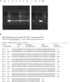
Human Papillomavirus polimerase chain reaction identification and typing. The presence of HPV 16 DNA was confirmed by HPV 16 genotype specific primer pair 5′- AAAGCCACTGTGTCCTGAAGA-3′ and 5′-CTGGGTTTCTCTACGTGTTCT-3′ able to amplify a 130 bps long fragment (424–553 nt, ref Seq Human Papillomavirus 16 type NC_001526). B, Blank, (1) Tumor mass, (2) Proximal margin, (3) Distal margin, (4) Lymph node, (5) Tumor-free mucosa of the surgical specimen, (6) Descending colon biopsies, (7) Caecum/ascending colon biopsies, (8) HPV DNA negative cervical cytological specimen, (9) HPV DNA positive cervical cytological specimen, M, Molecular weight marker (A). HPV 16 was present in the tumor mass as it is evident by the sequence alignment of the 460 bps L1 sequence obtained by MY11/09 PCR primers (B). HPV, Human Papillomavirus.
Figure 3
Figure 3
Human Papillomavirus detection and integration. In the neoplastic cells multiple black signals were shown by CISH with a “punctated” pattern characterized by multiple distinct dot-like intranuclear signals indicating viral integration (A); the normal mucosa of the resected specimen demonstrated scattered cells with HPV infection in a “diffuse” pattern with only few completely black stained nuclei corresponding to the episomal status of the virus (B). RNAscope assay detected E6 and E7 transcripts in the neoplastic (C) and normal mucosa cells of the surgical specimen (D, D inset) as multiple, strong red dot-like signals in the tumor and only few weak red signals in the cancer-free mucosa, corresponding the first pattern to viral integration and the second one to episomal infection. HPV-infected cells OCT3/4-positive (B inset). (A,B) CISH; (C,D) RNAscope; (A,C) original magnification (O.M.) 20x; (B,D) O.M. 40x; (B,D) inset, O.M. 63x. CISH, chromogenic in situ hybridization; HPV, Human Papillomavirus.
Figure 4
Figure 4
Human papillomavirus circulation. Electrophoresis showed the amplified HPV DNA obtained from plasma and Caski cells supernatant-derived exosomes. B, Blank; (1) Plasma-derived exosomes; (2) Caski cells; (3) Positive control (cervical HPV DNA positive cytological samples); M, Marker. The PCR assay was performed with MY11/09 primers (A). Nucleotide blast of HPV DNA amplified product obtained from plasma-derived exosomes by MY11/09 primers (B). HCT116 and NCM460 cell lines were exposed to CasKi supernatant derived exosomes and analyzed by Digital PCR (QuantStudio® 3D Digital PCR System, Life Technologies) to determine the capability of exosomes to transfer viral genetic material to recipient cells. Caski cell line was established from a metastasis in the small bowel mesentery and contains HPV16 DNA integrated into the genome. The reaction was performed using TaqMan Assay specific for HPV 16 E1 (Vi03453396_s1, Invitrogen, Milan, Italy). After 6 h of exposure the load of HPV DNA was greater than at 20 h in the normal human colon epithelial cell line (NCM460). In the human colon cancer cell line (HCT116) we report an HPV DNA burst at 6 h and a rapid decay, indeed it was absent at 20 h post exposure. (1) HCT116 at 3 h; (2) HCT116 at 6 h; (3) HCT116 at 20 h; (4) NCM460 at 3 h; (5) NCM460 at 6 h; (6) NCM4 60 at 20 h (C). The specimens illustrated in (C) were also amplified by conventional PCR with E6 HPV 16 specific primer pair showing concordant results with Digital PCR. B, Blank, (1) not exposed HCT116; (2) HCT116 at 3 h; (3) HCT116 at 6 h; (4) HCT116 at 20 h; (5) not exposed NCM460; (6) NCM460 at 3 h; (7) NCM460 at 6 h; (8) NCM460 at 20 h; (9) blank; (10) HPV DNA negative cervical specimen; (11) HPV DNA positive cervical specimen (D). The neoplastic and the neighbor cells demonstrated HPV DNA in the endothelial cells (red arrow) and in the fibroblasts (black arrow) by both CISH (brown chromogen) (E) and p16 IHC (E, inset). E, (E)-inset, original magnification 20x.
Figure 5
Figure 5
Microenvironment characterization. An increase of PDL-1 and PDL-2 expression in neoplastic cells was observed (A,B) whereas the reactive background showed a higher percentage of PD-1 positive elements (C). The tumor infiltrating lymphocytes were represented mainly by CD8/PD-1 positive T-cells (D) which were absent in normal mucosa (E). An increase of CD163/CD68-positive macrophages ratio was detected in the neoplastic component (F) as compared to the normal one (G). (A) PDL-1 staining; (B) PDL-2 staining; (C) PD-1 staining; (D,E) CD8/PD-1 double staining (CD8 brown, PD-1 red); (F,G) CD163/CD68 double staining (CD163 brown, CD68 red). (A–G), original magnification 20x.

References

    1. Kurelac I, Salfi NC, Ceccarelli C, Alessandrini F, Cricca M, Caliceti U, et al. . Human papillomavirus infection and pathogenic mitochondrial DNA mutation in bilateral multinodular oncocytic hyperplasia of the parotid. Pathology (2014) 46:250–3. 10.1097/PAT.0000000000000079 - DOI - PubMed
    1. Ambrosio MR, Onorati M, Rocca BJ, Santopietro R. Vulvar cancer and HPV infection: analysis of 22 cases. Pathologica (2008) 100:405–7. - PubMed
    1. Vidone M, Alessandrini F, Marucci G, Farnedi A, De Biase D, Ricceri F, et al. . Evidence of association of human papillomavirus with prognosis worsening in glioblastoma multiforme. Neuro Oncol. (2014) 16:298–302. 10.1093/neuonc/not140 - DOI - PMC - PubMed
    1. Chen XZ, Waterboer T, Castro FA, Brenner H. Viral infections and colorectal cancer: a systematic review of epidemiological studies. Int J Cancer (2015) 137:12–24. 10.1002/ijc.29180 - DOI - PubMed
    1. Jeannot E, Becette V, Campitelli M, Calméjane MA, Lappartient E, Ruff E, et al. . Circulating human papillomavirus DNA detected using droplet digital PCR in the serum of patients diagnosed with early stage human papillomavirus-associated invasive carcinoma. J Pathol Clin Res. (2016) 2:201–9. 10.1002/cjp2.47 - DOI - PMC - PubMed

Publication types