. 2019 Feb 25;4(1):1-14.
doi: 10.1016/j.jacbts.2018.10.008. eCollection 2019 Feb.

Neutrophil Subsets, Platelets, and Vascular Disease in Psoriasis

Affiliations

Neutrophil Subsets, Platelets, and Vascular Disease in Psoriasis

Heather L Teague et al. JACC Basic Transl Sci. .

Abstract

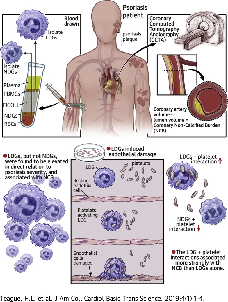
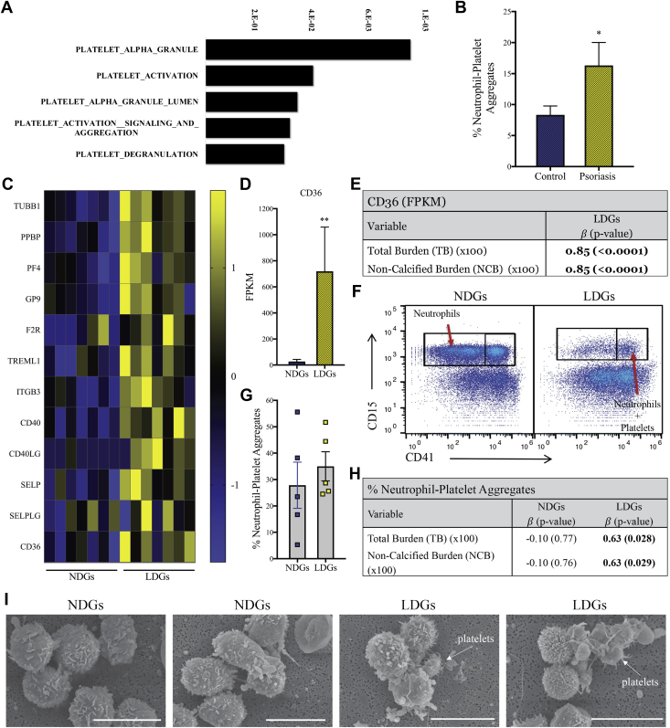
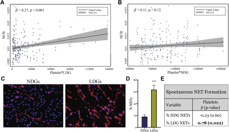
Psoriasis is an inflammatory skin disease associated with increased cardiovascular risk and serves as a reliable model to study inflammatory atherogenesis. Because neutrophils are implicated in atherosclerosis development, this study reports that the interaction among low-density granulocytes, a subset of neutrophils, and platelets is associated with a noncalcified coronary plaque burden assessed by coronary computed tomography angiography. Because early atherosclerotic noncalcified burden can lead to fatal myocardial infarction, the low-density granulocyte-platelet interaction may play a crucial target for clinical intervention.

Keywords: CCTA, coronary computed tomography angiography; CVD, cardiovascular disease; FDR, false discovery rate; HAoEC, human aortic endothelial cell; LDG, low-density granulocyte; MI, myocardial infarction; NCB, noncalcified coronary plaque burden; NDG, normal-density granulocyte; NET, neutrophil extracellular trap; PASI, psoriasis area severity index; SLE, systemic lupus erythematosus; TB, total coronary plaque burden; cardiovascular disease; low-density granulocytes; neutrophils; platelets; psoriasis.


Figures

None
Graphical abstract
Figure 1
Figure 1
LDGs Are Elevated in Psoriasis Patients and Are Associated With Psoriasis Severity (A) Normal-density granulocyte (NDG) and low-density granulocyte (LDG) frequencies were determined by flow cytometry and are elevated in psoriasis patients (n = 81) compared with healthy control subjects (n = 36). Data are represented as mean ± SEM. The Mann-Whitney test was performed, and significance was set at *p < 0.05* and ***p < 0.001. Regression analyses between (B) LDGs but not (C) NDGs are associated with the psoriasis area severity index (PASI) score for the psoriasis cohort (n = 81). (D) Surface marker expression of NDGs and LDGs was analyzed by flow cytometry and (E) showed a significant elevation in CD15 on psoriasis LDGs compared with healthy control and psoriasis NDGs, as well as lower CD11b and CD62L expression on psoriasis LDGs compared with healthy control LDGs. Data are represented as mean ± SEM. Significance was established by 1-way analysis of variance (ANOVA) and a Tukey’s multiple comparisons test set at *p < 0.05, ****p < 0.001, and ****p < 0.0001. MFI = median fluorescence intensity.
Figure 2
Figure 2
LDGs and Their NETs Induce Endothelial Cell Damage (A) Psoriasis LDGs but not (B) psoriasis NDGs are associated with noncalcified coronary plaque burden (NCB) in psoriasis (n = 81). (C) Representative flow cytometry plots from the cytotoxicity assay show (D) psoriasis LDGs (n = 7) increase apoptosis of human aortic endothelial cells (HAoECs) compared with psoriasis NDGs (n = 5), an effect abrogated by DNase treatment (n = 4). Data are represented as mean ± SEM. Significance established by a 1-way ANOVA and a Tukey’s multiple comparisons test set at *p < 0.05, **p < 0.01, and ***p < 0.0001. (E) Cytotoxicity of HAoECs pre-treated with tumor necrosis factor-α and interferon-γ is further increased by LDGs. Data are represented as means ± SEM. Significance established by 1-way ANOVA and a Tukey’s multiple comparisons test and set at *p < 0.05 and **p < 0.01. (F) HAoECs were incubated for 18 h with NDG neutrophil extracellular trap (NET) associated (n =5) or LDG-NET associated proteins (n =5), and apoptosis was quantified using flow cytometry. Data are represented as mean ± SEM. Significance established by unpaired 2-tailed Student’s t-test and set at **p < 0.01. (G) Scanning electron microscopy images of the formation of NETs from NDGs and LDGs over time subsequent to purification. CI = confidence interval; T/I = tumor necrosis factor alpha/interferon gamma; other abbreviations as in Figure 1.
Figure 3
Figure 3
Granule Proteins and Adhesion Molecules Are Upregulated in LDGs Compared With NDGs at the Gene Level RNA sequencing analysis was completed on 50,000 LDGs relative to NDGs from psoriasis patients. (A) Differentially expressed genes (n = 1,076) were identified between NDGs and LDGs from psoriasis patients (n = 7). NDGs are the reference, as are upregulated genes in LDGs. (B) The volcano plot shows clear separation between NDGs and LDGs. The upregulated (red) and downregulated (green) transcriptomes are NDGs, as LDGs are the reference sample. (C) The gene ontology biological process analysis highlighted biological processes that were differentially expressed between NDGs and LDGs. Significance was established by false discovery rate (FDR). (D) Granule proteins are upregulated in all patients (n = 7) and when normalized and by (E) FPKM values (n = 7). Data are represented as means ± SEM. Significance was established by the unpaired Mann-Whitney Student’s t-test and set at *p < 0.05. (F) Transmission electron microscopy images of LDGs and NDGs show LDGs had more electron dense granules. AZU1 = azurocidin 1; CTSG = cathepsin G; ELANE = neutrophil elastase; FPKM = fragments per kilobase of transcripts per million; ICAM2 = intercellular adhesion molecule 2; ITGAL = integrin subunit alpha L; ITGAM = integrin subunit alpha M; MPO = myeloperoxidase; P = patient; PRTN3 = proteinase 3; other abbreviations as Figures 1 and 2.
Figure 4
Figure 4
Upregulated Genes in LDGs Show Increased Binding to Platelets RNA sequencing analysis shows an (A) upregulation of platelet-specific biological pathways in psoriasis LDGs versus psoriasis NDGs. Significance was established by FDR. (B) Neutrophil platelet aggregates were increased in psoriasis patients (n = 12) compared with matched control subjects (n = 10). Data are represented as mean ± SEM. Significance was established by the unpaired Mann-Whitney Student’s t-test and set at *p < 0.05. (C) The platelet-specific transcriptomes were upregulated in LDGs compared with NDGs (n = 7). (D) The platelet receptor, CD36, was highly upregulated in LDGs, and the FPKM values were associated with (E) NCB (n = 7). (F) Flow cytometry plots show (G) aggregates of NDGs or LDGs with platelets and the percentages of platelets LDG aggregates are (H) highly associated with NCB. (I) Scanning electron microscopy images of NDGs and LDGs demonstrate platelet LDG aggregates. Scale bar: 10 μm. CD40LG = CD40 ligand; F2R = coagulation factor II thrombin receptor; GP9 = glycoprotein IX platelet; ITGB3 = integrin subunit beta 3; PF4 = platelet factor 4; PPBP = pro-platelet basic protein; SELP = selectin P; SELPLG = selectin P ligand; TREML1 = triggering receptor expressed on myeloid cells like 1; TUBB1 = tubulin beta 1 class VI; other abbreviations as in Figures 1, 2, and 3.
Figure 5
Figure 5
Percentage of LDGs to Spontaneously Form NETs Is Associated With Circulating Platelet Counts (A) The product of platelets and LDGs in circulation is associated with NCB; however, (B) this relationship is absent with the product of platelets and NDGs. (C) LDGs spontaneously form NETs at a (D) higher frequency than NDGs (n = 9). Data are represented as mean ± SEM. Significance was established by an unpaired Mann-Whitney Student’s t-test and set at ***p < 0.001. (E) The percentage of LDGs (n = 8) to undergo spontaneous NETosis is associated with the frequency of circulating platelets. Abbreviation as in Figures 1, 2, and 3.

References

    1. Prodanovich S., Kirsner R.S., Kravetz J.D., Ma F., Martinez L., Federman D.G. Association of psoriasis with coronary artery, cerebrovascular, and peripheral vascular diseases and mortality. Arch Dermatol. 2009;145:700–703. - PubMed
    1. Mehta N.N., Azfar R.S., Shin D.B., Neimann A.L., Troxel A.B., Gelfand J.M. Patients with severe psoriasis are at increased risk of cardiovascular mortality: cohort study using the General Practice Research Database. Eur Heart J. 2010;31:1000–1006. - PMC - PubMed
    1. Rachakonda T.D., Schupp C.W., Armstrong A.W. Psoriasis prevalence among adults in the United States. J Am Acad Dermatol. 2014;70:512–516. - PubMed
    1. Meyer N., Paul C., Feneron D. Psoriasis: an epidemiological evaluation of disease burden in 590 patients. J Eur Acad Dermatol Venereol. 2010;24:1075–1082. - PubMed
    1. Brauchli Y.B., Jick S.S., Miret M., Meier C.R. Psoriasis and risk of incident myocardial infarction, stroke or transient ischaemic attack: an inception cohort study with a nested case-control analysis. Br J Dermatol. 2009;160:1048–1056. - PubMed