. 2019 Feb 25;4(1):83-94.
doi: 10.1016/j.jacbts.2018.10.007. eCollection 2019 Feb.

Connective Tissue Growth Factor Inhibition Enhances Cardiac Repair and Limits Fibrosis After Myocardial Infarction

Affiliations

Connective Tissue Growth Factor Inhibition Enhances Cardiac Repair and Limits Fibrosis After Myocardial Infarction

Laura E Vainio et al. JACC Basic Transl Sci. .

Abstract

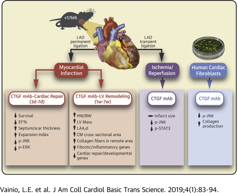
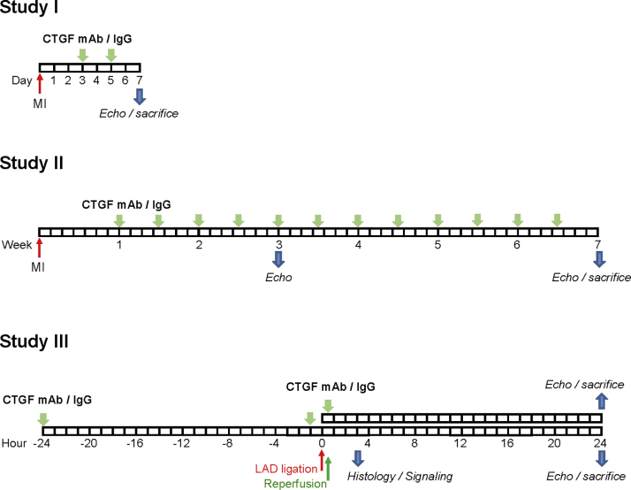
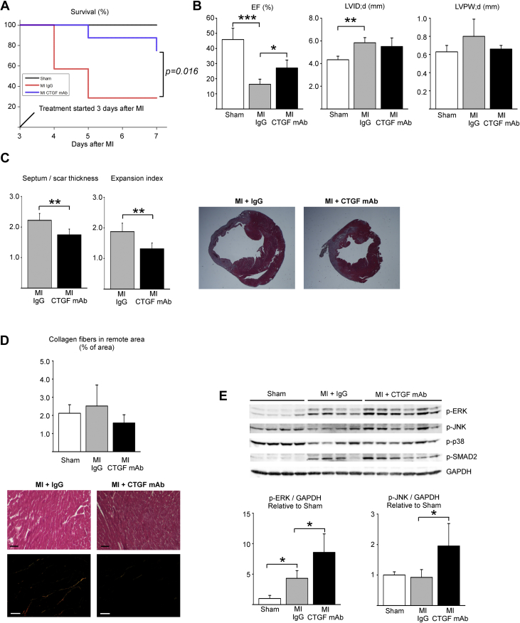
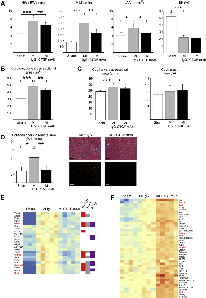
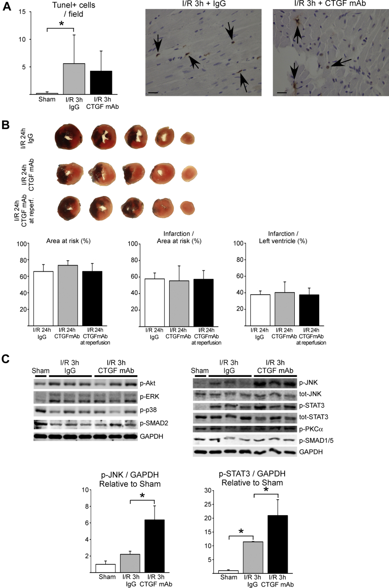
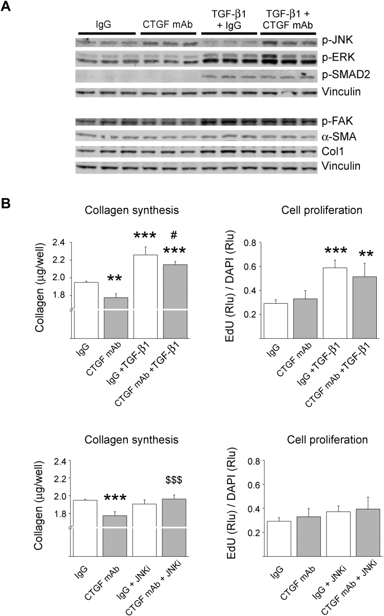
Myocardial infarction (MI)-induced cardiac fibrosis attenuates cardiac contractile function, and predisposes to arrhythmias and sudden cardiac death. Expression of connective tissue growth factor (CTGF) is elevated in affected organs in virtually every fibrotic disorder and in the diseased human myocardium. Mice were subjected to treatment with a CTGF monoclonal antibody (mAb) during infarct repair, post-MI left ventricular (LV) remodeling, or acute ischemia-reperfusion injury. CTGF mAb therapy during infarct repair improved survival and reduced LV dysfunction, and reduced post-MI LV hypertrophy and fibrosis. Mechanistically, CTGF mAb therapy induced expression of cardiac developmental and/or repair genes and attenuated expression of inflammatory and/or fibrotic genes.

Keywords: CTGF, connective tissue growth factor; ECM, extracellular matrix; ERK, extracellular signal-regulated kinase; FB, fibroblast; HF, heart failure; I/R, ischemia−reperfusion; Ig, immunoglobulin; JNK, c-Jun N-terminal kinase; LV, left ventricular; MI, myocardial infarction; TGF, transforming growth factor; connective tissue growth factor monoclonal antibody; fibrosis; heart failure; ischemia−reperfusion injury; left ventricle; mAb, monoclonal antibody; myocardial infarction.


Figures

None
Graphical abstract
Figure 1
Figure 1
Summary of Experimental Protocols Study I, protocol for inhibition of connective tissue growth factor (CTGF) during post—myocardial infarction (MI) cardiac repair (treatment initiation on day 3, treatment completion on day 7). Study II, protocol for CTGF inhibition during post-MI cardiac remodeling (treatment initiation on day 7, treatment completion at the end of week 7). Study III, protocol for CTGF inhibition during and/or after acute ischemia (treatment 24 and 1 h before ischemia or only at reperfusion). IgG = immunoglobulin-G; LAD = left anterior descending; mAb = monoclonal antibody.
Figure 2
Figure 2
CTGF mAb Enhances LV Function (Study I) Mice were subjected to MI, and 3 days after surgery randomly divided to receive either IgG vehicle or CTGF mAb for 4 days. (A) Survival of animals during the experiment. (B) Left ventricular (LV) ejection fraction (EF), end-diastolic dimension (LVID;d), and posterior wall thickness (LVPW;d) were analyzed by echocardiography at 7 days after MI injury. (C) Ratio of thickness of septum versus thickness of infarct and the infarct expansion index. (D) Analysis of interstitial fibrosis from picrosirius red−stained LV sections under polarized light. Masson’s trichrome-stained sections from the same tissue block are also shown. Scale bar: 50 μm. (E) Western blot analysis of LV samples from infarct areas for phosphorylated extracellular signal-regulated kinase (p-ERK), c-Jun N-terminal kinase (p-JNK), p38, and SMAD2. Glyceraldehyde 3-phosphate dehydrogenase (GAPDH) was used as loading control. Data are presented as mean ± SD; number of animals was sham (n = 4), IgG (n = 5), and CTGF mAb (n = 8). *p < 0.05; **p < 0.01; ***p < 0.001. Abbreviations as in Figure 1.
Figure 3
Figure 3
CTGF mAb Protects Against Post-MI LV Hypertrophy and Fibrosis (Study II) Mice were subjected to MI and 1 week after surgery randomly divided to receive IgG vehicle or CTGF mAb for 6 weeks. (A) Analysis for heart weight to body weight (HW/BW) ratio, LV mass, left atrial end-diastolic area (LAA;d), and EF. (B) Analysis of cardiomyocyte cross-sectional area from Masson trichrome−stained LV sections. (C) Analysis of mean capillary cross-sectional size and the number of capillaries per cardiomyocyte in the nonischemic myocardium from CD31 staining, (D) Analysis of interstitial fibrosis from picrosirius red−stained LV sections under polarized light. Scale bar: 50 μm. (A to D) Data are presented as mean ± SD; number of animals was sham (n = 5), IgG (n = 7), and CTGF mAb (n = 8). *p < 0.05; **p < 0.01; ***p < 0.001. (E) Hierarchical clustering of RNAseq data for transcripts that were altered by MI and at least partially normalized by CTGF mAb, indicating that many of these genes are regulated by transforming growth factor-β1 (TGF-β1), tumor necrosis factor (TNF)-α, or interleukin (IL)-1β. Red highlights indicate genes associated with various fibrotic disorders. (F) Hierarchical clustering of RNAseq data showing transcripts whose expression was increased in hearts of mice treated with CTGF mAb. Known cardiac development and/or repair related genes are highlighted (red). (E and F) Number of animals was sham (n = 3), IgG (n = 5), and CTGF mAb (n = 5). Abbreviations as in Figures 1 and 2.
Figure 4
Figure 4
Antagonizing the Function of CTGF mAb During Cardiac I/R Injury (Study III) Mice were treated with IgG vehicle or CTGF mAb and subjected to ischemia−reperfusion injury (I/R). (A) Quantitative analysis of TUNEL-positive cells in hearts subjected to 30 min of ischemia and 3 h of reperfusion is shown. TUNEL-positive cells are marked with arrows; scale bar: 20 μm. (B) Mice were treated with IgG vehicle or CTGF mAb 24 h before ischemia and at reperfusion, or with CTGF mAb at reperfusion only. Infarct size and area at risk were analyzed from triphenyl tetrazolium chloride (TTC)−stained myocardial sections. (C) Western blot analysis of samples from infarct area 3 h after I/R injury. Analysis of phosphorylated protein kinase B (Akt), ERK, JNK, signal transducer and activator of transcription 3 (STAT3), p38, protein kinase C alpha (PKCα), SMAD2, and SMAD1/5 is shown. GAPDH was used as a loading control. Ratio of p-JNK to GAPDH and p-STAT3 to GAPDH data in the bar graphs are presented as mean ± SD. Number of animals in 3-h I/R experiment, including TUNEL labeling, was sham (n = 3), IgG (n = 6), and CTGF mAb (n = 6). Number of animals in 24-h I/R experiment including TTC staining was IgG (n = 11), CTGF mAb (n = 12), and CTGF in reperfusion (n = 12). TUNEL = terminal deoxynucleotidyl transferase dUTP nick end labeling; other abbreviations as in Figures 1 and 2.
Figure 5
Figure 5
CTGF mAb Activates JNK and Reduces Collagen Production in Cultured Human Cardiac Fibroblasts Cultured human fibroblasts were treated with 10 μg/ml CTGF mAb or control IgG, and co-treated with TGF-β1 (1 ng/ml) or inhibitor of JNK inhibitor (JNKi) [(L)-Form, 2 μM)], where indicated. (A) Western blot analysis for phosphorylated JNK2, p-ERK, phosphorylated SMAD2, phosphorylated focal adhesion kinase (p-FAK), smooth muscle alpha actin (α-SMA), and collagen 1 (Col1) is shown. Vinculin was used as loading control. (B) Quantitative analysis for collagen production and fibroblast proliferation. Data are presented as mean ± SD. **p < 0.01; ***p < 0.001 versus IgG; #p < 0.05 versus IgG + TGF-β1; $$$p < 0.001 versus CTGF mAb. N = 5 per group. Abbreviations as in Figures 1, 2, and 3.

References

    1. Writing Group M. Mozaffarian D., Benjamin E.J. Heart Disease and Stroke Statistics-2016 Update: A Report From the American Heart Association. Circulation. 2016;133:e38–e360. - PubMed
    1. Heusch G., Libby P., Gersh B. Cardiovascular remodelling in coronary artery disease and heart failure. Lancet. 2014;383:1933–1943. - PMC - PubMed
    1. Leask A., Abraham D.J. All in the CCN family: essential matricellular signaling modulators emerge from the bunker. J Cell Sci. 2006;119:4803–4810. - PubMed
    1. Jun J.I., Lau L.F. Taking aim at the extracellular matrix: CCN proteins as emerging therapeutic targets. Nat Rev Drug Discov. 2011;10:945–963. - PMC - PubMed
    1. Koitabashi N., Arai M., Niwano K. Plasma connective tissue growth factor is a novel potential biomarker of cardiac dysfunction in patients with chronic heart failure. Eur J Heart Fail. 2008;10:373–379. - PubMed