Review
. 2019 May;137(5):757-783.
doi: 10.1007/s00401-019-01980-7. Epub 2019 Mar 7.

The contribution of astrocytes to the neuroinflammatory response in multiple sclerosis and experimental autoimmune encephalomyelitis

Affiliations
Review

The contribution of astrocytes to the neuroinflammatory response in multiple sclerosis and experimental autoimmune encephalomyelitis

Roberta Brambilla. Acta Neuropathol. 2019 May.

Abstract

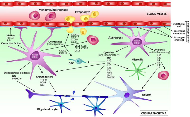
Neuroinflammation is the coordinated response of the central nervous system (CNS) to threats to its integrity posed by a variety of conditions, including autoimmunity, pathogens and trauma. Activated astrocytes, in concert with other cellular elements of the CNS and immune system, are important players in the modulation of the neuroinflammatory response. During neurological disease, they produce and respond to cellular signals that often lead to dichotomous processes, which can promote further damage or contribute to repair. This occurs also in multiple sclerosis (MS), where astrocytes are now recognized as key components of its immunopathology. Evidence supporting this role has emerged not only from studies in MS patients, but also from animal models, among which the experimental autoimmune encephalomyelitis (EAE) model has proved especially instrumental. Based on this premise, the purpose of the present review is to summarize the current knowledge of astrocyte behavior in MS and EAE. Following a brief description of the pathological characteristics of the two diseases and the main functional roles of astrocytes in CNS physiology, we will delve into the specific responses of this cell population, analyzing MS and EAE in parallel. We will define the temporal and anatomical profile of astroglial activation, then focus on key processes they participate in. These include: (1) production and response to soluble mediators (e.g., cytokines and chemokines), (2) regulation of oxidative stress, and (3) maintenance of BBB integrity and function. Finally, we will review the state of the art on the available methods to measure astroglial activation in vivo in MS patients, and how this could be exploited to optimize diagnosis, prognosis and treatment decisions. Ultimately, we believe that integrating the knowledge obtained from studies in MS and EAE may help not only better understand the pathophysiology of MS, but also uncover new signals to be targeted for therapeutic intervention.

Keywords: Astroglia; Demyelinating disorder; Experimental autoimmune encephalomyelitis; Multiple sclerosis; Neuroimmune disease; Neuroinflammation.


Conflict of interest statement

Conflict of interest: The author declares no conflicts of interest.

Figures

Figure 1.
Figure 1.. Astroglial reactivity and axonal damage in the mouse optic nerve at the chronic stage of EAE.
EAE was induced by MOG35–55 immunization in C57BL/6 mice. Optic nerves of mice sacrificed at chronic disease (40 dpi) were compared to naïve optic nerves. Astrocytes were immunostained for GFAP, and axons for non-phosphorylated neurofilament H (clone SMI32). In naïve conditions, GFAP expression was low, indicative of negligible astroglial activation. In parallel, SMI32 expression, which is elevated in correlation with axonal damage, was mild and diffuse to indicate axonal integrity. At chronic EAE, GFAP was highly upregulated demonstrating increased astroglial activation. This was accompanied by increased reactivity for SMI32, which concentrated in axon retraction bulbs (white arrows), demonstrating ongoing wallerian degeneration of axons. Scale bar: 20 μm.
Figure 2.
Figure 2.. Astroglial reactivity in the mouse retina at the chronic stage of EAE.
EAE was induced by MOG35–55 immunization in C57BL/6 mice. Retinas of mice sacrificed at chronic disease (40 dpi) (b, d) were compared to naïve retinas (a, c). Astrocytes were immunostained for GFAP (a, b), or AQP4 (c, d). Nuclei of retinal ganglion cells (RGCs) were identified by DAPI staining. In naïve conditions, GFAP expression was low (a), and AQP4 diffuse and uniformly distributed (c), indicative of negligible astroglial activation and intact glia limitans. The RGC layer was compact and uninterrupted (a, c). At chronic EAE, GFAP was highly upregulated indicating ongoing astroglial activation (b). Labelling was evident throughout the RGC layer (b, yellow arrows), which appeared disrupted. In parallel, AQP4 labeling was reduced and redistributed, absent in certain areas (d, white arrows), suggesting alterations of the glia limitans and potential BBB damage. Scale bar: 50 μm.
Figure 3.
Figure 3.. Astroglial reactivity in the mouse spinal cord at the pre-symptomatic stage of EAE.
EAE was induced by MOG35–55 immunization in C57BL/6 mice. In spinal cord sections of mice sacrificed at 10 dpi, before onset of symptoms, astrocytes were immunostained for GFAP and AQP4. Strong GFAP labeling was observed, indicative of astroglial activation already occurring. Intense labeling for AQP4 was also observed at the astrocyte end-feet (b, blue arrows) as well as on the cell body of hypertrophic astrocytes highly expressing GFAP (b, pink arrow). AQP4 and GFAP clearly colocalized within the glia limitans surrounding the blood vessels (a, white arrows), where no gaps in AQP4 labeling were observed, indicating that the structural integrity of the glia limitans was maintained at this early EAE stage. *Blood vessel. Scale bar: 10 μm.
Figure 4.
Figure 4.. Schematic representation of the soluble mediators released by astrocytes in MS and EAE.
Astrocytes respond to neuroimmune disease with a process of cellular activation that leads to the production of soluble mediators. These signal in a cell autonomous and non-autonomous manner to promote both neurotoxic, and neuroprotective functions. Activated astrocytes release pro-inflammatory cytokines (e.g. TNF, BAFF, TWEAK, IL1β, IL6) that act on immune cells infiltrated in the CNS, as well as resident microglia to induce their activation. They also interact with neurons and oligodendrocytes to cause cellular damage. In parallel, astrocytes can also secrete anti-inflammatory cytokines and mediators (e.g. IL10, IL4, IL27, Shh) that behave as immuno-suppressants and contribute to turn off the pro-inflammatory activation of leukocytes and microglia, favouring repair and neuroprotection. Additionally, in MS and EAE astrocytes are a powerful source of chemokines (e.g. CCL2, CXCL10, CCL20, CXCL1), which serve as signals for the continued recruitment of immune cells into the CNS. Select chemokines (e.g. CXCL1 and CXCL12) have been shown to favour the migration of OPCs to the site of demyelination, thus exerting a reparative role. Some of the growth factors released by astrocytes (e.g. VEGF-A) can cause pathological alterations to the BBB, others can promote neuroprotective functions (e.g. BDNF, NGF). Finally, astrocytes participate in the oxidative stress response by producing neurotoxic radicals (e.g. NO), or neutralizing them, specifically via the production of peroxiredoxins (PRDX2–6).

References

    1. Abbott NJ, Ronnback L, Hansson E (2006) Astrocyte-endothelial interactions at the blood-brain barrier. Nat Rev Neurosci 7: 41–53 Doi 10.1038/nrn1824 - DOI - PubMed
    1. Abdelhak A, Huss A, Kassubek J, Tumani H, Otto M (2018) Serum GFAP as a biomarker for disease severity in multiple sclerosis. Sci Rep 8: 14798 Doi 10.1038/s41598-018-33158-8 - DOI - PMC - PubMed
    1. Agrawal S, Anderson P, Durbeej M, van Rooijen N, Ivars F, Opdenakker G, Sorokin LM (2006) Dystroglycan is selectively cleaved at the parenchymal basement membrane at sites of leukocyte extravasation in experimental autoimmune encephalomyelitis. J Exp Med 203: 1007–1019 Doi 10.1084/jem.20051342 - DOI - PMC - PubMed
    1. Alvarez JI, Cayrol R, Prat A (2011) Disruption of central nervous system barriers in multiple sclerosis. Biochim Biophys Acta 1812: 252–264 Doi 10.1016/j.bbadis.2010.06.017 - DOI - PubMed
    1. Alvarez JI, Dodelet-Devillers A, Kebir H, Ifergan I, Fabre PJ, Terouz S, Sabbagh M, Wosik K, Bourbonniere L, Bernard M et al (2011) The Hedgehog pathway promotes blood-brain barrier integrity and CNS immune quiescence. Science 334: 1727–1731 Doi 10.1126/science.1206936 - DOI - PubMed

Publication types

MeSH terms