Review
. 2019 Mar;7(2):10.1128/microbiolspec.bai-0001-2019.
doi: 10.1128/microbiolspec.BAI-0001-2019.

Mycobacterium tuberculosis: Bacterial Fitness within the Host Macrophage

Affiliations
Review

Mycobacterium tuberculosis: Bacterial Fitness within the Host Macrophage

Lu Huang et al. Microbiol Spectr. 2019 Mar.

Abstract

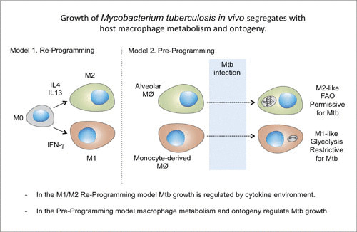
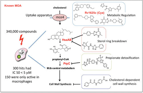
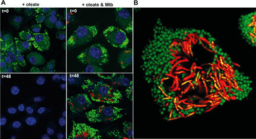
Mycobacterium tuberculosis has evolved to become the single greatest cause of death from an infectious agent. The pathogen spends most of its infection cycle in its human host within a phagocyte. The bacterium has evolved to block the normal maturation and acidification of its phagosome and resides in a vacuole contiguous with the early endosomal network. Cytokine-mediated activation of the host cell can overcome this blockage, and an array of antimicrobial responses can limit its survival. The survival of M. tuberculosis in its host cell is fueled predominantly by fatty acids and cholesterol. The ability of M. tuberculosis to degrade sterols is an unusual metabolic characteristic that was likely retained from a saprophytic ancestor. Recent results with fluorescent M. tuberculosis reporter strains demonstrate that bacterial survival differs with the host macrophage population. Tissue-resident alveolar macrophages, which are biased towards an alternatively activated, M2-like phenotype, are more permissive to bacterial growth than monocyte-derived, inflammatory, M1-like interstitial macrophages. The differential growth of the bacterium in these different phagocyte populations appears to be linked to host cell metabolism.


Figures

FIGURE 1
FIGURE 1
(A) Schematic illustration of the potential outcomes of infection with M. tuberculosis. In most hosts, M. tuberculosis exhibits rapid expansion of the bacterial burden during the first 3 to 4 weeks of infection. At this point, the acquired immune response has developed and controls the bacterial burden at a subclinical level but is unable to clear the infection. In vaccinated hosts, this transition to control of the bacterial burden is achieved at around 1 log fewer bacilli. While resolution of infection is theoretically possible, it is virtually impossible to demonstrate. Progression from latent disease to active disease appears to occur in the face of a robust systemic immune response that is Th1 dominant. While there are candidate indicators of early disease progression, the field lacks immunological markers to detect vaccine-induced protection. Published previously in reference . (B) The main features of the human TB granuloma. A fully formed human TB granuloma is an extremely stratified structure. The center of the granuloma is caseous and rich in lipids, thought to be derived from the lipids present in foamy macrophages. The caseum is surrounded by a macrophage-rich layer that contains foamy macrophages, multinucleated giant cells, and epithelioid macrophages. M. tuberculosis bacilli are observed in many of these cells. This structure is frequently encased in a fibrous capsule of collagen and other extracellular matrix proteins. Lymphocytes tend to be restricted to the periphery of the granuloma outside the fibrous outer layer. Published previously in reference .
FIGURE 2
FIGURE 2
Usefulness of the hspX′::GFP reporter strain in assessing and reporting on the localized induction of inducible nitric oxide synthase at the site of infection. Phosphate-buffered saline-immunized (naïve) mice and mice vaccinated with heat-killed M. tuberculosis (vac) were infected with an hspX′::GFP smyc′:: mCherry Erdman M. tuberculosis reporter strain. Fluorescence induction of the hspX promoter-dependent GFP is higher at 14 days in the vaccinated animals, as assessed by confocal microscopy of thick tissue sections (A), which were scored subsequently by Volocity (B). (C) The thick tissue sections were probed with antibodies against murine NOS2 (magenta), demonstrating the colocalization between GFP induction and NOS2 expression at the site(s) of infection. N.S., not significant. Data are from reference 5).
FIGURE 3
FIGURE 3
Selective depletion of AMs and IMs results in a decrease and an increase in bacterial burden, respectively. Mice were treated with clodronate (Clodro.) liposomes delivered either intranasally (i.n.) (A) or intravenously (i.v.) (B) to deplete the AMs or the circulating monocytes, which depleted the recruited IMs. Neither treatment impacted the neutrophil population within the infected lung tissue. Interestingly, depletion of AMs led to a reduction in bacterial burden, while depletion of IMs led to an increase in bacterial burden. The data demonstrate how modulation of the relative dimensions of the permissive (AM) and controller (IM) macrophage populations directly impacts bacterial burden. Data are from reference .
FIGURE 4
FIGURE 4
Models of macrophage reprogramming and preprogramming. How macrophages function in the reprogramming model (Model 1) is determined by immune signaling within the tissue niche. In the proposed preprogramming model (Model 2), the function of coexisting macrophage lineages in the lung in M. tuberculosis infection is determined, in large part, by the origin of the macrophage. Mtb, M. tuberculosis; IL, interleukin; FAO, fatty acid oxidation. Published previously in reference .
FIGURE 5
FIGURE 5
Major classes of cholesterol-dependent anti-M. tuberculosis compounds identified in a screen against intracellular M. tuberculosis (M.tb). The primary screen of 340,000 compounds identified 300 hits with 50% inhibitory concentrations (IC 50) less than 5 μM, 50% of which showed activity only against intracellular bacteria and had no activity against M. tuberculosis in rich broth. However, the majority of these compounds recovered their activity when M. tuberculosis was grown in medium with cholesterol or fatty acids as the limiting carbon source. Major targets or functions inhibited by the compounds are shown. Activators of an adenylate cyclase (rv1625c [Cya]) were shown to be involved in regulation of cholesterol utilization, as well as specific inhibitors of the enzymes HsaAB and PrpC, which are involved in cholesterol breakdown or propionyl coenzyme A (propionyl-CoA) detoxification. Data are from reference .
FIGURE 6
FIGURE 6
M. tuberculosis infection leads to retention of the foamy macrophage phenotype and facilitates bacterial access to host-derived lipids. (A) Murine bone marrow-derived macrophages were induced to form foamy cells through incubation with 400 μM oleate for 24 h. The cells were subsequently infected with M. tuberculosis or left uninfected. At 0 h and 48 h after infection (t=0 and t=48), cells were fixed and stained with BODIPY 493/503. M. tuberculosis organisms are displayed in red, BODIPY 493/503 is displayed in green, and DAPI (4′,6-diamidino-2-phenylindole)-stained nuclei are shown in blue. The absence of green stain in uninfected cells at 48 h indicates loss of oleate-induced lipid droplets. (B) Visualization of trafficking of host-acquired lipids into intracellular M. tuberculosis. Murine bone marrow-derived macrophages were infected with M. tuberculosis for 5 days and treated with 400 μM oleate for 24 h. The cells were incubated with the fluorescent fatty acid BODIPY FL-C16 for 60 min prior to analysis by confocal microscopy. M. tuberculosis organisms are displayed in red, BODIPY FL-C16 is displayed in green, and colocalization of M. tuberculosis with the fluorescent lipid appears in yellow. Data from reference .

Similar articles

Cited by

References

    1. Aldridge BB, Fernandez-Suarez M, Heller D, Ambravaneswaran V, Irimia D, Toner M, Fortune SM. 2012. Asymmetry and aging of mycobacterial cells lead to variable growth and antibiotic susceptibility. Science 335:100–104 10.1126/science.1216166. [PubMed] - DOI - PMC - PubMed
    1. Manina G, Dhar N, McKinney JD. 2015. Stress and host immunity amplify Mycobacterium tuberculosis phenotypic heterogeneity and induce nongrowing metabolically active forms. Cell Host Microbe 17:32–46 10.1016/j.chom.2014.11.016. [PubMed] - DOI - PubMed
    1. Rego EH, Audette RE, Rubin EJ. 2017. Deletion of a mycobacterial divisome factor collapses single-cell phenotypic heterogeneity. Nature 546:153–157 10.1038/nature22361. [PubMed] - DOI - PMC - PubMed
    1. Abramovitch RB, Rohde KH, Hsu FF, Russell DG. 2011. aprABC: a Mycobacterium tuberculosis complex-specific locus that modulates pH-driven adaptation to the macrophage phagosome. Mol Microbiol 80:678–694 10.1111/j.1365-2958.2011.07601.x. [PubMed] - DOI - PMC - PubMed
    1. Sukumar N, Tan S, Aldridge BB, Russell DG. 2014. Exploitation of Mycobacterium tuberculosis reporter strains to probe the impact of vaccination at sites of infection. PLoS Pathog 10:e1004394 10.1371/journal.ppat.1004394. [PubMed] - DOI - PMC - PubMed

Publication types

MeSH terms