Review
. 2019 Mar;7(2):10.1128/microbiolspec.bai-0006-2019.
doi: 10.1128/microbiolspec.BAI-0006-2019.

The Intracellular Life Cycle of Brucella spp

Affiliations
Review

The Intracellular Life Cycle of Brucella spp

Jean Celli. Microbiol Spectr. 2019 Mar.

Abstract

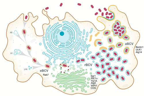
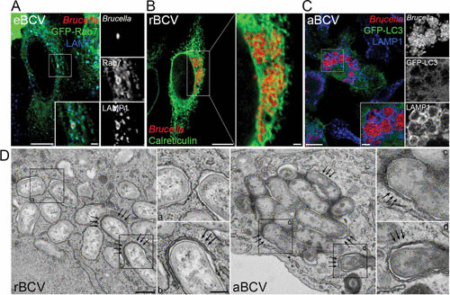
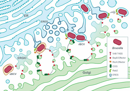
Bacteria of the genus Brucella colonize a wide variety of mammalian hosts, in which their infectious cycle and ability to cause disease predominantly rely on an intracellular lifestyle within phagocytes. Upon entry into host cells, Brucella organisms undergo a complex, multistage intracellular cycle in which they sequentially traffic through, and exploit functions of, the endocytic, secretory, and autophagic compartments via type IV secretion system (T4SS)-mediated delivery of bacterial effectors. These effectors modulate an array of host functions and machineries to first promote conversion of the initial endosome-like Brucella-containing vacuole (eBCV) into a replication-permissive organelle derived from the host endoplasmic reticulum (rBCV) and then to an autophagy-related vacuole (aBCV) that mediates bacterial egress. Here we detail and discuss our current knowledge of cellular and molecular events of the Brucella intracellular cycle. We discuss the importance of the endosomal stage in determining T4SS competency, the roles of autophagy in rBCV biogenesis and aBCV formation, and T4SS-driven mechanisms of modulation of host secretory traffic in rBCV biogenesis and bacterial egress.


Figures

FIGURE 1
FIGURE 1
Model of the Brucella intracellular cycle in macrophages. Following phagocytic uptake by macrophages, Brucella spp. reside during the first 8 to 12 h postinfection within a membrane-bound vacuole that undergoes endosomal maturation via sequential interactions with early (EE) and late (LE) endosomes and lysosomes (LYS) to become an acidified eBCV. The host small GTPase Rab7 contributes to eBCV maturation, which provides physicochemical cues promoting expression of the VirB T4SS, which translocates effector proteins (red) that mediate eBCV interactions with the ER exit site and acquisition of ER and Golgi apparatus-derived membranes. These events lead to the biogenesis of replication permissive, ER-derived BCVs, called rBCVs. The host proteins Sar1, IRE1α, Yip1A, Atg9, and WIPI1 and the COG complex contribute to rBCV biogenesis. Bacteria then undergo extensive replication in rBCVs between 12 and 48 h postinfection, after which rBCVs are captured within autophagosome-like structures in a VirB T4SS-dependent manner to become aBCVs. aBCV formation requires the host autophagy proteins beclin1, ULK1, and Atg14. aBCVs harbor features of autolysosomes and are required for bacterial egress and new cycles of intracellular infections.
FIGURE 2
FIGURE 2
Structure and membrane composition of BCVs during the Brucella intracellular cycle. (A) Confocal fluorescence micrograph of HeLa cells expressing green fluorescent protein (GFP)-Rab7 and infected with DsRedm-expressing B. abortus strain 2308 for 6 h. The inset shows an eBCV with the typical accumulation of the late endosomal/lysosomal markers Rab7 and LAMP1. Bars, 10 and 2 μm. (B) Confocal fluorescence micrograph of a HeLa cell infected with DsRedm-expressing B. abortus strain 2308 for 24 h and stained for the ER marker calreticulin. The inset shows a cluster of calreticulin-positive rBCVs containing replicating bacteria and associated with the ER network. Bars, 10 and 1 μm. (C) Confocal fluorescence micrograph of a primary murine bone marrow-derived macrophage expressing the autophagy marker GFP-LC3 and infected with DsRedm-expressing B. abortus strain 2308 for 72 h. The inset shows a group of aBCVs with the typical accumulation of the late endosomal/lysosomal LAMP1 but not LC3. Bars, 10 and 2 μm. (D) Transmission electron micrographs of bone marrow-derived macrophages infected with B. abortus strain 2308 for 72 h and showing the ultrastructures of rBCVs (left, single-membrane-bound vacuoles [inset a]), of forming aBCVs (inset b and arrows), and of completed double-membrane-bound aBCVs (right [insets c and d and arrows]). Bars, 500 and 200 nm. Reprinted from reference with permission.
FIGURE 3
FIGURE 3
Model of VirB T4SS-dependent biogenesis of the rBCV. Bacteria in eBCVs induce expression of the VirB T4SS, which delivers effector proteins into the host cell. Among these, BspB traffics to Golgi membranes via the ER-to-Golgi intermediate compartment (ERGIC) and binds to the COG complex to promote redirection of Golgi apparatus-derived vesicular traffic to BCVs. RicA binds the small host secretory GTPase Rab2, which contributes to its recruitment on maturing eBCVs and role in rBCV biogenesis. Additionally, eBCV interaction with ERES is accompanied by the upregulation of COPII coat components, induction of IRE1α, and Yip1A-dependent formation of ER-derived vesicles, which are also thought to contribute to rBCV biogenesis. T4SS-dependent acquisition of ER- and Golgi apparatus-derived secretory membranes by BCVs is thought to mediate eBCV-to-rBCV conversion.

References

    1. Moreno E. 2014. Retrospective and prospective perspectives on zoonotic brucellosis. Front Microbiol 5:213 10.3389/fmicb.2014.00213. [PubMed] - DOI - PMC - PubMed
    1. Pappas G, Akritidis N, Bosilkovski M, Tsianos E. 2005. Brucellosis. N Engl J Med 352:2325–2336 10.1056/NEJMra050570. [PubMed] - DOI - PubMed
    1. Pappas G, Papadimitriou P, Akritidis N, Christou L, Tsianos EV. 2006.The new global map of human brucellosis. Lancet Infect Dis 6:91–99 10.1016/S1473-3099(06)70382-6. - DOI - PubMed
    1. Atluri VL, Xavier MN, de Jong MF, den Hartigh AB, Tsolis RM. 2011. Interactions of the human pathogenic Brucella species with their hosts. Annu Rev Microbiol 65:523–541 10.1146/annurev-micro-090110-102905. [PubMed] - DOI - PubMed
    1. Celli J. 2015. The changing nature of the Brucella-containing vacuole. Cell Microbiol 17:951–958 10.1111/cmi.12452. [PubMed] - DOI - PMC - PubMed

Publication types

Substances

LinkOut - more resources