. 2020 Mar;45(3):580-590.
doi: 10.1007/s11064-019-02754-z. Epub 2019 Mar 8.

Targeted Oligodendrocyte Apoptosis in Optic Nerve Leads to Persistent Demyelination

Affiliations

Targeted Oligodendrocyte Apoptosis in Optic Nerve Leads to Persistent Demyelination

Ahdeah Pajoohesh-Ganji et al. Neurochem Res. 2020 Mar.

Abstract

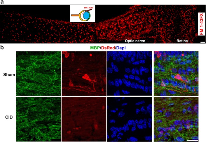
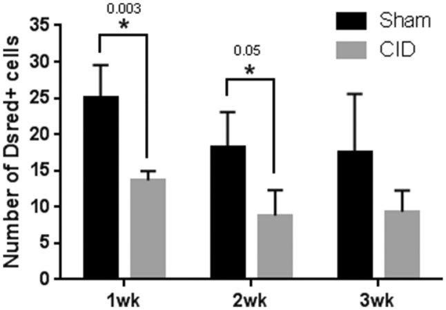
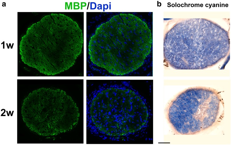
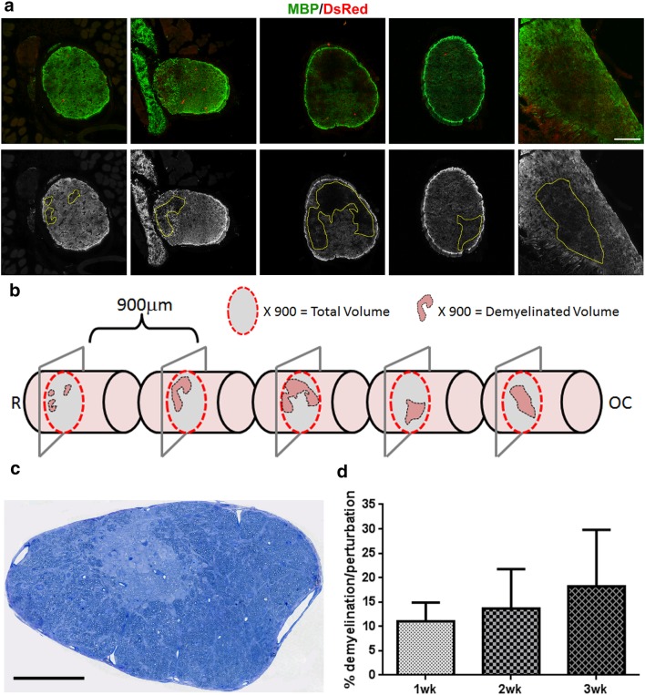
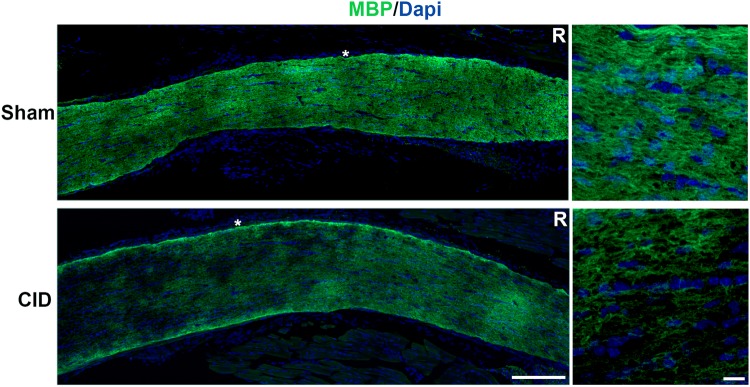
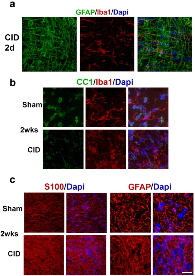
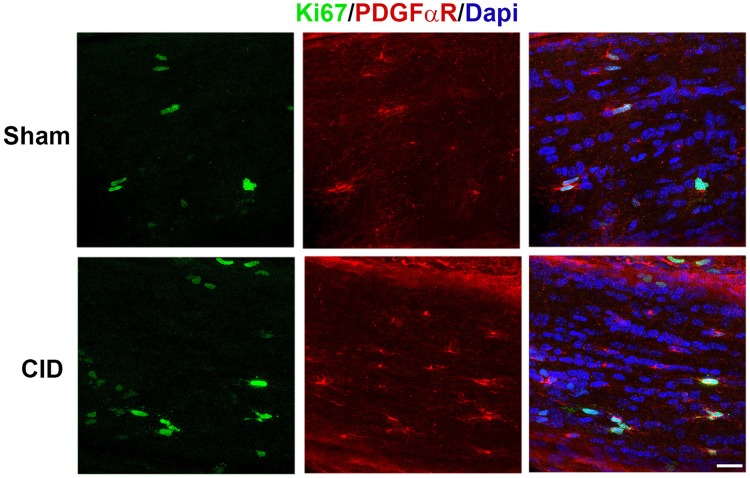
The optic nerve represents one of the simplest regions of the CNS and has been useful in developing an understanding of glial development and myelination. While the visual system is frequently affected in demyelinating conditions, utilizing the optic nerve to model demyelination/remyelination studies has been difficult due to its accessibility, relatively small size, and dense nature that makes direct injections challenging. Taking advantage of the lack of oligodendrocytes and myelination in the mouse retina, we have developed a model in which the induction of apoptosis in mature oligodendrocytes allows for the selective, non-invasive generation of demyelinating lesions in optic nerve. Delivery of an inducer of oligodendrocyte apoptosis by intravitreous injection minimizes trauma to the optic nerve and allows for the assessment of oligodendrocyte death in the absence of injury related factors. Here we show that following induction of apoptosis, oligodendrocytes are lost within 3 days. The loss of oligodendrocytes is associated with limited microglial and astrocyte response, is patchy along the nerve, and results in localized myelin loss. Unlike in other regions of the murine CNS, where local demyelination stimulates activation of local oligodendrocyte precursors and remyelination, optic nerve demyelination induced by oligodendrocyte apoptosis fails to recover and results in persistent areas of myelin loss. Over time these chronic lesions change cellular composition and ultimately become devoid of GFAP+ astrocytes and OPCs. Why the optic nerve lesions fail to repair may reflect the lack of early immune responsiveness and provide a novel model of chronic demyelination.

Keywords: Apoptosis; Demyelination; Oligodendrocytes; Optic nerve.


Figures

Fig. 1
Fig. 1
CID intravitreous injection leads to oligodendrocyte apoptosis in the optic nerve. a Intravitreous delivery of small molecules results in broad dispersal along the optic nerve. Injection of the dye (FM 1-43FX) demonstrates rapid penetration of the dye from the retina into the optic nerve within 1 h. The inset depicts the angle of injection into the vitreous at the limbus (Bar = 50 µm). b Intravitreous delivery of CID into MBP-iCP9 transgenic animals results in loss of DsRed oligodendrocytes. Animals were injected with CID in the right eye and the left eye was the sham control. Mice were sacrificed 2 days after the first injection and longitudinal sections are stained with MBP (green) and DsRed (red) to visualize myelin and oligodendrocytes, respectively; Dapi (blue) stains the nuclei. In control (sham) nerves extensive DsRed+ oligodendrocyte cell bodies and processes where detectable. By contrast following CID injection only fragments of DsRed+ cell bodies and processes remain. Bar = 10 µm
Fig. 2
Fig. 2
Persistent loss of DsRed+ cells following CID injection. Quantitation of the relative number of DsRed+ oligodendrocytes taken from cross sections of the optic nerve following CID injection. Significant reductions in the number of DsRed+ oligodendrocytes were observed at 1 (p value = 0.003) and 2 (p value = 0.05) weeks after CID injection compared to controls. The same trend is observed at 3 weeks after treatment, suggesting a lack of rapid recovery of the DsRed+ cells
Fig. 3
Fig. 3
Demyelination of the optic nerve is detectable 1 week after CID treatment. a Cross sections of optic nerve were labeled with antibodies against myelin basic protein. Areas of demyelination were detectable as reduced MBP expression (green) at 1 and 2 weeks after CID injection. At 1 week post CID, loss of myelin was relatively diffuse in both a MBP immunohistochemistry and b solochorme cyanine labeling. At 2 weeks post CID, the nerves appeared thinner, the areas of demyelination were more pronounced and an enhanced cellularity was present in regions of myelin loss. Bar = 50 µm
Fig. 4
Fig. 4
Demyelination induced by oligodendrocyte apoptosis does not repair rapidly and lesion volumes expands over time. MBP-iCP9 transgenic mice were injected with CID and sacrificed at 1, 2, or 3 weeks after the first injection. a Cross sections from CID-treated nerves 3 weeks after CID injection were cut using stereological methods and stained with MBP (green) and DsRed (red) to quantify change in demyelination. The MBP images were then converted to grayscale and the total nerve and demyelinated areas were measured using image J software and volumes were calculated. b 3D cartoon based on the sections depicts the patchy nature of the demyelination. c Toluidine blue staining of semi-thin section demonstrates distinct regions of myelin loss in CID treated nerves. d Percent demyelination was calculated at different time points indicating that the lesion does not repair up to 3 weeks after oligodendrocyte ablation. Bar = 100 µm in a and c
Fig. 5
Fig. 5
Demyelination persists 4 weeks after CID injection. Longitudinal sections were cut and stained with anti-MBP (green) and Dapi (blue) 4 weeks after CID injection. In the left panel, loss of myelin is apparent as dark patches in the CID injected optic nerve, which increases with distance from the retina (R). Regions marked with the asterisks are shown at higher magnification in the right panels. Bar = 10 µm
Fig. 6
Fig. 6
Oligodendrocytes ablation fails to stimulate an early robust immune response. a Sections labeled for microglia (Iba1, red) and Dapi (blue) 2 days after CID injection showed little microglial activation at this time point. b At 2 weeks after CID injections, there was a decrease in the number of mature oligodendrocytes and an increase in microglia compared to controls. Cross sections of optic nerve were labeled for mature oligodendrocytes (CC1, green) and microglia (Iba1, red). c At 2 weeks after CID injection, there is an increase in S100 and GFAP+ astrocytes (GFAP, red) and nuclei (Dapi, blue) compared to controls. Bar = 10 µm
Fig. 7
Fig. 7
Oligodendrocyte progenitor cells have a muted response after CID treatment. Sections from 1 week post CID and sham nerves double labeled with antibodies to Ki67, green, and PDGFαR, red, show only a slight increase in the proliferation of OPCs following oligodendrocyte ablation. The total number of cells is also not substantially increased as shown by Dapi (blue). Bar = 20 µm

Similar articles

Cited by

References

    1. Akassoglou K, Bauer J, Kassiotis G, Pasparakis M, Lassmann H, Kollias G, Probert L. Oligodendrocyte apoptosis and primary demyelination induced by local TNF/p55TNF receptor signaling in the central nervous system of transgenic mice: models for multiple sclerosis with primary oligodendrogliopathy. Am J Pathol. 1998;153(3):801–813. doi: 10.1016/S0002-9440(10)65622-2. - DOI - PMC - PubMed
    1. Schwab N, Schneider-Hohendorf T, Wiendl H. Therapeutic uses of anti-alpha4-integrin (anti-VLA-4) antibodies in multiple sclerosis. Int Immunol. 2015;27(1):47–53. doi: 10.1093/intimm/dxu096. - DOI - PubMed
    1. Compston A, Coles A. Multiple sclerosis. Lancet. 2008;372(9648):1502–1517. doi: 10.1016/S0140-6736(08)61620-7. - DOI - PubMed
    1. Rangachari M, Kuchroo VK. Using EAE to better understand principles of immune function and autoimmune pathology. J Autoimmun. 2013;45:31–39. doi: 10.1016/j.jaut.2013.06.008. - DOI - PMC - PubMed
    1. Sachs HH, Bercury KK, Popescu DC, Narayanan SP, Macklin WB. A new model of cuprizone-mediated demyelination/remyelination. ASN Neuro. 2014 doi: 10.1177/1759091414551955. - DOI - PMC - PubMed

LinkOut - more resources