. 2019 Oct;129(10):E355-E364.
doi: 10.1002/lary.27909. Epub 2019 Mar 8.

Methodology for the establishment of primary porcine vocal fold epithelial cell cultures

Affiliations

Methodology for the establishment of primary porcine vocal fold epithelial cell cultures

Elizabeth Erickson-DiRenzo et al. Laryngoscope. 2019 Oct.

Abstract

Objective: A current lack of methods for epithelial cell culture significantly hinders our understanding of the role of the epithelial and mucus barriers in vocal fold health and disease. Our first objective was to establish reproducible techniques for the isolation and culture of primary porcine vocal fold epithelial cells. Our second objective was to evaluate the functional significance of cell cultures using an in vitro exposure to an inflammatory cytokine.

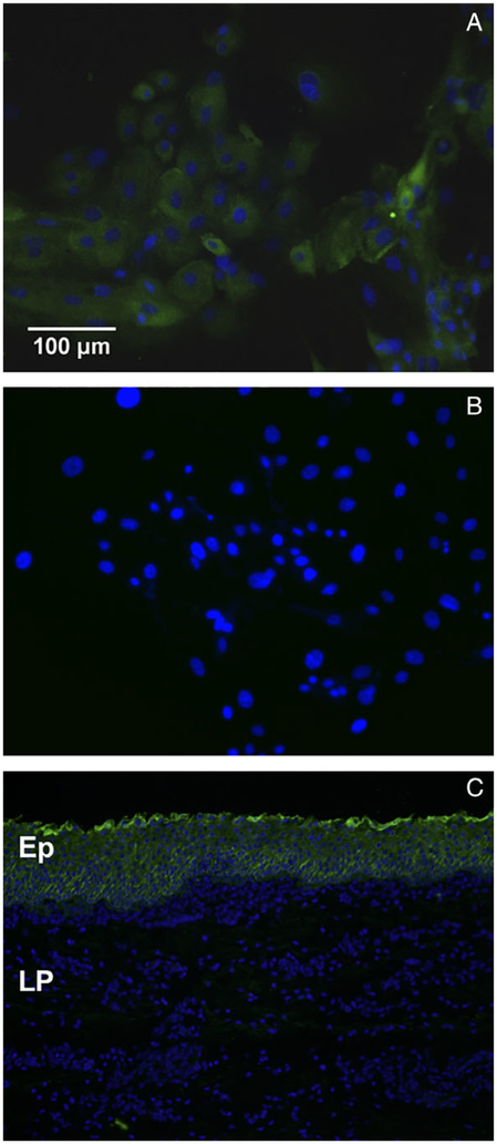
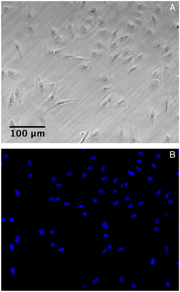
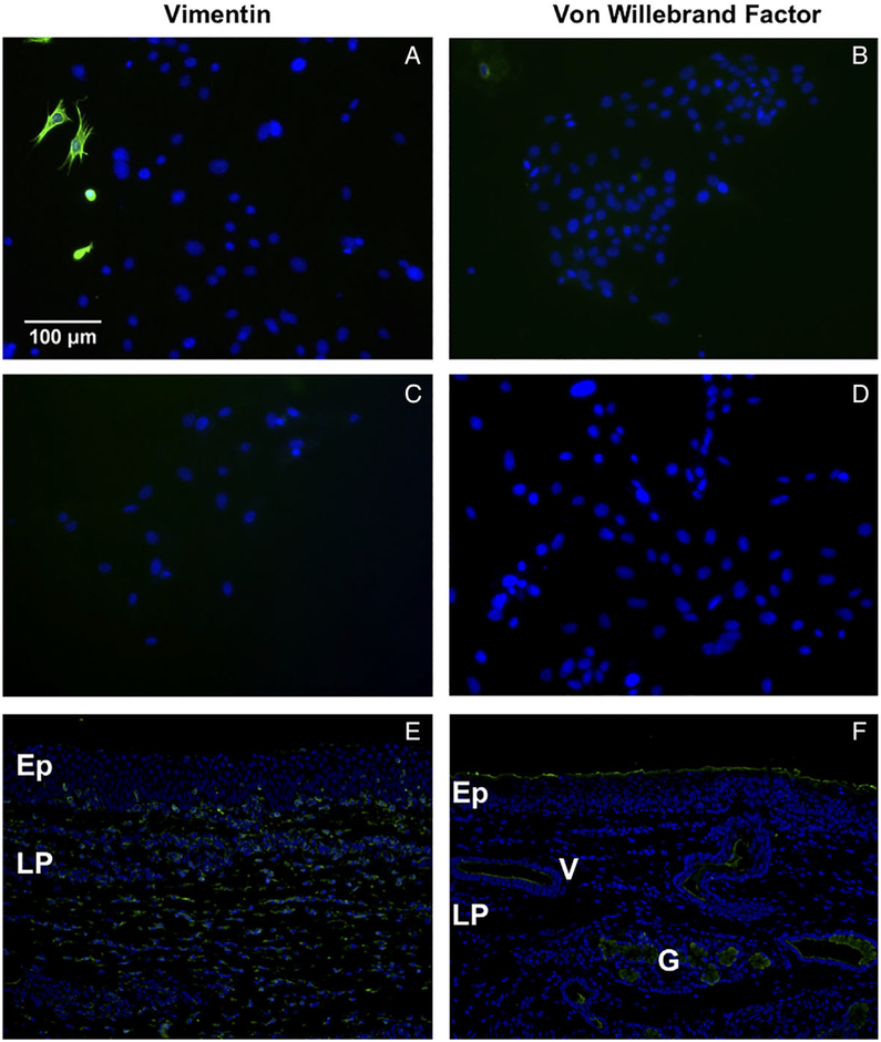
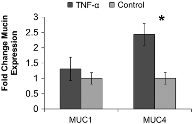
Methods: Epithelial cells were isolated from porcine vocal folds and expanded in culture. Characterization of cultures was completed by immunostaining with markers for pan-cytokeratin (epithelial cells), vimentin (stromal cells), von Willebrand factor (endothelial cell), and MUC1 and MUC4 (mucin) glycoproteins. Established epithelial cell cultures were then exposed to the inflammatory cytokine tumor necrosis factor alpha (TNF-α) for 24-hours, and transcript expression of MUC1 and MUC4 was evaluated.

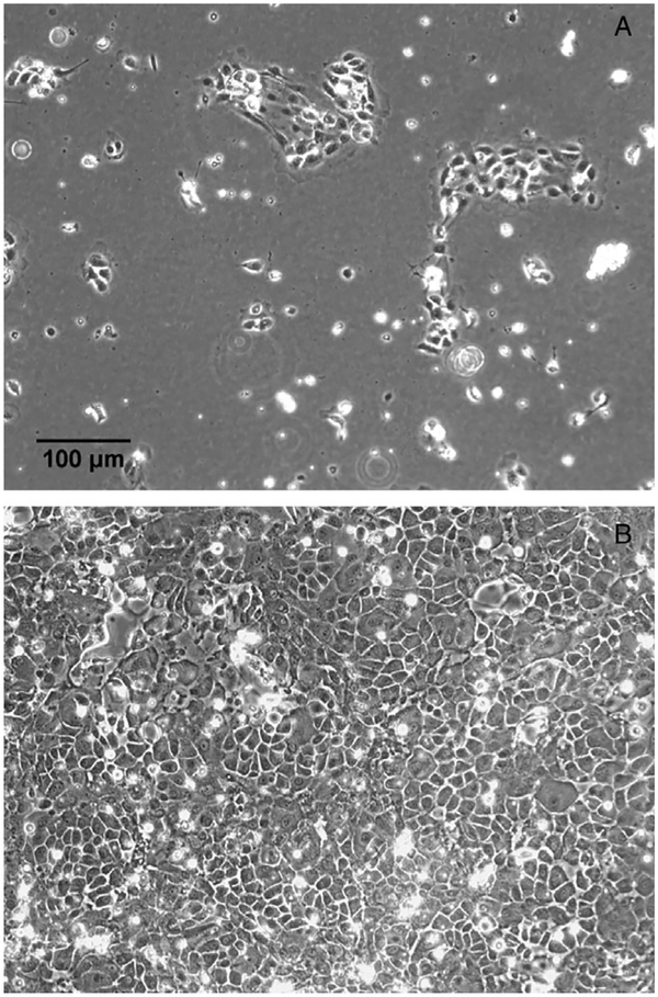
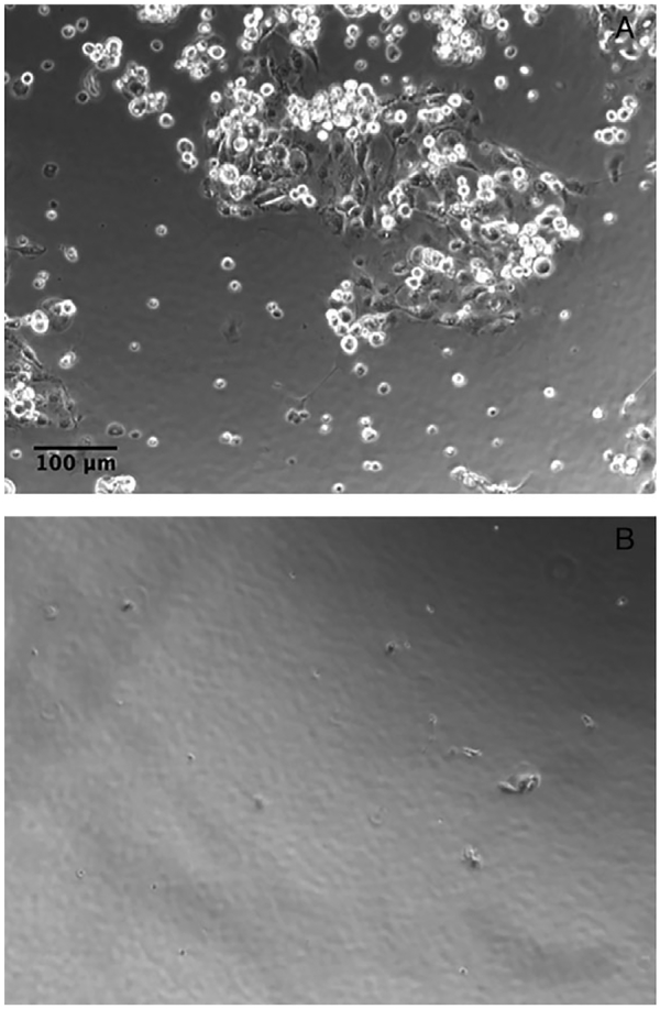
Results: Reproducible, porcine vocal fold epithelial cell cultures, demonstrating cobblestone appearance characteristic of the typical morphology of epithelial cell cultures were created. Cells showed positive staining for pan-cytokeratin with limited expression of vimentin and von Willebrand factor. Epithelial cells also expressed MUC1 and MUC4. TNF-α significantly increased transcript expression of MUC4.

Conclusion: Here, we present the first report of successful culture of primary porcine vocal fold epithelial cells. Cultures will provide researchers with a valuable new in vitro tool to investigate vocal fold epithelium and mucus as well as the effects of common challenges, including inflammatory cytokines, on these barriers.

Level of evidence: NA Laryngoscope, 129:E355-E364, 2019.

Keywords: Vocal fold; cell culture; epithelium; mucin; porcine.


Figures

Fig. 1.
Fig. 1.
Porcine vocal fold epithelial cells following culture for 2 (A) and 5 (B) days demonstrate cobblestone appearance consistent with epithelial cells.
Fig. 2.
Fig. 2.
Immunofluorescence confirmed that vocal fold epithelial cells stained positive (green) for pan-cytokeratin (A). No staining was observed in cells treated with goat anti-mouse secondary antibody only (B). Porcine vocal fold tissue was utilized as a positive control for epithelial pan-cytokeratin expression. Tissue demonstrates positive staining (green) for pan-cytokeratin in the Ep, but not LP (C). DAPI (blue) was used as a nuclear stain. Ep = epithelium; LP = lamina propria.
Fig. 3.
Fig. 3.
Porcine vocal fold fibroblasts demonstrated a spindle-shaped morphology (A). Immunofluorescence exhibited that porcine vocal fold fibroblasts were negative for pan-cytokeratin expression (B). DAPI (blue) was used as a nuclear stain.
Fig. 4.
Fig. 4.
Immunofluorescence demonstrated some isolated positive staining (green) of cell cultures with the stromal cell marker vimentin (A) and endothelial cell marker vWf (B). No staining was observed in cells treated with goat anti-mouse (C) and goat anti-rabbit (D) secondary antibody only. Porcine vocal fold tissue was utilized as positive control for vimentin and vWf expression. Tissue demonstrates positive staining (green) for vimentin primarily in the LP (E). Tissue demonstrates positive staining (green) for vWf in V and G (F). DAPI (blue) was used as a nuclear stain. Ep = epithelium; G = mucus glands; LP = lamina propria; V = vessels; vWf = von Willebrand factor.
Fig. 5.
Fig. 5.
Immunofluorescence exhibited that vocal fold epithelial cells stained positive (green) for MUC1 (A) and MUC4 (B). No staining was observed in cells treated with goat anti-rabbit (C) and goat anti-mouse (D) secondary antibody only. DAPI (blue) was used as a nuclear stain. Porcine vocal fold tissue was utilized as positive control for epithelial MUC1 and MUC4 expression. Tissue demonstrates isolated positive staining (brown) in the Ep for MUC1 (E) and diffuse positive staining (brown) in the Ep for MUC4 (F). Hematoxylin (purple) was used as a nuclear stain. Ep = epithelium; LP = lamina propria; MUC = mucin.
Fig. 6.
Fig. 6.
Fold change in MUC1 and MUC4 gene expression in TNF-α relative to vehicle-challenged cell cultures. Positive fold-change indicates an increase in gene expression. Expression of MUC4 significantly (*) increased following the TNF-α challenge. Error bars represent standard error. MUC = mucin; TNF-α = tumor necrosis factor alpha.
Fig. 7.
Fig. 7.
Porcine vocal fold epithelial subcultures following 2 (A) and 5 (B) days. Cellular growth was observed at day 2, but not at day 5.

Similar articles

Cited by

References

    1. Levendoski EE, Leydon C, Thibeault SL. Vocal fold epithelial barrier in health and injury: a research review. J Speech Lang Hear Res 2014;57: 1679–1691. - PMC - PubMed
    1. Samuels T, Handler E, Syring N, Blumin J, Kerschner J, Johnston N. Mucin gene expression in human laryngeal epithelia: effect of laryngopharyngeal reflux. Ann Otol Rhinol Laryngol 2008;117:688–695. - PubMed
    1. Rousseau B, Suehiro A, Echemendia N, Sivasankar M. Raised intensity phonation compromises epithelial barrier integrity. Laryngoscope 2011; 121:346–351. - PMC - PubMed
    1. Gill G, Johnston N, Buda A, et al. Laryngeal epithelial defenses against laryngopharyngeal reflux: Investigations of E-cadherin, carbonic anhydrase isoenzyme III, and pepsin. Ann Otol Rhinol Laryngol 2005;114:913–921. - PubMed
    1. Dworkin JP. Laryngitis: types, causes, and treatments. Otolaryngol Clin North Am 2008;41:419–436, ix. - PubMed

Publication types