. 2019 Sep;61(3):322-331.
doi: 10.1165/rcmb.2018-0326OC.

SMAD Signaling Restricts Mucous Cell Differentiation in Human Airway Epithelium

Affiliations

SMAD Signaling Restricts Mucous Cell Differentiation in Human Airway Epithelium

Michael B Feldman et al. Am J Respir Cell Mol Biol. 2019 Sep.

Abstract

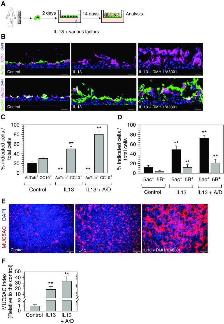
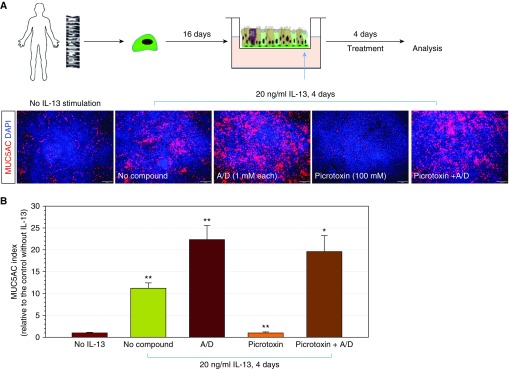
Mucin-secreting goblet cell metaplasia and hyperplasia (GCMH) is a common pathological phenotype in many human respiratory diseases, including asthma, chronic obstructive pulmonary disease, cystic fibrosis, primary ciliary dyskinesia, and infections. A better understanding of how goblet cell quantities or proportions in the airway epithelium are regulated may provide novel therapeutic targets to mitigate GCMH in these devastating diseases. We identify canonical SMAD signaling as the principal pathway restricting goblet cell differentiation in human airway epithelium. Differentiated goblet cells express low levels of phosphorylated SMAD. Accordingly, inhibition of SMAD signaling markedly amplifies GCMH induced by mucous mediators. In contrast, SMAD signaling activation impedes goblet cell generation and accelerates the resolution of preexisting GCMH. SMAD signaling inhibition can override the suppressive effects imposed by a GABAergic receptor inhibitor, suggesting the GABAergic pathway likely operates through inhibition of SMAD signaling in regulating mucous differentiation. Collectively, our data demonstrate that SMAD signaling plays a determining role in mucous cell differentiation, and thus raise the possibility that dysregulation of this pathway contributes to respiratory pathophysiology during airway inflammation and pulmonary diseases. In addition, our study also highlights the potential for SMAD modulation as a therapeutic target in mitigating GCMH.

Keywords: GABAergic pathway; SMAD signaling; airway diseases; goblet cell; human airway epithelium.


Figures

Figure 1.
Figure 1.
SMAD signaling activity is suppressed in differentiated goblet cells in human airway epithelium. (A and B) Costaining of phosphorylated (p)-SMAD1/5/8 (A) and p-SMAD2/3 (B) with goblet cell marker (MUC5AC), ciliated cell marker (FOXJ1), and basal cell marker (p63) in human bronchial epithelium. The nucleus is stained by DAPI. Scale bars: 10 μm. Insets show a fourfold-enlarged portion of the staining image to highlight finer detail. (C) Costaining of p-SMAD1/5/8 and p-SMAD2/3 (mixed antibodies) with goblet cell marker (MUC5AC), club cell marker (CCSP), ciliated cell marker (FOXJ1), and basal cell marker (CK5) in human airway epithelium differentiated on air–liquid interface (ALI). The nucleus is stained by DAPI. Scale bars: 10 μm. The dotted lines outline the shape of club cells or goblet cells. The staining shown was performed from bronchial sections of one representative donor. Staining has been replicated in n = 4 donors and similar results were observed.
Figure 2.
Figure 2.
Model of differential SMAD signaling activity in human respiratory epithelial cells. In a simplified model of human airway epithelium, basal cells give rise to club cells, which subsequently differentiate into ciliated cells or goblet cells, depending on environmental signals. SMAD signaling is suppressed in immature basal cells. During the differentiation of basal cells into club cells and ciliated cells, SMAD signaling increases in parallel with increasing maturity. During the differentiation of goblet cells from club cells, SMAD signaling is decreased. This model proposes that SMAD signaling changes in club cells drive the determination of whether club cells will give rise to ciliated cells or goblet cells.
Figure 3.
Figure 3.
Inhibition of bone morphogenetic protein (BMP)/transforming growth factor (TGF)-β/SMAD signaling amplifies IL-13– and IL-17A–induced goblet cell metaplasia and hyperplasia (GCMH) in human airway epithelium. (A) Human airway basal cells derived from healthy donors (n = 4) were cultured on ALI for 16 days to establish differentiated airway epithelium. IL-13 (20 ng/ml) alone or together with BMP4 (100 ng/ml), TGF-β (10 ng/ml), DMH-1 (1 μM), or A-8301 (1 μM) were added to the ALI medium in the basolateral chamber and ALIs were cultured for another 4 days. (B) ALI membranes were fixed and stained for MUC5AC. Representative images shown here were from one representative experiment. Scale bars: 100 μm. (C) Quantification of MUC5AC production in ALI membranes generated from the experiment shown in B. MUC5AC index was scored using ImageJ software (National Institutes of Health) based on the total area of MUC5AC immunopositivity out of the imaging area normalized to the control without IL-13 stimulation (mean ± SD; n = 3 independent imaging fields; **P ≤ 0.001 and ***P ≤ 0.0001). (D) Immunoblot analysis of FOXA3 and SAM pointed domain containing ETS transcription factor (SPDEF) of ALI cultures generated as described in B. β-actin was used as loading control. (E) Human airway basal cells from donors with asthma (n = 2), cystic fibrosis (CF) (n = 3), and chronic obstructive pulmonary disease (COPD; n = 2) were cultured on ALI for 16 days and then treated with IL-13 (20 ng/ml), IL-13 with BMP (100 ng/ml) and TGF-β (10 ng/ml), or IL-13 with DMH-1 (1 μM) and A8301 (1 μM) for 4 days. After treatment, ALI membranes were fixed and stained for MUC5AC. Representative images shown here were from one representative experiment on representative donors. Scale bars: 100 μm. (F) Quantification of MUC5AC in the experiment described in E. A total of three independent areas were imaged for each sample and quantified (mean ± SD; n = 3 independent imaging fields; **P ≤ 0.001 and ***P ≤ 0.0001) (G) Human airway basal cells derived from a healthy donor and a donor with asthma were cultured on ALI for 16 days and then treated with IL-17A (50 ng/ml), IL-17A with BMP (100 ng/ml) and TGF-β (10 ng/ml), or IL-17A with DMH-1 (1 μM) and A8301 (1 μM) for 4 days. ALI membranes were fixed and stained with MUC5AC. Representative images shown here are from one representative experiment on representative donors. Scale bars: 100 μm. (H) Quantification of MUC5AC in the experiment described in G. MUC5AC index was scored as described previously here. A total of three independent areas were imaged and quantified (mean ± SD; n = 3 independent imaging fields; ***P ≤ 0.0001 and **P ≤ 0.001). DMH-1 = a BMP-SMAD pathway inhibitor.
Figure 4.
Figure 4.
Inhibition of BMP/TGF-β/SMAD signaling amplifies GCMH from early committed progenitor cells. (A) Human airway basal cells derived from a healthy donor were seeded on ALI. At Day 2, cells were cultured in control ALI medium alone, or ALI medium with IL-13 (20 ng/ml), or ALI medium with IL-13 (20 ng/ml) with A8301 (1 μM)/DMH-1 (1 μM) for another 14 days. ALI membranes were fixed to examine various airway epithelial cell markers. (B) Staining of AcTub (ciliated cells), CCSP (club cells), and MUC5AC and MUC5B (goblet cells) on ALI trans-sections. Scale bars: 10 μm. (C and D) Quantification of AcTub+ cells and CCSP+ cells (C) and MUC5AC+ cells and MUC5B+ cells (D) out of the total cells (total DAPI number) based on staining described in B. A total of three independent areas were imaged and quantified (mean ± SD; n = 3 independent imaging fields; **P ≤ 0.001). (E) Whole-mount staining of MUC5AC on ALI membranes. Scale bars: 100 μm. (F) Quantification of MUC5AC production in the experiment described in E. MUC5AC index was scored using ImageJ software based on the total area of MUC5AC immunopositivity out of the imaging area and calculated relative to the control without IL-13 stimulation. A total of three independent areas were imaged and quantified (mean ± SD; n = 3 independent imaging fields; **P ≤ 0.001). A/D = A8301 + DMH-1.
Figure 5.
Figure 5.
Activation of SMAD signaling facilitates the resolution of GCMH. (A and B) Human airway basal cells derived from a representative healthy donor were cultured on ALI for 16 days and treated with IL-13 (20 ng/ml) for 4 days to induce GCMH. ALI cultures were then rinsed gently and cultured further in various conditions (ALI medium only; ALI medium + BMP4 [100 ng/ml] + TGF-β [10 ng/ml]; or ALI medium + BMP4 [100 ng/ml] + TGF-β [10 ng/ml] + Y-27632 ROCK inhibitor [10 μM]) for another 7 days. ALI membranes were fixed and stained with MUC5AC antibody. Scale bars: 100 μm. (C) Quantification of MUC5AC production in the experiment described in A and B. MUC5AC index was scored based on the total area of MUC5AC immunopositivity out of the imaging area and calculated relative to Day 0 (the day when IL-13 stimulation was stopped and washed off). A total of three independent areas were imaged and quantified (mean ± SD; n = 3 independent imaging fields; **P ≤ 0.001). ROCKi = ROCK inhibitor.
Figure 6.
Figure 6.
SMAD signaling inhibition overrides the suppressive effect of γ-aminobutyric acid (GABA) receptor inhibitor in goblet cell production. (A) Human airway basal cells derived from a representative healthy donor were cultured on ALI for 16 days and treated with IL-13 (20 ng/ml) for 4 days to induce goblet cell hyperplasia. SMAD signaling inhibitors (1 μM A8301 + 1 μM DMH-1) and GABAergic system receptor (100 μM picrotoxin), or in combination, were included in ALI culture to examine their effects on goblet cell generation. ALI membranes were fixed and stained with MUC5AC antibody. Scale bars: 100 μm. (B) Quantification of MUC5AC production in the experiment shown in A. MUC5AC index was calculated based on the total area of MUC5AC positivity out of total imaging area. The score was relative to the control (without IL-13 stimulation; mean ± SD; n = 3 independent imaging fields; *P ≤ 0.01 and **P ≤ 0.001).

Comment in

Similar articles

Cited by

References

    1. Tanabe T, Rubin BK. Airway goblet cells secrete pro-inflammatory cytokines, chemokines, and growth fa2ctors. Chest. 2016;149:714–720. - PubMed
    1. Hogg JC. Pathophysiology of airflow limitation in chronic obstructive pulmonary disease. Lancet. 2004;364:709–721. - PubMed
    1. Parulekar AD, Kao CC, Diamant Z, Hanania NA. Targeting the interleukin-4 and interleukin-13 pathways in severe asthma: current knowledge and future needs. Curr Opin Pulm Med. 2018;24:50–55. - PubMed
    1. Rael EL, Lockey RF. Interleukin-13 signaling and its role in asthma. World Allergy Organ J. 2011;4:54–64. - PMC - PubMed
    1. Danahay H, Pessotti AD, Coote J, Montgomery BE, Xia D, Wilson A, et al. Notch2 is required for inflammatory cytokine-driven goblet cell metaplasia in the lung. Cell Reports. 2015;10:239–252. - PubMed

Publication types

LinkOut - more resources