

High-resolution contrast-enhanced vessel wall imaging in patients with suspected cerebral vasculitis: Prospective comparison of whole-brain 3D T1 SPACE versus 2D T1 black blood MRI at 3 Tesla
- PMID: 30849127
- PMCID: PMC6407784
- DOI: 10.1371/journal.pone.0213514
High-resolution contrast-enhanced vessel wall imaging in patients with suspected cerebral vasculitis: Prospective comparison of whole-brain 3D T1 SPACE versus 2D T1 black blood MRI at 3 Tesla
Abstract
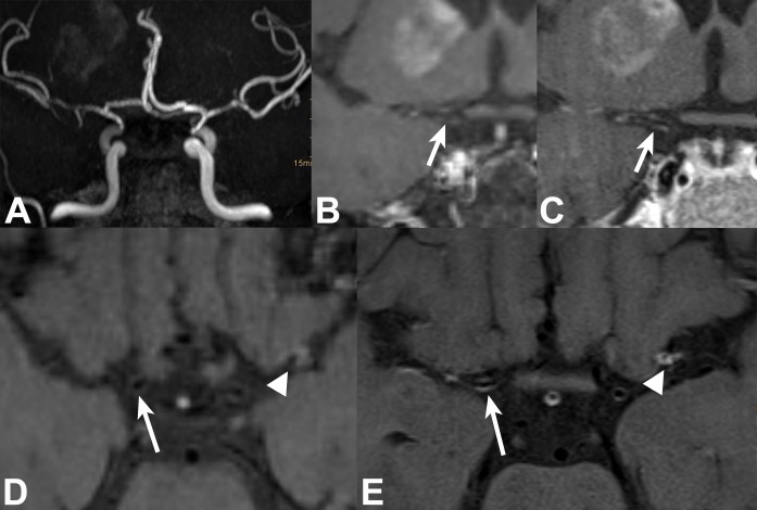
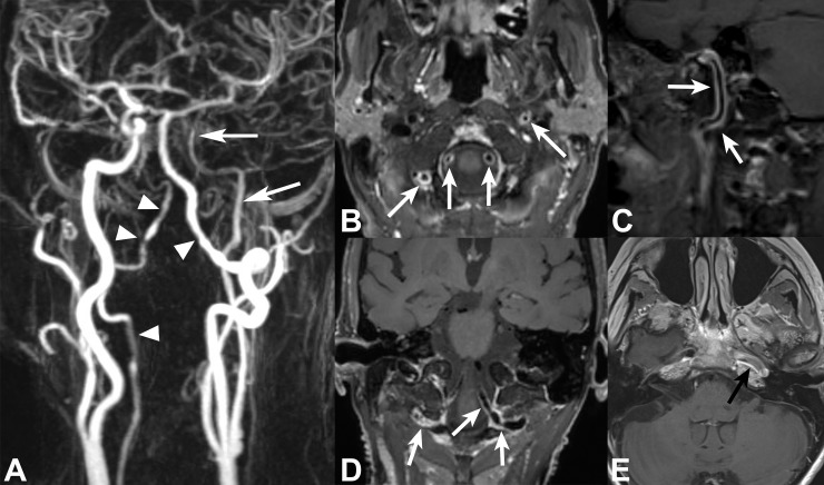
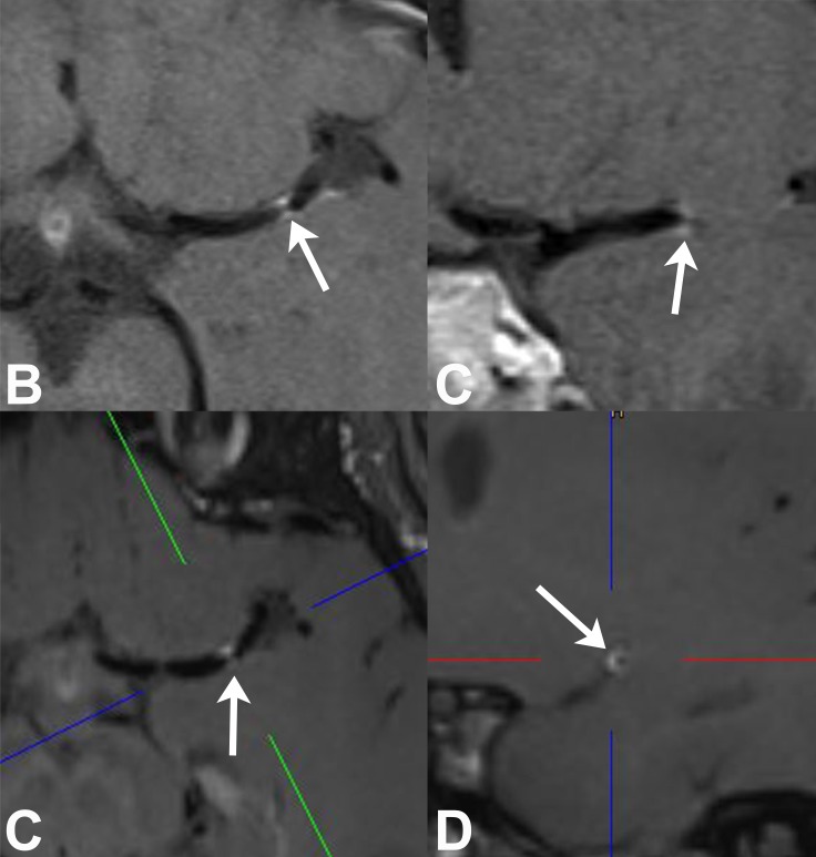
Purpose: Vessel wall imaging (VWI) using T1 dark blood MRI can depict inflammation of intracranial arteries in patients with cerebral vasculitis. Recently, 3D VWI sequences were introduced at 3 Tesla. We aimed to compare 2D and 3D VWI for detection of intracranial vessel wall enhancement (VWE) in patients suspected of cerebral vasculitis.
Methods: 44 MRI scans of 39 patients were assessed that included bi-planar 2D T1 and whole-brain 3D T1 SPACE dark blood VWI pre and post contrast. Visibility and VWE were analyzed in 31 pre-specified intracranial artery segments. Additionally, leptomeningeal and parenchymal contrast enhancement was assessed.
Results: Overall, more arterial segments were visualized with 3D VWI (p<0.0001). Detection of VWE showed fair agreement between 2D and 3D VWI (κ = 0.583). On segmental level, more VWE was detected in intradural ICA by 2D VWI (p<0.001) and in VA V4 segment by 3D VWI (p<0.05). 3D VWI showed more leptomeningeal (p<0.05) and parenchymal (p<0.01) contrast enhancement. In patients with positive diagnosis of cerebral vasculitis, sensitivity was of 67% (2D and 3D VWI) and specificity was 44% (2D VWI) and 48% (3D VWI); more VWE was seen in arteries distal to VA and ICA compared to non-vasculitic patients.
Conclusion: 2D and 3D VWI differed in the ability to detect VWE. Whole brain coverage with better evaluability of VAs and distal intracranial artery segments, and depiction of more parenchymal and leptomeningeal enhancement make 3D VWI more favorable. As VWE in arteries distal to VA and ICA may be used for discrimination of vasculitic and non-vasculitic patients, future increase in spatial resolution of 3D VWI sequences may be beneficial.
Conflict of interest statement
The senior author of this manuscript (SM) states the following competing interests: Acandis GmbH: consultant and member of the scientific advisory board, received honoraria and travel grants. Medtronic: received speaker honorarium (modest), travel grant, and non-financial support for video case production. Microvention; Stryker: received travel grants. Bracco S.p.A.: received research grant (money paid to institution). Novartis Pharma GmbH: received consultant fee. This does not alter our adherence to PLOS ONE policies on sharing data and materials.
Figures
References
-
- Calabrese LH, Mallek JA. Primary angiitis of the central nervous system. Report of 8 new cases, review of the literature, and proposal for diagnostic criteria. Medicine. 1988;67(1):20–39. Epub 1988/01/01. . - PubMed
-
- Cupps TR, Moore PM, Fauci AS. Isolated angiitis of the central nervous system. Prospective diagnostic and therapeutic experience. The American journal of medicine. 1983;74(1):97–105. Epub 1983/01/01. . - PubMed
-
- Lie JT. Primary (granulomatous) angiitis of the central nervous system: a clinicopathologic analysis of 15 new cases and a review of the literature. Human pathology. 1992;23(2):164–71. Epub 1992/02/01. . - PubMed
-
- Duna GF, Calabrese LH. Limitations of invasive modalities in the diagnosis of primary angiitis of the central nervous system. The Journal of rheumatology. 1995;22(4):662–7. Epub 1995/04/01. . - PubMed
Publication types
MeSH terms
Substances
LinkOut - more resources
Full Text Sources
Research Materials
Miscellaneous
