. 2019 Jul 15;200(2):235-246.
doi: 10.1164/rccm.201808-1599OC.

IL4Rα Signaling Abrogates Hypoxic Neutrophil Survival and Limits Acute Lung Injury Responses In Vivo

Affiliations

IL4Rα Signaling Abrogates Hypoxic Neutrophil Survival and Limits Acute Lung Injury Responses In Vivo

Alison J Harris et al. Am J Respir Crit Care Med. .

Abstract

Rationale: Acute respiratory distress syndrome is defined by the presence of systemic hypoxia and consequent on disordered neutrophilic inflammation. Local mechanisms limiting the duration and magnitude of this neutrophilic response remain poorly understood. Objectives: To test the hypothesis that during acute lung inflammation tissue production of proresolution type 2 cytokines (IL-4 and IL-13) dampens the proinflammatory effects of hypoxia through suppression of HIF-1α (hypoxia-inducible factor-1α)-mediated neutrophil adaptation, resulting in resolution of lung injury. Methods: Neutrophil activation of IL4Ra (IL-4 receptor α) signaling pathways was explored ex vivo in human acute respiratory distress syndrome patient samples, in vitro after the culture of human peripheral blood neutrophils with recombinant IL-4 under conditions of hypoxia, and in vivo through the study of IL4Ra-deficient neutrophils in competitive chimera models and wild-type mice treated with IL-4. Measurements and Main Results: IL-4 was elevated in human BAL from patients with acute respiratory distress syndrome, and its receptor was identified on patient blood neutrophils. Treatment of human neutrophils with IL-4 suppressed HIF-1α-dependent hypoxic survival and limited proinflammatory transcriptional responses. Increased neutrophil apoptosis in hypoxia, also observed with IL-13, required active STAT signaling, and was dependent on expression of the oxygen-sensing prolyl hydroxylase PHD2. In vivo, IL-4Ra-deficient neutrophils had a survival advantage within a hypoxic inflamed niche; in contrast, inflamed lung treatment with IL-4 accelerated resolution through increased neutrophil apoptosis. Conclusions: We describe an important interaction whereby IL4Rα-dependent type 2 cytokine signaling can directly inhibit hypoxic neutrophil survival in tissues and promote resolution of neutrophil-mediated acute lung injury.

Keywords: IL-4; acute respiratory distress syndrome; hypoxia; hypoxia-inducible factor-1α; neutrophil.


Figures
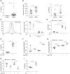
Figure 1.
Figure 1.
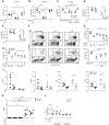
IL-4 is present in human and mouse models of acute respiratory distress syndrome (ARDS) with IL-4Ra expression found in both human and mouse neutrophils. (A) IL-4 levels were measured in BAL samples obtained from patients with ARDS and healthy control subjects by ultrasensitive ELISA. (B) Neutrophils from whole blood were identified by flow cytometry from patients with ARDS and healthy control subjects and levels of IL-4Ra surface protein and transcript measured. (C) Representative histogram (dashed black line = normoxia isotype, dashed gray = hypoxia isotype, black solid line = normoxia, gray solid line = hypoxia) and summary data of IL4Rα expression on human peripheral blood neutrophils after 4 hours of culture with or without IL-4. (C) Cell surface protein was determined by flow cytometry, with freshly isolated (T0) cells for comparison, and (D) mRNA was quantified relative to ACTB by qRT-PCR. (E) IL-4 levels from BAL from LPS-treated mice or naive mice (C57BL/6) obtained at 24 hours was measured by multiplex assay and (F) levels of IL-4Ra expression on lung neutrophils, (G) alveolar macrophages, and (H) T cells were measured by flow cytometry. (I) Apoptosis rates of inflammatory BAL neutrophils harvested from normoxic C57BL/6 mice 24 hours after LPS nebulization (neb) and then cultured ex vivo for 6 hours in hypoxia with or without 100 ng/ml IL-4 as determined by morphology. Data are expressed as individual data points with median and interquartile range (A, B, E, and I) or mean ± SEM (C, D, and FH). Statistical significance was determined by Mann-Whitney U test (A, B [IL4R mRNA], E, and I), unpaired Student’s t test (B [IL4Rα gMFI]), or two-way ANOVA with Holm-Sidak post hoc tests comparing each condition with unstimulated control within normoxic and hypoxic groups, respectively (C, D, F, and H). *P < 0.05, **P < 0.01, and ***P < 0.001. gMFi = geometric mean fluorescence intensity.
Figure 2.
Figure 2.
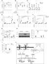
Modulation of human neutrophil hypoxic survival and inflammatory responses by IL-4. (A–F) Apoptosis of cytokine-treated neutrophils was assessed by morphology at 6 (A) and 20 hours (B and C) or by flow cytometry with annexin V at 20 hours (n = 3–6) (DF). (G and H) Neutrophil apoptosis after preincubation with IL4Rα-neutralizing antibody (α-IL4RA) before culture with IL-4 or IL-13 for 20 hours in hypoxia (n = 3–4). (I) Effect of IL-4 and IL-13 on LPS-induced survival in normoxia at 20 hours (n = 3) as determined by morphology. (J) Effects of IL-4 on LPS-mediated proinflammatory gene induction in human peripheral blood neutrophils cultured for 4 hours in normoxia or hypoxia, determined by qRT-PCR (relative to ACTB) (n = 4). (K) CCL17 expression relative to ACTB was determined by qRT-PCR, after 4 hours of culture in normoxia or hypoxia in the presence of IFN-γ, IL-4, and/or LPS (n = 4). (L) Reactive oxygen species (ROS) production was determined by flow cytometry using the fluorescent oxygen sensor DCF-DA. Neutrophils were incubated with IL-4 or IFN-γ + LPS for 4 hours with or without 100 nM N-formyl-met-leu-phe (fMLP) for a further 45 minutes (n = 4). Data are expressed as mean ± SEM. Significance was determined by repeated measures two-way ANOVA with Holm-Sidak post hoc test comparing cytokine treatments with unstimulated control within the normoxic and hypoxic groups, respectively (AE, and K), or LPS control (J), repeated measures one-way ANOVA with post hoc test for linear trend (G and H) or Holm-Sidak post hoc test (I), or separate two-way ANOVAs for baseline and fMLP-stimulated ROS production, with Holm-Sidak post hoc tests comparing IFN-γ + LPS and IL-4 with unstimulated control within normoxic and hypoxic groups, respectively (L). *P < 0.05, **P < 0.01, ***P < 0.001, and ****P < 0.0001. HU = hypoxia unstimulated; NU = normoxia unstimulated; PE = phycoerythrin.
Figure 3.
Figure 3.
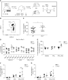
IL-4 regulates NF-κB (nuclear factor-κB) at a post-transcriptional level without altering metabolic flux. (A) Expression of glycolytic genes in human neutrophils after 8 hours of culture in normoxia or hypoxia with IL-4 or IFN-γ + LPS, determined by qRT-PCR relative to ACTB (n = 4). (B) Flux through glycolysis and fatty acid oxidation were quantified by 3H2O release after uptake of [5-3H]-glucose and [9,10-3H]-palmitic acid, respectively, in normoxia or hypoxia with or without IL-4. Energy status (ATP/ADP ratio) after 4 hours was determined by reverse phase HPLC (n = 3). (C) NF-κB p65 (RELA) protein levels after 6 hours of culture in normoxia or hypoxia with or without IL-4 were assessed by Western blot, levels quantified relative to p38 by densitometry, and fold change calculated relative to normoxia (n = 4). (D) Gene expression of RELA (NF-κB) and CHUK (IKKα) relative to ACTB was determined by qRT-PCR after 4 hours of culture in normoxia or hypoxia with or without IL-4 (10 ng/ml) (n = 4). Data are expressed as mean ± SEM. Statistical significance was determined by repeated measures two-way ANOVA with Holm-Sidak post hoc test comparisons with unstimulated control subjects within the normoxic and hypoxic groups, respectively (A, B, and D) or Mann-Whitney U test (C). *P < 0.05 and **P < 0.01.
Figure 4.
Figure 4.
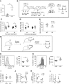
IL-4–mediated STAT/PPARγ signaling upregulates PHD2 transcript and reduces HIF-1α (hypoxia-inducible factor-1α) protein levels, resulting in loss of hypoxic neutrophil survival. (A and B) Effect of STAT3 and STAT6 inhibitors (5,15-DPP and AS 1517499, respectively) on cytokine-induced human neutrophil apoptosis in hypoxia at 20 hours (determined by morphology) (n = 3–4). (CE) Neutrophil expression of genes involved in PPARγ signaling and (F) of EGLN1/PHD2, after 4 hours of cytokine/LPS treatment in normoxia or hypoxia, determined by qRT-PCR (relative to ACTB) (n = 4). (G) HIF1A gene expression, after 4 hours of cytokine/LPS treatment in normoxia or hypoxia, determined by qRT-PCR (relative to ACTB) (n = 4). (H) HIF-1α protein levels after 6 hours of culture with IL-4 in normoxia or hypoxia were assessed by Western blot and levels relative to p38 quantified by densitometry (n = 4). (I) Phd2−/− mice or their littermate control subjects were treated with nebulized LPS and BAL neutrophils treated with IL-4 (100 ng/ml) in hypoxia ex vivo for 6 hours. Data normalized to apoptosis rates in unstimulated hypoxia control. (J) Illustration of IL-4 signaling pathway leading to loss of hypoxic survival in neutrophils. Data are expressed as mean ± SEM (AH) or median and interquartile range (I). Statistical significance was determined by one-way ANOVA with Holm-Sidak post hoc test comparing cytokine-only group with every other group (A and B), repeated measures two-way ANOVA with Holm-Sidak post hoc test comparisons with unstimulated control subjects within normoxic and hypoxic groups, respectively (CH), or Mann-Whitney U test of log-transformed data (I). *P < 0.05, **P < 0.01, ***P < 0.001, and ****P < 0.0001. NF-κB = nuclear factor-κB; WT = wild-type.
Figure 5.
Figure 5.
Il4ra−/− neutrophils have a competitive advantage in bone marrow chimeras in a liver injury model. (A) Cd45.2+/− Cd45.1+/− mice were irradiated to deplete host bone marrow, then reconstituted with Cd45.2+/+ Cd45.1−/− donor marrow, either wild-type (WT) or Il4ra−/−, as depicted. Mice were exposed to a single dose of CCl4 to induce liver damage, or olive oil vehicle control. (B) Chimerism (% CD45.1 donor neutrophils) was determined by flow cytometry. (C) Blood chimerism at Day 0 (pre-CCl4) in mice receiving WT or Il4ra−/− marrow. (D) Blood chimerism at Day 0 (pre-CCl4) and Day 3 in mice treated with CCl4 with or without IL4c and in olive oil vehicle control subjects. (E) Liver chimerism normalized to blood at Day 3 after CCl4. Total numbers of donor (F) and host (G) neutrophils per gram of liver at Day 3 after CCl4. Statistical significance was determined by unpaired Student’s t test (C), multiple t tests with Holm-Sidak correction (Day 0 vs. Day 3 for each treatment/genotype combination) (D), or two-way ANOVA with Holm-Sidak post hoc tests (WT vs. Il4rα−/−) (EG). Data shown as individual mice with mean ± SEM. *P < 0.05, ***P < 0.001, and ****P < 0.0001.
Figure 6.
Figure 6.
Exogenous IL-4c treatment accelerates the resolution of neutrophilic lung inflammation in hypoxia. (A) IL-4 levels from BAL from naive mice or mice treated with nebulized LPS and placed in hypoxia (10%) for 24 hours was measured by multiplex assay. (B) Cd45.2+/− Cd45.1+/− mice were irradiated to deplete host bone marrow (BM), then injected with donor marrow comprising 50% wild-type (WT) Cd45.2+/− Cd45.1+/− mixed with either 50% Il4ra+/+ Cd45.2+/+ Cd45.1−/− (WT) or 50% Il4ra−/− Cd45.2+/+ CD45.1−/− (Il4ra−/−), as depicted. Mice were nebulized with LPS and placed in hypoxia 25 days after transplant. Neutrophil chimerism (% of Ly6G+ cells lacking CD45.1) was determined by flow cytometry. Chimerism was normalized to WT control chimerism at each time point. Blood (C) and BAL neutrophil chimerism (D) after LPS and hypoxia. (E) Proportion of donor (Cd45.2+/+ Cd45.1−/−) BM CXCR4+ neutrophils. (F) C57BL/6 mice were treated with intratracheal IL-4c or PBS 6 hours after LPS nebulization and returned to hypoxia for a further 18 hours. BM neutrophil expression of CXCR4 (G) and CXCR2 (H) was determined by flow cytometry (representative histograms shown: light gray = FMO control, dark gray = PBS, and dotted line = IL-4c). (I) Proportion of circulating neutrophils in whole-blood leukocytes. (J) Total BAL neutrophils were determined by flow cytometry. (K) Proportion of BAL apoptotic (Annexin V+) neutrophils was measured by flow cytometry. (L) Total BAL IgM release was measured at 24 hours. Data shown as individual points with median and interquartile range (A, E, G, I, and L) or mean ± SD (C, D, J, and K). Statistical significance was determined by two-way ANOVA with Holm-Sidak post hoc test (WT vs. Il4ra−/− for each time point) (C, D, J, and K), Mann-Whitney U test (A, E, and L), or unpaired Student’s t test (GI). *P < 0.05, **P < 0.01, and ***P < 0.001. gMFi = geometric mean fluorescence intensity; KO = knockout; WT = wild-type.

Comment in

References

    1. Eltzschig HK, Carmeliet P. Hypoxia and inflammation. N Engl J Med. 2011;364:656–665. - PMC - PubMed
    1. Harris AJ, Thompson AR, Whyte MKB, Walmsley SR. HIF-mediated innate immune responses: cell signaling and therapeutic implications. Hypoxia (Auckl) 2014;2:47–58. - PMC - PubMed
    1. Galdiero MR, Bonavita E, Barajon I, Garlanda C, Mantovani A, Jaillon S. Tumor associated macrophages and neutrophils in cancer. Immunobiology. 2013;218:1402–1410. - PubMed
    1. Garlichs CD, Eskafi S, Cicha I, Schmeisser A, Walzog B, Raaz D, et al. Delay of neutrophil apoptosis in acute coronary syndromes. J Leukoc Biol. 2004;75:828–835. - PubMed
    1. Uddin M, Nong G, Ward J, Seumois G, Prince LR, Wilson SJ, et al. Prosurvival activity for airway neutrophils in severe asthma. Thorax. 2010;65:684–689. - PubMed

Publication types

MeSH terms

Substances