. 2019 Jul:127:163-177.
doi: 10.1016/j.nbd.2019.03.001. Epub 2019 Mar 5.

Systemic peptide mediated delivery of an siRNA targeting α-syn in the CNS ameliorates the neurodegenerative process in a transgenic model of Lewy body disease

Affiliations

Systemic peptide mediated delivery of an siRNA targeting α-syn in the CNS ameliorates the neurodegenerative process in a transgenic model of Lewy body disease

Brian Spencer et al. Neurobiol Dis. 2019 Jul.

Erratum in

Abstract

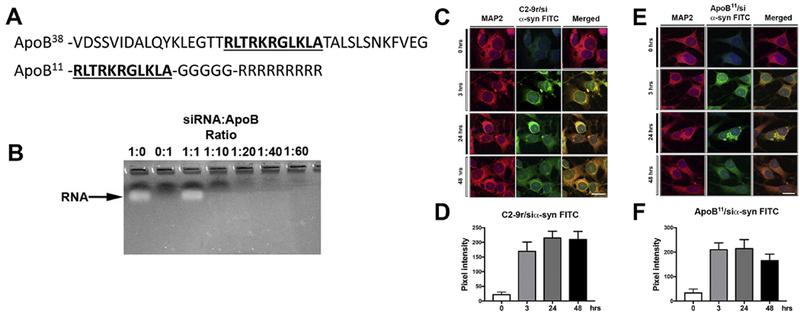
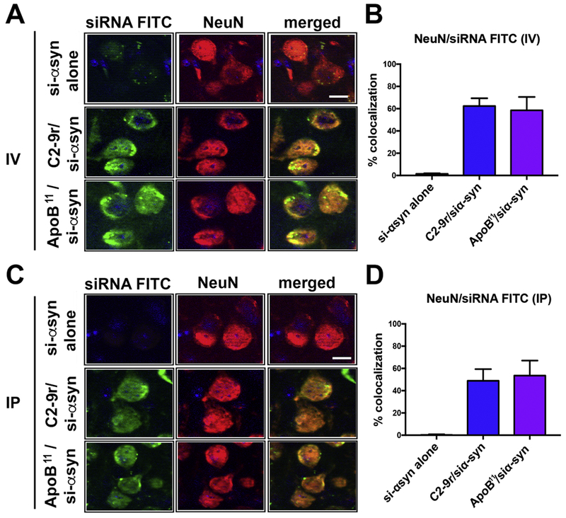
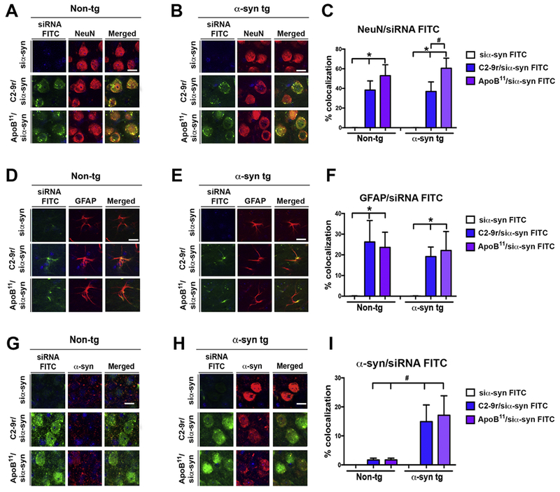
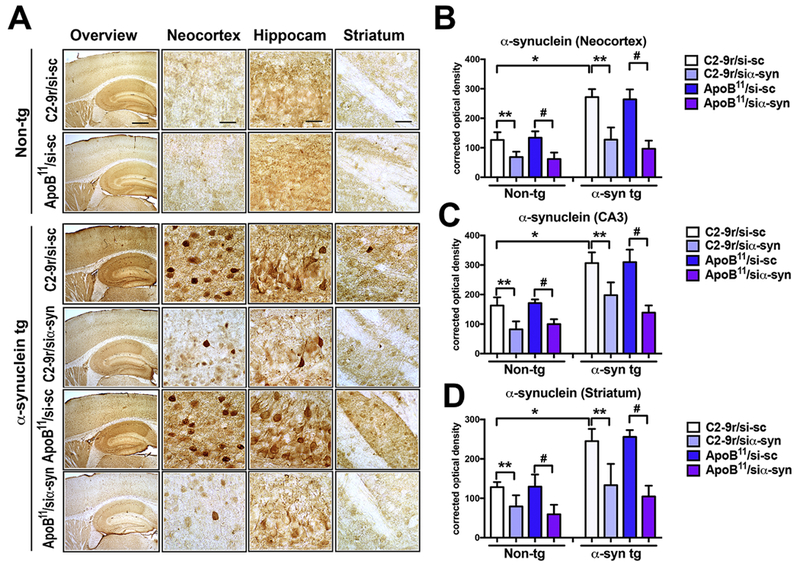
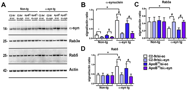
Neurodegenerative disorders of the aging population are characterized by progressive accumulation of neuronal proteins such as α-synuclein (α-syn) in Parkinson's Disease (PD) and Amyloid ß (Aß) and Tau in Alzheimer's disease (AD) for which no treatments are currently available. The ability to regulate the expression at the gene transcription level would be beneficial for reducing the accumulation of these proteins or regulating expression levels of other genes in the CNS. Short interfering RNA molecules can bind specifically to target RNAs and deliver them for degradation. This approach has shown promise therapeutically in vitro and in vivo in mouse models of PD and AD and other neurological disorders; however, delivery of the siRNA to the CNS in vivo has been achieved primarily through intra-cerebral or intra-thecal injections that may be less amenable for clinical translation; therefore, alternative approaches for delivery of siRNAs to the brain is needed. Recently, we described a small peptide from the envelope protein of the rabies virus (C2-9r) that was utilized to deliver an siRNA targeting α-syn across the blood brain barrier (BBB) following intravenous injection. This approach showed reduced expression of α-syn and neuroprotection in a toxic mouse model of PD. However, since receptor-mediated delivery is potentially saturable, each allowing the delivery of a limited number of molecules, we identified an alternative peptide for the transport of nucleotides across the BBB based on the apolipoprotein B (apoB) protein targeted to the family of low-density lipoprotein receptors (LDL-R). We used an 11-amino acid sequence from the apoB protein (ApoB11) that, when coupled with a 9-amino acid arginine linker, can transport siRNAs across the BBB to neuronal and glial cells. To examine the value of this peptide mediated oligonucleotide delivery system for PD, we delivered an siRNA targeting the α-syn (siα-syn) in a transgenic mouse model of PD. We found that ApoB11 was effective (comparable to C2-9r) at mediating the delivery of siα-syn into the CNS, co-localized to neurons and glial cells and reduced levels of α-syn protein translation and accumulation. Delivery of ApoB11/siα-syn was accompanied by protection from degeneration of selected neuronal populations in the neocortex, limbic system and striato-nigral system and reduced neuro-inflammation. Taken together, these results suggest that systemic delivery of oligonucleotides targeting α-syn using ApoB11 might be an interesting alternative strategy worth considering for the experimental treatment of synucleinopathies.

Keywords: Apolipoprotein B; Blood-brain barrier; Low density lipoprotein receptor; Parkinson's disease; Receptor mediated transytosis; siRNA; α-Synuclein.


Figures

Figure 1.
Figure 1.. Characteristics of a non-viral peptide derived from ApoB that delivers a short RNA targeting α-syn to neuronal cells.
(A) Diagrammatic illustration of the original 38-amino acid peptide derived from the Apolipoprotein B protein for delivery of proteins and peptides for receptor mediated BBB transcytosis to the CNS and the novel shorter, 11 amino acid (underlined) fragment coupled to a 5 Glycine linker and a 9 Arginine positively charged tail utilized for delivering siRNA into neuronal cells. (B) The siRNA targeting α-syn was incubated with different molar ratios of the ApoB11 peptide for 30 minutes. Complexed RNA and protein were run through an agarose gel and unconjugated RNA was observed by ethidium bromide staining. (C, E) Immunocytochemical and laser scanning microscopy analysis of N2A mouse neuronal cells incubated with FITC labeled siRNA targeting α-syn added at a ratio of 1:40 siRNA:C2-9r (derived from rhabdovirus positive control) or 1:10 siRNA:ApoB11 (respectively) and analyzed at different time points for uptake of the FITC labeled siRNA. Neuronal cells were labeled with an antibody against MAP2 and visualized with Tyramide Red. (D, F) Fluorescence levels representative of FITC tagged siRNA:C2-9r or siRNA:ApoB11 (respectively) trafficking into neuronal cells were estimated after 3, 24 and 48 hrs post incubation and expressed as pixel intensity. Levels rapidly increased and plateau after 3 hrs. Scale bar represents 15μm.
Figure 2.
Figure 2.. Analysis of levels of α-syn following delivery of ApoB11siRNA targeting α-syn in neuronal cells in vitro.
(A) N2A neuronal cells were treated with 100pmol siα-syn conjugated to the (A) C2-9r peptide (derived from rhabdovirus, positive control) or to the (C) ApoB11 peptide and double immunostained with antibodies against α-syn (red) and the neuronal marker MAP2 (FITC) and analyzed with the laser confocal microscope. (B, D) Fluorescence levels representative of α-syn immunoreactivity expressed as pixel intensity following 24, 48 and 72 hrs. Levels of α-syn immunostaining started to decrease after 24 hrs reaching maximum effects after 48hrs with the C2-9r/siα-syn and 72 hrs with the ApoB11/siα-syn. (E) Representative immunoblot from replicate from N2A neuronal cells treated with siα-syn conjugated to the C2-9r peptide or the ApoB11 peptides after 24 and 48 hrs. (F) Image analysis of the levels of α-syn immunoreactive band at 14 kDa as a ratio to ß-actin after 24 and 48 hrs. Scale bar represents 10 μm.
Figure 3.
Figure 3.. Comparison of the levels of the peptide-siRNA α-syn in the mouse brain following intra-venous or intra-peritoneal delivery.
Non-tg mice received a single intravenous (IV) or intra-peritoneal (IP) injection of FITC labeled α-syn siRNA (si-αsyn) conjugated to either C2-9r, ApoB11 or naked siRNA (si-αsyn alone), 4 hours after injection, mice were sacrificed, whole brain removed, fixed and sections immunolabeled with an antibody against the neuronal marker NeuN (red). (A) Representative images from the frontal cortex following IV injection of FITC tagged si-αsyn alone or conjugated to C2-9r or ApoB11 in sections immunolabeled with NeuN and analyzed with the laser scanning confocal microscope. (B) Image analysis of the sections to estimate the co-localization with between the FITC labeled siRNA (green) and NeuN (red) expressed as % showing very little or no trafficking with the naked siRNA while comparable levels of penetration into neurons in vivo with the C2-9r, ApoB11 peptides. (C) Representative images from the frontal cortex following IP injection of FITC tagged si-αsyn. (D) Image analysis of the % co-localization between the FITC labeled siRNA (green) and NeuN (red) showing no trafficking of the naked siRNA while high levels of penetration into neurons in vivo with the C2-9r, ApoB11 peptides. n=3 non-tg (4m/o) mice per group. Scale bar represents 10 μm.
Figure 4.
Figure 4.. Cellular distribution of the peptide-siRNA α-syn in the brains of non-transgenic and α-syn transgenic mice following intra-peritoneal injections.
Non-tg and α-syn tg mice received a single IP injection of FITC labeled α-syn siRNA (si-αsyn) (green) conjugated to either C2-9r, ApoB11 or naked siRNA (si-αsyn alone), 4 hours after injection, mice were sacrificed, whole brain removed fixed and sections immunolabeled with antibodies against the neuronal marker NeuN, astroglial cells GFAP and (red) α-syn. (A, B) Representative images of the frontal cortex of non-tg and α-syn tg mice (respectively) following IP injection of FITC tagged si-αsyn alone or conjugated to C2-9r or ApoB11 in sections immunostained with NeuN (red) and analyzed with the laser scanning confocal microscope. (C) Image analysis of the sections to estimate the co-localization between the FITC labeled siRNA (green) and NeuN (red) expressed as % showing comparable high levels in neurons in vivo with the C2-9r, ApoB11 peptides in non-tg and α-syn tg mice. (D, E) Representative images of the frontal cortex of non-tg and α-syn tg mice (respectively) following IP injection of FITC tagged si-αsyn alone or conjugated to C2-9r or ApoB11 in sections immunostained with GFAP (red) and analyzed with the laser scanning confocal microscope. (F) Image analysis of the sections to estimate the co-localization between the FITC labeled siRNA (green) and GFAP (red) expressed as % showing comparable colocalization in astroglial cells in vivo between the C2-9r or ApoB11 peptides in non-tg and α-syn tg mice. (G, H) Representative images of the frontal cortex of non-tg and α-syn tg mice (respectively) following IP injection of FITC tagged si-αsyn alone or conjugated to C2-9r or ApoB11 in sections immunostained with α-syn (red) and analyzed with the laser scanning confocal microscope. (F) Image analysis of the sections to estimate the co-localization between the FITC labeled siRNA (green) and α-syn (red) expressed as % showing comparable colocalization in cells with α-syn aggregates in vivo between the C2-9r or ApoB11 peptides in non-tg and α-syn tg mice. One-way ANOVA with poshoc Dunnet when comparing to control and Tukey-Kramer when comparing in between groups. * indicates statistical significance p < 0.05 compared to siRNA alone. # indicates statistical significance p < 0.05 compared to C2-9r siRNA. n=3 non-tg (4m/o) mice per group. Scale bar represents 10 μm.
Figure 5.
Figure 5.. Immunocytochemical analysis of the effects of systemic delivery of the siα-syn conjugated to C2-9r or ApoB11 peptides on accumulation of α-syn in transgenic mice.
Four-month-old α-syn tg and non-tg mice received repeated intraperitoneal injections of scrambled siRNA (si-sc) or α-syn siRNA (si-αsyn) conjugated to either C2-9r, ApoB11 and were sacrificed 4 weeks later for analysis. Sections were immunolabled with an antibody against FL-α-syn and analyzed by bright field microscopy. (A) Representative images of the neocortex, hippocampus and striatum of non-tg and α-syn tg mice treated with either si-sc or si-αsyn conjugated with C2-9r or ApoB11. Image analysis of levels of expressed as corrected optical density for α-syn immunoreactivity in the (B) neocortex, (C) hippocampus (CA3), and (D) striatum. One-way ANOVA with poshoc Dunnet when comparing to control and Tukey-Kramer when comparing among groups. Statistical significance p < 0.05 compared to: * non-tg mice; ** C2-9r/si-sc; # ApoB11/si-sc. Non-tg (n=16) and α-syn tg (n=24). Scale bar represents 250 μm for low magnification panels and 25 μm for higher magnification panels.
Figure 6.
Figure 6.. Immunoblot analysis of the effects of systemic delivery of the siα-syn conjugated to C2-9r or ApoB11 peptides of α-syn and endosomal markers in α-syn tg mice.
Total homogenates from the cortex were fractioned by ultracentrifugation, the membrane fraction was run for immunoblot analysis. (A) Representative, immunoblot with brain homogenates from non-tg and α-syn tg mice treated with scrambled siRNA (sc-si) or α-syn siRNA (si-αsyn) conjugated to either C2-9r or ApoB11 probed with antibodies against α-syn, endosomal markers (Rab3a, Rab5) and β-actin. Image analysis of levels of immunoreactivity for the bands corresponding to (B) α-syn (14kDa), (C) Rab3a (26kDa) and (D) Rab5 (24kDa) analyzed as ratio to β-actin signal. One-way ANOVA with poshoc Dunnet when comparing to control and Tukey-Kramer when comparing among groups. Statistical significance p < 0.05 compared to: * C2-9r/si-sc non-tg; & ApoB11/si-sc non-tg; * ApoB11/si-sc α-syn tg mice. Non-tg (n=16) and α-syn tg (n=24).
Figure 7.
Figure 7.. Immunocytochemical analysis of the effects of systemic delivery of the siα-syn conjugated to C2-9r or ApoB11 peptides on total neurons in the brain of α-syn tg mice.
Four-month-old α-syn tg and non-tg mice received repeated intraperitoneal injections of scrambled siRNA (si-sc) or α-syn siRNA (si-αsyn) conjugated with either C2-9r or ApoB11 and were sacrificed 4 weeks later for analysis. Sections were immunolabeled with an antibody against the neuronal marker NeuN and analyzed by bright field microscopy and stereology (dissector method). (A) Representative images from the neocortex, hippocampus and striatum of non-tg and α-syn tg mice treated with either si-sc or si-αsyn conjugated with C2-9r or ApoB11. Stereological estimates of total NeuN-positive neuronal counts in the hemibrain in the (B) neocortex, (C) hippocampus (CA3), and (D) striatum. One-way ANOVA with poshoc Dunnet when comparing to control and Tukey-Kramer when comparing in between groups. Statistical significance p < 0.05 compared to: * non-tg mice; ** C2-9r/si-sc α-syn-tg mice; # ApoB11/si-sc α-syn-tg mice. Scale bar represents 250 μm for low magnification panels and 25 μm for higher magnification panels. Non-tg (n=16) and α-syn tg (n=24).
Figure 8.
Figure 8.. Immunocytochemical analysis of the effects of systemic delivery of the siα-syn conjugated to C2-9r or ApoB11 peptides on tyrosine hydroxylase dopaminergic neurons and terminals in the brain of α-syn tg mice.
Four-month-old α-syn tg and non-tg mice received repeated intraperitoneal injections of scrambled siRNA (si-sc) orα-syn siRNA (si-αsyn) conjugated to either C2-9r, ApoB11 and were sacrificed 4 weeks later for analysis. Sections were immunolabeled with an antibody against the dopaminergic neuronal marker tyrosine hydroxylase (TH) and analyzed by bright field microscopy for optical density (striatum) and stereology (S. Nigra pars compacta, dissector method). (A) Representative images of TH immunostaining in the striatum and S. Nigra of non-tg and α-syn tg mice treated with either si-sc or si-αsyn conjugated with C2-9r or ApoB11. (B) Optical density analysis of TH positive fibers in the striatum. (C) Stereological analysis estimates (hemibrain) of numbers of TFI positive neurons in the S. Nigra. One-way ANOVA with poshoc Dunnet when comparing to control and Tukey-Kramer when comparing in between groups. Statistical significance p < 0.05 compared to: *non-tg mice; ** C2-9r/si-sc α-syn-tg mice; & ApoB11/si-sc α-syn-tg mice. Scale bar represents 250 μm for low magnification panels and 25 μm for higher magnification panels. Non-tg (n=16) and α-syn tg (n=24).
Figure 9.
Figure 9.. Immunocytochemical analysis of the effects of systemic delivery of the siα-syn conjugated to C2-9r or ApoB11 peptides on astrogliosis in the brains of α-syn tg mice.
Four-month-old α-syn tg and non-tg mice received repeated intraperitoneal injections of scrambled siRNA (si-sc) or α-syn siRNA (si-αsyn) conjugated to either C2-9r or ApoB11 and were sacrificed 4 weeks later for analysis. Sections were immunolabeled with an antibody against the astroglial cell marker glial fibrillary acidic protein (GFAP). (A) Representative images of the patters of GFAP immunoreactivity in the neocortex, hippocampus and striatum of non-tg and α-syn tg mice treated with either si-sc or si-αsyn conjugated with C2-9r or ApoB11. Image analysis of levels of GFAP immunoreactivity expressed as corrected optical density in the (B) neocortex, (C) hippocampus (CA3), and (D) striatum. One-way ANOVA with poshoc Dunnet when comparing to control and Tukey-Kramer when comparing in between groups. * indicates statistical significance p < 0.05 compared to non-tg mice. ** p < 0.05 compared to C2-9r/si-sc. # indicates statistical significance p < 0.05 compared to α-syn-tg mice treated with ApoB11/si-sc. Scale bar represents 250 μm for low magnification panels and 25 μm for higher magnification panels. Non-tg (n=16) and α-syn tg (n=24).
Figure 10.
Figure 10.. Immunocytochemical analysis of the effects of systemic delivery of the siα-syn conjugated to C2-9r or ApoB11 peptides on microgliosis in the brains of α-syn tg mice.
Four-month-old α-syn tg and non-tg mice received repeated intraperitoneal injections of scrambled siRNA (si-sc) or α-syn siRNA (si-αsyn) conjugated to either C2-9r or ApoB11 and were sacrificed 4 weeks later for analysis. Sections were immunolabeled with an antibody against the microglia cell marker-Iba1. (A) Representative images of the neocortex, hippocampus and striatum of non-tg and α-syn tg mice treated with either si-sc or si-αsyn conjugated with C2-9r or ApoB11 of sections immunostained with the Iba1 antibody. Image analysis of the relative numbers of Iba1 immunoreactive cells per 102 in the (B) neocortex, (C) hippocampus (CA3), and (D) striatum. One-way ANOVA with poshoc Dunnet when comparing to control and Tukey-Kramer when comparing in between groups. * indicates statistical significance p < 0.05 compared to non-tg mice. ** p < 0.05 compared to C2-9r/si-sc. # indicates statistical significance p < 0.05 compared to α-syn-tg mice treated with ApoB11/si-sc. Scale bar represents 250 μm for low magnification panels and 25 μm for higher magnification panels. Non-tg (n=16) and α-syn tg (n=24).

Similar articles

Cited by

References

    1. Abbott NJ, et al., 2006. Astrocyte-endothelial interactions at the blood-brain barrier. Nat Rev Neurosci. 7,41–53. - PubMed
    1. Abeliovich A, et al., 2000. Mice lacking alpha-synuclein display functional deficits in the nigrostriatal dopamine system. Neuron. 25,239–52. - PubMed
    1. Alafuzoff I, Hartikainen P, 2017. Alpha-synucleinopathies. Handb Clin Neurol. 145, 339–353. - PubMed
    1. Alarcon-Aris D, et al., 2018. Selective alpha-Synuclein Knockdown in Monoamine Neurons by Intranasal Oligonucleotide Delivery: Potential Therapy for Parkinson’s Disease. Mol Ther. 26, 550–567. - PMC - PubMed
    1. Alves S, et al., 2016. Gene Therapy Strategies for Alzheimer’s Disease: An Overview. Hum Gene Ther. 27, 100–7. - PubMed

Publication types

MeSH terms