Review
. 2019 Feb:34:117-129.
doi: 10.1016/j.coviro.2019.01.004. Epub 2019 Mar 5.

Virus recognition of glycan receptors

Affiliations
Review

Virus recognition of glycan receptors

Andrew J Thompson et al. Curr Opin Virol. 2019 Feb.

Abstract

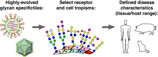
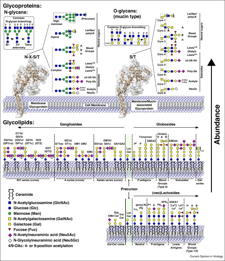
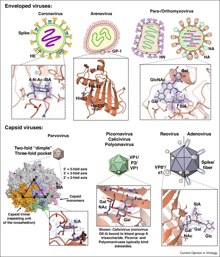
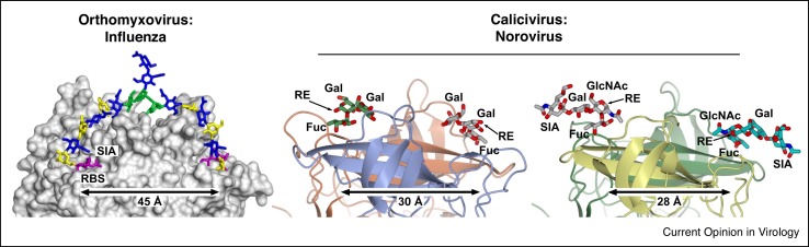
Attachment of viruses to cell-surface receptors is the initial step in infection. Many mammalian viruses have evolved to recognize receptors that are glycans on cell-surface glycoproteins or glycolipids. Although glycans are a ubiquitous component of mammalian cells, the types of terminal structures expressed vary among different cell-types and tissues, and even between comparable cells and tissues from different species, frequently leading to specific tissue and species tropisms as a direct consequence of glycan receptor recognition. Covering the majority of known virus families, this review provides an overview of mammalian viruses that use glycans as receptors, and their roles in determining in host recognition and tropism.


Figures

None
Graphical abstract
Figure 1
Figure 1
Common glycan receptors for viruses found on mammalian host cells, including: protein N-linked glycans (upper left panel), protein O-linked glycans (upper right panel), and glycolipids (lower panel). N-glycans and O-glycans are assembled by combinations of specific glycosidases and glycosyl transferases from one or several shared cores. Combinations of branching (inset) and various terminal groups leads to huge variation and near-infinite possible receptor structures. Conversely, glycolipids maintain defined, and thus far fewer, individual structures, many of which are shown in the lower panel. For simplicity, linkage information has been omitted; however, common scaffolds include lactose (Galβ1-4Glc) and LacNAc (Galβ1-3/4GlcNAc; type 1/2), while terminal sialic acids are typically found in α2-3 (NeuAcα2-3Gal), α2-6 (NeuAcα2-6Gal), or α2-8 (NeuAcα2-8NeuAc) configurations.
Figure 2
Figure 2
Surface structures and glycan-binding profiles of mammalian viruses that use glycan receptors. Membrane-enveloped viruses (upper panels) typically coordinate glycans via a viral-surface glycoprotein, with examples shown for rat coronavirus (New Jersey strain) [133] bound to a non-hydrolysable 4-O-Ac-NeuAc analogue (PDB ID: 5JIF; left panel); the predicted αDG/LAMP1 binding domain of murine LFV [134] (PDB ID: 4ZJF; center panel); and LSTc bound to the 2009 pandemic H1N1 A/California/04/2009 [135] (PDB ID: 3UBE; right). Capsid viruses (lower panels) coordinate glycans either in shallow pockets directly on the outer shell, or within evolved glycan-binding domains that protrude from the surface. Examples are shown for an icosahedral pentamer of AAV1 [80] which binds SA in pockets around the threefold axis (PDB ID: 5EGC; left panel); human norovirus (strain GII.4) [136] bound to blood group A trisaccharide (PDB ID: 3SLD; center); and the terminal σ1 domain of a type 1 (Lang) reovirus [100] bound to GM2 (PDB ID: 4GU3; right). For all panels, viral proteins are shown in coral; bound carbohydrates are shown as cylinders with carbons in grey, while electron density for bound ligands is depicted in blue mesh.
Figure 3
Figure 3
Several virus species show potential for multivalent interactions with glycan receptors. Adaptation to human receptor specificity by influenza viruses (left panel) alters the receptor binding to mode to one where incoming glycans have potential to bivalently engage two monomeric receptor binding sites (RBSs) within a single HA trimer (figure adapted from glycan docking MD simulations reported in Peng, de Vries et al. [19••]). Similarly, the P2 glycan-binding domain of many caliciviruses typically present as dimers, likely sufficient to permit similar bivalent interactions. Structures depict P2 from human noroviruses VA387 bound to HBGA B trisaccharide [106] (PDB ID: 2OBT; center panel) and VA207 bound to sialyl-Lewis X [118] (PDB ID: 3PVD). For influenza, the location of sialic acid and the RBS are marked, other sugar residues are colored according to CFG nomenclature. For calicivirus, all sugar residues are labelled, together with the location of respective sugar reducing ends (RE). Interestingly, the two different binding modes for HBGAs and Lewis antigens in caliciviruses lead to reducing-end sugars pointing in opposite directions within the receptor binding site.

References

    1. Stroh L.J., Stehle T. Glycan engagement by viruses: receptor switches and specificity. Annu Rev Virol. 2014;1:285–306. - PubMed
    1. de Graaf M., Fouchier R.A. Role of receptor binding specificity in influenza A virus transmission and pathogenesis. EMBO J. 2014;33:823–841. - PMC - PubMed
    2. Comprehensive review on roles of glycan specificity in tissue and species tropism of influenza virus.

    1. Olofsson S., Bergstrom T. Glycoconjugate glycans as viral receptors. Ann Med. 2005;37:154–172. - PubMed
    1. Horvath C.A., Boulet G.A., Renoux V.M., Delvenne P.O., Bogers J.P. Mechanisms of cell entry by human papillomaviruses: an overview. Virol J. 2010;7:11. - PMC - PubMed
    1. Akiyama H., Ramirez N.G., Gudheti M.V., Gummuluru S. CD169-mediated trafficking of HIV to plasma membrane invaginations in dendritic cells attenuates efficacy of anti-gp120 broadly neutralizing antibodies. PLoS Pathog. 2015;11 - PMC - PubMed

Publication types

LinkOut - more resources