. 2019 Mar 8;38(1):120.
doi: 10.1186/s13046-019-1117-z.

Cysteine-rich intestinal protein 1 suppresses apoptosis and chemosensitivity to 5-fluorouracil in colorectal cancer through ubiquitin-mediated Fas degradation

Affiliations

Cysteine-rich intestinal protein 1 suppresses apoptosis and chemosensitivity to 5-fluorouracil in colorectal cancer through ubiquitin-mediated Fas degradation

Lanzhi Zhang et al. J Exp Clin Cancer Res. .

Abstract

Background: Cysteine-rich intestinal protein 1 (CRIP1) is highly expressed in human intestine and aberrantly expressed in several types of tumor. However, studies on CRIP1 are limited and its role on tumor development and progression remains controversial and elusive.

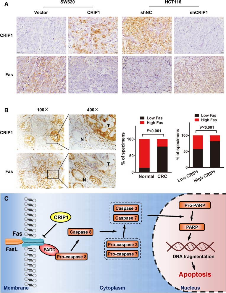
Methods: Immunohistochemistry was performed to evaluate the expression of CRIP1 in paired normal and colorectal tumor specimens, as well as colorectal cell lines. Functional assays, such as CCK8, TUNEL assay and in vivo tumor growth assay, were used to detect the proliferation, apoptosis and response to 5-FU of CRIP1. Western blot was used to analyze Fas-mediated pathway induced by CRIP1. Rescue experiments were performed to evaluate the essential role of CRIP1 for Fas-mediated apoptosis.

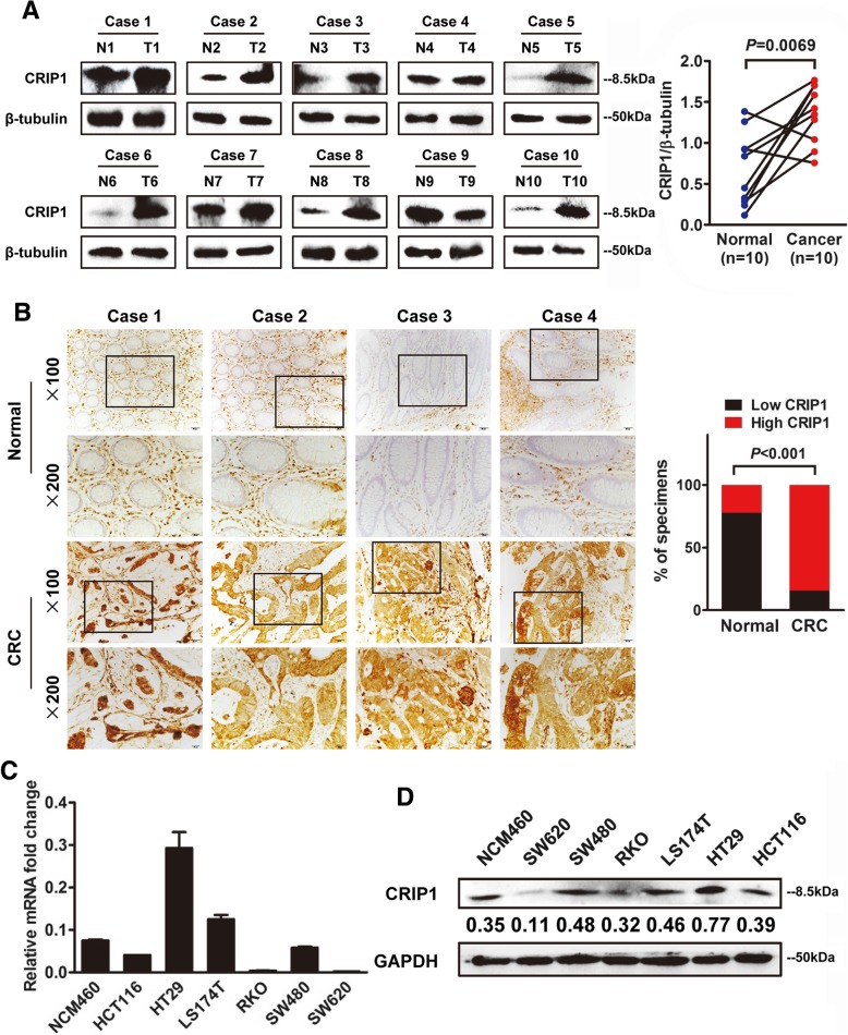
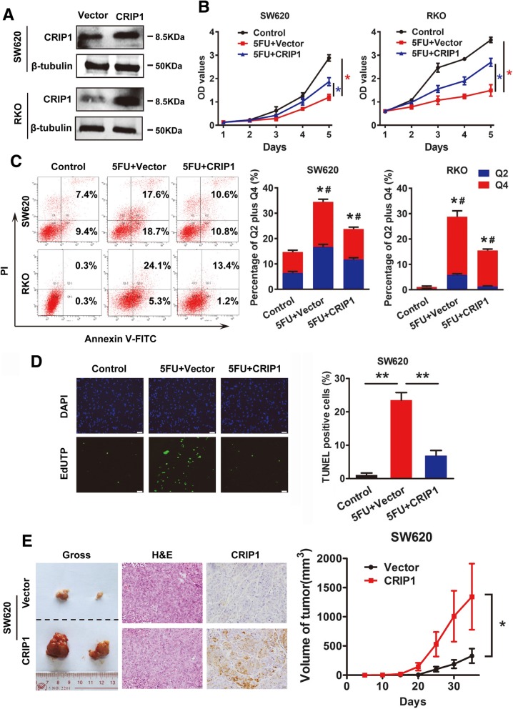
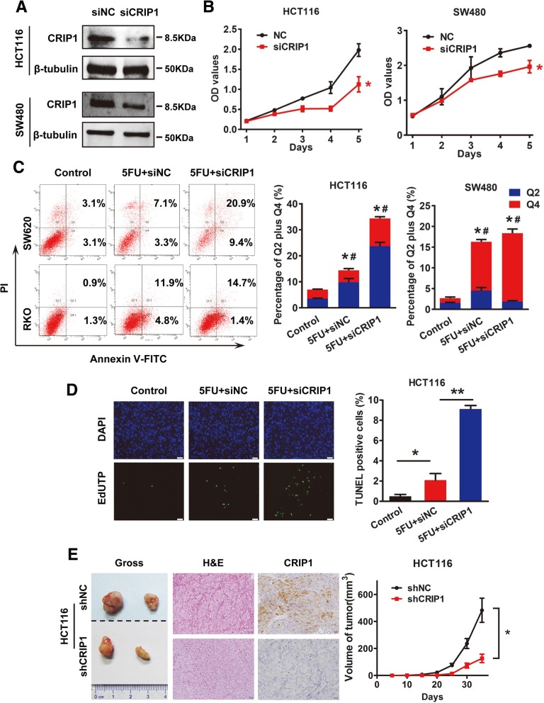
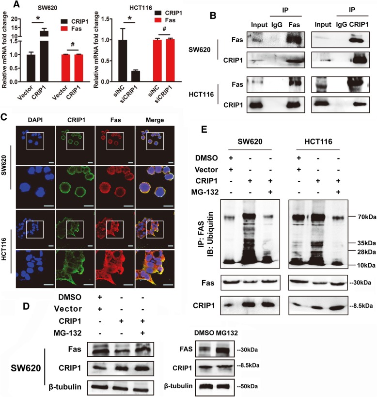
Results: We demonstrated that CRIP1 is overexpressed in CRC tissues compared with adjacent normal mucosa. CRIP1 could dramatically recover the 5-Fluorouracil (5-FU) inhibited CRC cell proliferation in vitro and stimulate the tumor formation of CRC in vivo, probably through inhibiting CRC cell apoptosis. Moreover, CRIP1 also dramatically recovered the 5-Fluorouracil (5-FU) induced tumor cell apoptosis in vitro. Further study demonstrated that CRIP1 down-regulated the expression of Fas protein and proteins related to Fas-mediated apoptosis. CRIP1 could interact with Fas protein and stimulate its ubiquitination and degradation. In addition, a negative correlation was detected between the expression of CRIP1 and Fas protein in most of the clinical human CRC samples.

Conclusion: The current research reveals a vital role of CRIP1 in CRC progression, which provide a novel target for clinical drug resistance of colorectal cancer and undoubtedly contributing to the therapeutic strategies in CRC.

Keywords: Apoptosis; Chemoresistant; Colorectal cancer; Cysteine-rich intestinal protein 1; FAS.


Conflict of interest statement

Ethics approval

All experiments involving patients are endorsed by the Ethics Committee of Southern Medical University and complied with the Declaration of Helsinki. No informed consent was required because data were going to be analyzed anonymously. All animal experiments involved ethical and humane treatment under a license from the Guangdong Provincial Bureau of Science.

Consent for publication

Not applicable.

Competing interests

The authors declare that they have no competing interests.

Publisher’s Note

Springer Nature remains neutral with regard to jurisdictional claims in published maps and institutional affiliations.

Figures

Fig. 1
Fig. 1
CRIP1 is overexpressed in both CRC tissues and cell lines. a. Left panel: Western blot assay for CRIP1 expression in ten CRC tissues (T) and matched normal adjacent normal tissues (N) from the same patient. Right panel: Quantification of CRIP1 expression in CRC tissues and adjacent normal tissues of ten patients. b. Left panel: IHC staining indicated that CRIP1 protein expression was increased in human CRC compared with normal intestinal epithelium (four representative slides was shown). Right panel: Graphical illustration of statistical CRIP1 distribution in CRC tissues. CRIP1 is significantly highly expressed in CRC tissues compared with that in adjacent non-tumorous tissue. c. Real-time PCR analyzed the expression of CRIP1 in NCM460 and six different CRC cell lines. The values represented relative CRIP1 level after normalization to the expression of β-tubulin (means ±SD, N = 3) d. Western blot analysis for the expression of CRIP1 in NCM460 and CRC cell lines. Immunosignals were quantified by densitometric scanning
Fig. 2
Fig. 2
Overexpression of CRIP1 recovers 5-FU suppressed CRC cell proliferation both in vitro and in vivo. a. Western blot assay was used to detect the overexpression of CRIP1 in SW620 and RKO cells. b. CCK8 assay was used to study the effects of CRIP1 on 5-FU inhibited CRC cell proliferation. c. Annexin V-FITC/PI flow cytometry assay showed the effects of CRIP1 on 5-FU induced apoptosis. Bars on the right panel represent percentage of cells in Q2 + Q4. d. TUNEL assay showed the effects of CRIP1 on 5-FU induced apoptosis of CRC cells. Bars on the right panel represent the percentage of TUNEL positive cells. e. IHC staining showed the expression of CRIP1 in subcutaneous tumors formed by CRIP1 overexpressed CRC cells. Right panel represents the growth pattern of subcutaneous tumors
Fig. 3
Fig. 3
Knockdown of CRIP1 further suppresses 5-FU suppressed CRC cell proliferation both in vitro and in vivo. a. Western blot assay was used to detect the silencing of CRIP1 in HCT116 and SW480 cells. b. CCK8 assay was used to study the effects of siCRIP1 on 5-FU inhibited CRC cell proliferation. c. Annexin V-FITC/PI flow cytometry assay showed the effects of siCRIP1 on 5-FU induced apoptosis. Bars on the right panel represent percentage of cells in Q2 + Q4. d. TUNEL assay showed the effects of siCRIP1 on 5-FU induced apoptosis of CRC cells. Bars on the right panel represent the percentage of TUNEL positive cells. e. IHC staining showed the expression of CRIP1 in subcutaneous tumors formed by CRIP1 stable silencing CRC cells. Right panel represents the growth pattern of subcutaneous tumors
Fig. 4
Fig. 4
CRIP1 inhibits Fas induced apoptosis of CRC cells. a. Western blot analysis of apoptotic related proteins in CRIP1 overexpressed or silencing indicated cells. b. Western blot analysis of the effects of CRIP1 on Fas-mediated apoptosis. c. CCK8 assay shows the effects of CRIP1 on Fas-mediated apoptosis. d. Annexin V-FITC/PI flow cytometry assay showed the effects of CRIP1 on Fas-mediated apoptosis. Bars on the upper panel represent percentage of cells in Q2 + Q4. e. TUNEL assay showed the effects of CRIP1 on Fas-mediated apoptosis of CRC cells. Bars on the right panel represent the percentage of TUNEL positive cells
Fig. 5
Fig. 5
CRIP1 stimulates the ubiquitination and degradation of Fas. a. Real-time PCR shows the effects of CRIP1 on the expression of Fas at transcriptional level in both SW620 and HCT116 cells. b. Co-Ip shows the interaction between CRIP1 and Fas in both SW620 and HCT116 cells. c. The immunofluorescence staining demonstrated the co-localization of CRIP1 and Fas in both SW620 and HCT116 cells. d. Left panel: western blot assay shows that MG-132 recovered the inhibitory role of CRIP1 on Fas Protein. Right panel: the effects of MG-132 alone on the expression of Fas protein. e. CRIP1 stimulates the ubiquitination of Fas, while both siCRIP1 and MG-132 could recover the ubiquitin-mediated degradation of Fas
Fig. 6
Fig. 6
A negative correlation exists between the expression of CRIP1 and Fas in CRC tissues. a. Overexpression of CRIP1 is accompanied with the low expression of Fas, whereas silencing of CRIP1 is followed by the high expression of Fas in subcutaneous tumors. b. A reversed expression pattern of CRIP1 and Fas was detected in CRC tissues. Left panel: CRIP1 is usually overexpressed in CRC tissues while Fas highly expressed in its adjacent normal mucosa. The middle panel: the percentage of high or low Fas expression specimens in normal and cancer tissues. The right panel: the percentage of high or low Fas expression specimens in high or low CRIP1 expression specimens. c. A hypothetical model illustrating that CRIP1 interacts with Fas and stimulates the ubiquitin-mediated degradation of Fas to inhibit apoptosis of CRC cells

Similar articles

Cited by

References

    1. Bray F, Ferlay J, Soerjomataram I, Siegel RL, Torre LA, Jemal A. Global cancer statistics 2018: GLOBOCAN estimates of incidence and mortality worldwide for 36 cancers in 185 countries. CA Cancer J Clin. 2018;68:394–424. doi: 10.3322/caac.21492. - DOI - PubMed
    1. Center MM, Jemal A, Smith RA, Ward E. Worldwide variations in colorectal cancer. CA Cancer J Clin. 2009;59:366–378. doi: 10.3322/caac.20038. - DOI - PubMed
    1. Saltz LB, Cox JV, Blanke C, Rosen LS, Fehrenbacher L, Moore MJ, Maroun JA, Ackland SP, Locker PK, Pirotta N, et al. Irinotecan plus fluorouracil and leucovorin for metastatic colorectal cancer. Irinotecan Study Group. N Engl J Med. 2000;343:905–914. doi: 10.1056/NEJM200009283431302. - DOI - PubMed
    1. Hempe JM, Cousins RJ. Cysteine-rich intestinal protein binds zinc during transmucosal zinc transport. Proc Natl Acad Sci U S A. 1991;88:9671–9674. doi: 10.1073/pnas.88.21.9671. - DOI - PMC - PubMed
    1. Davis BA, Blanchard RK, Lanningham-Foster L, Cousins RJ. Structural characterization of the rat cysteine-rich intestinal protein gene and overexpression of this LIM-only protein in transgenic mice. DNA Cell Biol. 1998;17:1057–1064. doi: 10.1089/dna.1998.17.1057. - DOI - PubMed

MeSH terms