. 2019 Mar 8;9(1):3929.
doi: 10.1038/s41598-019-40501-0.

RNA-seq of Isolated Chromaffin Cells Highlights the Role of Sex-Linked and Imprinted Genes in Adrenal Medulla Development

Affiliations

RNA-seq of Isolated Chromaffin Cells Highlights the Role of Sex-Linked and Imprinted Genes in Adrenal Medulla Development

Wing Hei Chan et al. Sci Rep. .

Abstract

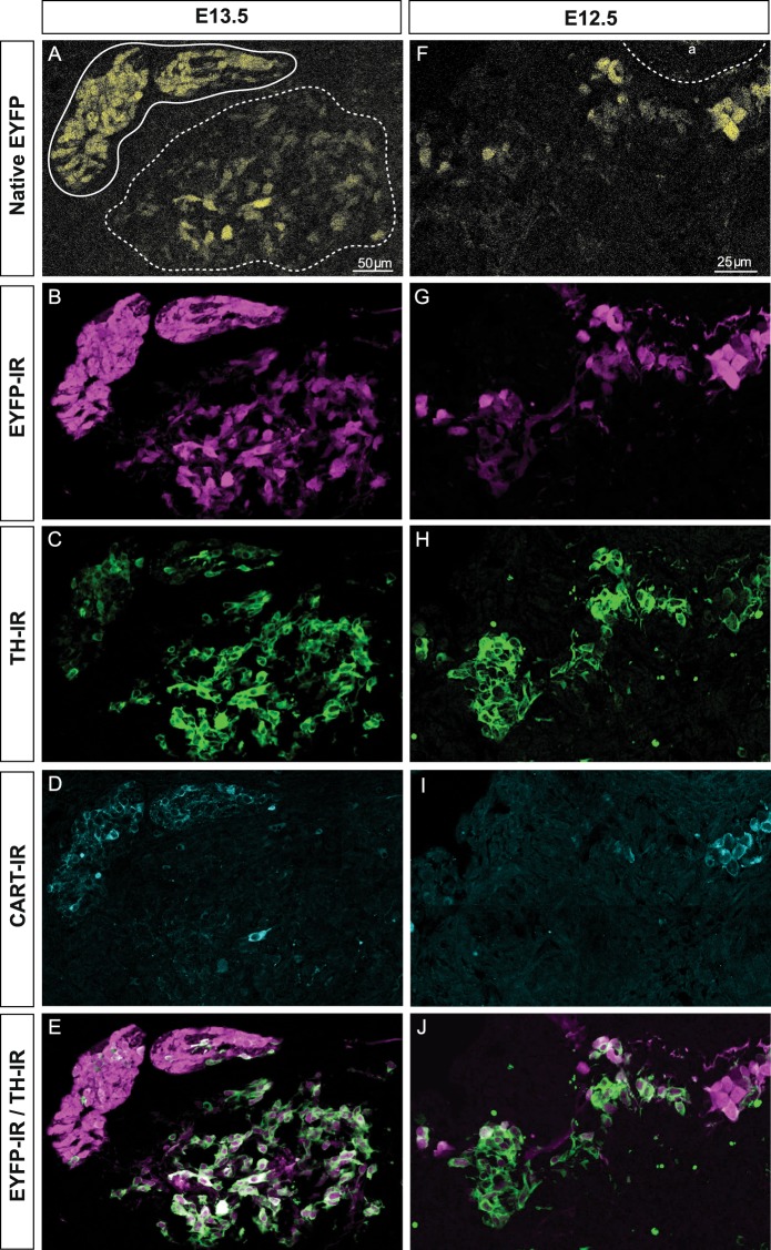
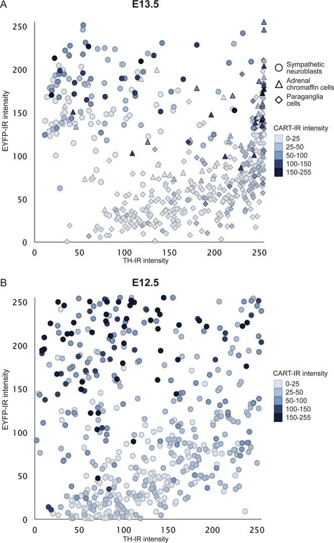
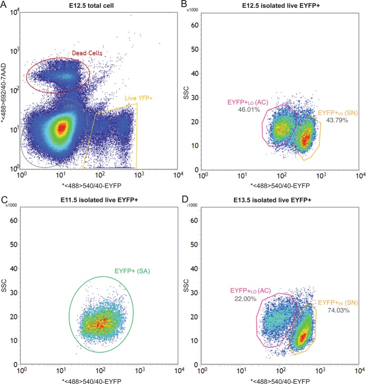
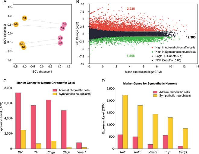
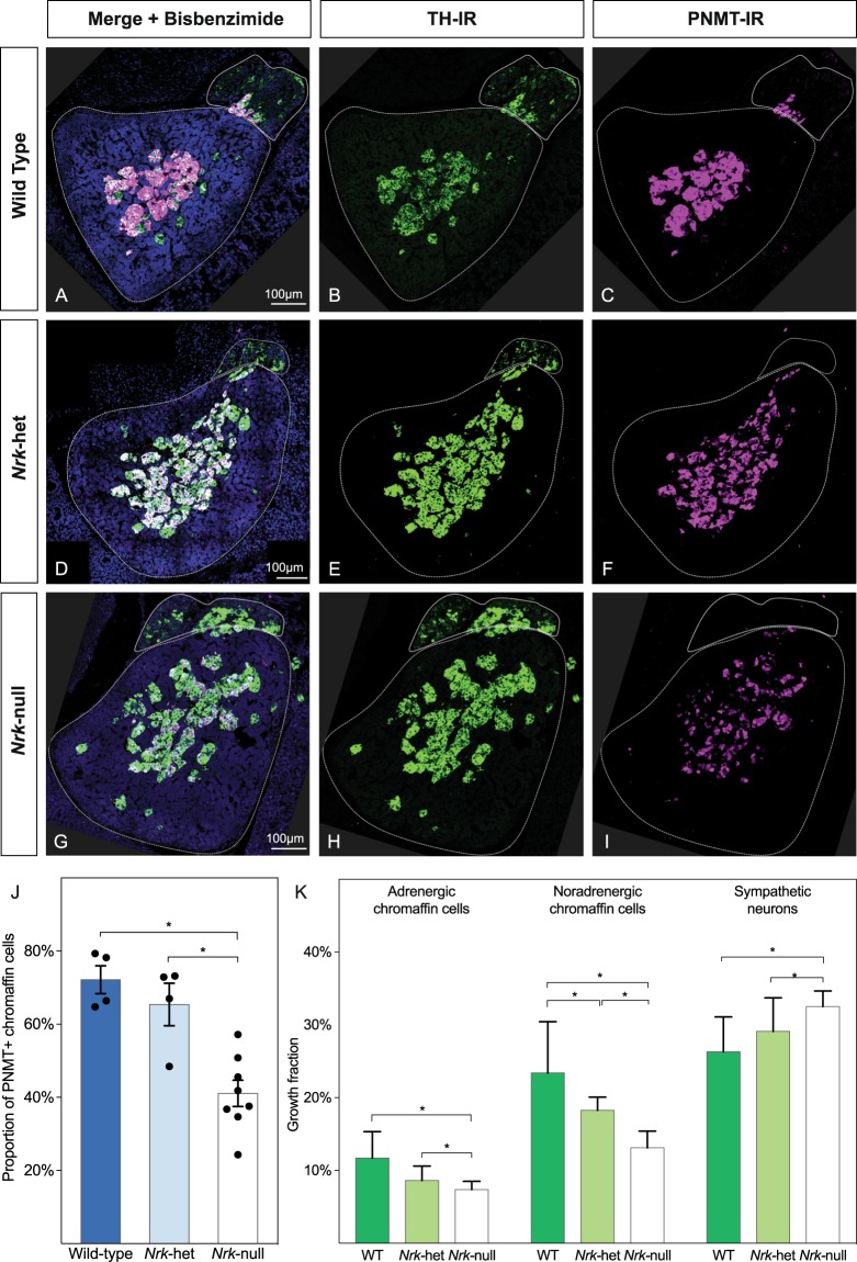
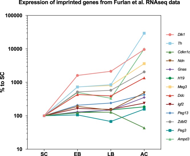
Adrenal chromaffin cells and sympathetic neurons synthesize and release catecholamines, and both cell types are derived from neural crest precursors. However, they have different developmental histories, with sympathetic neurons derived directly from neural crest precursors while adrenal chromaffin cells arise from neural crest-derived cells that express Schwann cell markers. We have sought to identify the genes, including imprinted genes, which regulate the development of the two cell types in mice. We developed a method of separating the two cell types as early as E12.5, using differences in expression of enhanced yellow fluorescent protein driven from the tyrosine hydroxylase gene, and then used RNA sequencing to confirm the characteristic molecular signatures of the two cell types. We identified genes differentially expressed by adrenal chromaffin cells and sympathetic neurons. Deletion of a gene highly expressed by adrenal chromaffin cells, NIK-related kinase, a gene on the X-chromosome, results in reduced expression of adrenaline-synthesizing enzyme, phenyl-N-methyl transferase, by adrenal chromaffin cells and changes in cell cycle dynamics. Finally, many imprinted genes are up-regulated in chromaffin cells and may play key roles in their development.


Conflict of interest statement

The authors declare no competing interests.

Figures

Figure 1
Figure 1
Immunostaining of transverse sections through the adrenal region of TH-Cre::R26R-EYFP mouse embryos at E13.5 (AE) and E12.5 (FJ). A shows the native EYFP (yellow) signal after fixation of TH-Cre::R26R-EYFP mouse embryos at E13.5, the prevertebral suprarenal ganglion (solid line) and the adrenal medulla (dashed line) marked. EYFP-immunoreactivity for the same section is shown in (B), TH-immunoreactivity in (C) and CART-immunoreactivity in (D). (E) Is a merge of images (B,C). Note that TH immunoreactivity shows the reverse pattern of intensity to both native EYFP and EYFP-immunoreactivity. (FJ) is an equivalent region from an E12.5 embryo as (AE). The dorsal aorta (a) is indicated. Note that differential expression of TH-driven EYFP was observed in that some TH-expressing cells were brighter in EYFP than the others, but there was no obvious anatomical segregation of cells differentially expressing EYFP.
Figure 2
Figure 2
Scatter plot of the relative fluorescent intensity of TH-IR versus that of EYFP-IR in individual sympathetic neuroblasts and adrenal chromaffin cells in sections through the abdomen of E13.5 (A) and E12.5 (B) TH-Cre::R26R-EYFP mice. CART-IR fluorescence intensity in each cell is shown by the shading of each symbol (darker is more intense CART-IR). Relative fluorescent intensity for TH-IR (X-axis), EYFP-IR (Y-axis) and CART-IR (shading) were measured on a 0–255 scale (0 = no fluorescence, 255 = maximum fluorescence). The upper left quadrant of each plot contained TH-IR Lo/EYFP-IRHi cells while lower right quadrant contained TH-IRHi/EYFP-IRLo cells. For E13.5, cells are identified and categorized into sympathetic neuroblasts (○), paraganglia (△) and adrenal chromaffin cells (◊) based on anatomical segregation into adrenal medulla or sympathetic ganglia. On E12.5, it was not possible to discriminate cell type by anatomical organisation and the cells are unclassified. However, the pattern observed in the plot is identical to that seen at E13.5, leading to the conclusion that cells that are TH-IRLo/EYFP-IRHi and which also express CART-IR are likely to be sympathetic neuroblasts while TH-IRHi/EYFP-IRLo cells lacking CART-IR are likely to be adrenal chromaffin cells. For E13.5, n = 3 embryos, 521 cells and for E12.5, n = 2 embryos, 505 cells.
Figure 3
Figure 3
Representative FACS plots of cells in TH-Cre::R26R-EYFP mice embryo at E11.5–E13.5. Cells dissociated after dissection were sorted by FACS for EYFP fluorescent intensity and viability (7-AAD). (A) Plot of all cells at E12.5, showing clear separation of living EYFP+ cells (yellow gate) from living EYFP− cells (grey gate) and dead cells (red gate). (B) Plot of living E12.5 EYFP+ cells showing the two cell clusters with similar population size separated based on EYFP fluorescent intensity and SSC. EYFP+Lo (pink gate) are presumptive adrenal chromaffin cells (46.01%) while EYFP+Hi (orange gate) are presumptive sympathetic neuroblasts cells (43.79%). (C) Plot of living E11.5 EYFP+ cells showing a homogeneous population (green gate). (D) Plot of living E13.5 EYFP+ cells showing a similar separation to the E12.5 cells except the population size of sympathetic neuroblasts were about 3 times more than adrenal chromaffin cells.
Figure 4
Figure 4
(A) Principal component analysis plot of biological coefficient of variation (BCV) showing the transcriptome profiles from each of 4 paired (connected by line) E12.5 samples separated clearly by the biological effect of interest i.e. cell types (Dimension 1, X-axis). The effect size of technical batch effect on replicates (Dimension 2, Y-axis) was small, about half of the biological effect. (B) MA-plot of transcriptomic profile in adrenal chromaffin precursor cells versus sympathetic neuroblasts. RNA sequencing analysis of adrenal chromaffin cells and sympathetic neuroblasts revealed 4,786 differential expressed genes with fold change >2 and false discovery rate (FDR) < 0.05 out of 17169 annotated genes, that 2,938 genes expressed higher in adrenal chromaffin cells (red) and 1,848 genes were expressed higher in sympathetic neuroblasts (green). (C,D) The mRNA expression levels of the ten representative cell type-specific marker genes are shown. Markers for chromaffin cells were all expressed at higher level in the adrenal chromaffin cell transcriptomes (C) while markers for neurons were all expressed at higher levels in the sympathetic neuroblast transcriptomes (D).
Figure 5
Figure 5
Temporal gene expression pattern of Dlk1, Foxq1, Nrk and Elf3 for sympathetic neuroblasts/neurons (orange) and adrenal chromaffin precursors/cells (pink) after FACS based on differential expression of EYFP in THCre::R26REYFP embryos. (A,B) Gene expression pattern of Dlk1 and Nrk were measured at E11.5 to P0. (C,D) Gene expression pattern of Foxq1 and Elf4 were measured at E11.5 to E14.5. Absolute concentration of mRNA copies per μL were measured and reported as individual measures (dot) and mean ± SEM (bar). In each case, 3 biological replicates were averaged, except on P0 for Dlk1 where only 1 biological replicate was measured. Asterisks indicate pairs of means that were significantly different using two-way ANOVA (*p < 0.05; **p < 0.01).
Figure 6
Figure 6
Nrk disruption in homozygous mutant led to a defect in adrenergic phenotype acquisition in the adrenal chromaffin cells. (A) Immunostaining of transverse sections through the adrenal region of E18.75 mouse embryos in wild type and Nrk mutant mice, showing TH (green) and PNMT (magenta) immunoreactivity. (AC) Is from a wild type E18.75 mouse. (DF) Show a section from an Nrk-het mutant mouse. (GI) Is from an Nrk-null mutant mouse. The prevertebral suprarenal ganglia and the adrenal glands are outlined in a solid line and dashed line respectively, based on bisbenzimide (BB, cyan) staining (A,D,G). (J) Loss of Nrk reduces the proportion of adrenergic chromaffin cell in the adrenal glands. The number of TH+ noradrenergic adrenal chromaffin cells and their PNMT+ adrenergic subpopulation cells were counted in sections from wild-type, Nrk-het and Nrk-null (both Nrk−/− female and Nrk−/Y male) mutant mice with n = 4. The proportion of PNMT+ adrenergic chromaffin cells were calculated by number of PNMT+ (adrenergic) cells/number PNMT−, TH+ (noradrenergic) adrenal chromaffin cells and shown here as mean proportion ± SEM along with individual measures for each embryo. (K) Growth fractions for adrenergic chromaffin cells, noradrenergic chromaffin cells and sympathetic neurons were examined using Ki67 immunostaining for cycling cells in and calculated as proportion of total cells of each type.
Figure 7
Figure 7
Data from Furlan et al., showing the expression of imprinted genes commonly expressed in differentiating cells. For each gene, values are normalised to expression levels in the Schwann cell precursor (SC). Note the logarithmic Y-axis. Nearly all genes are expressed at low levels in the Schwann cell precursor and in early (EB) and late (LB) bridge cells but at greatly increased levels in differentiating adrenal chromaffin cells.

Similar articles

Cited by

References

    1. Anderson DJ. Molecular control of cell fate in the neural crest: the sympathoadrenal lineage. Annual review of neuroscience. 1993;16:129–158. doi: 10.1146/annurev.ne.16.030193.001021. - DOI - PubMed
    1. Landis SC, Patterson PH. Neural crest cell lineages. Trends Neurosci. 1981;4:172–175. doi: 10.1016/0166-2236(81)90056-4. - DOI
    1. Schober A, Krieglstein K, Unsicker K. Molecular cues for the development of adrenal chromaffin cells and their preganglionic innervation. Eur J Clin Invest. 2000;30:87–90. doi: 10.1046/J.1365-2362.2000.0300s3087.X. - DOI - PubMed
    1. Furlan, A. et al. Multipotent peripheral glial cells generate neuroendocrine cells of the adrenal medulla. Science357, 10.1126/science.aal3753 (2017). - PMC - PubMed
    1. Jiang M, Stanke J, Lahti JM. The connections between neural crest development and neuroblastoma. Curr Top Dev Biol. 2011;94:77–127. doi: 10.1016/B978-0-12-380916-2.00004-8. - DOI - PMC - PubMed

Publication types

MeSH terms

Substances

LinkOut - more resources