Review
. 2019 Jul;18(7):501-526.
doi: 10.1038/s41573-019-0018-3.

Prospects for pharmacological targeting of pseudokinases

Affiliations
Review

Prospects for pharmacological targeting of pseudokinases

Jennifer E Kung et al. Nat Rev Drug Discov. 2019 Jul.

Abstract

Pseudokinases are members of the protein kinase superfamily but signal primarily through noncatalytic mechanisms. Many pseudokinases contribute to the pathologies of human diseases, yet they remain largely unexplored as drug targets owing to challenges associated with modulation of their biological functions. Our understanding of the structure and physiological roles of pseudokinases has improved substantially over the past decade, revealing intriguing similarities between pseudokinases and their catalytically active counterparts. Pseudokinases often adopt conformations that are analogous to those seen in catalytically active kinases and, in some cases, can also bind metal cations and/or nucleotides. Several clinically approved kinase inhibitors have been shown to influence the noncatalytic functions of active kinases, providing hope that similar properties in pseudokinases could be pharmacologically regulated. In this Review, we discuss known roles of pseudokinases in disease, their unique structural features and the progress that has been made towards developing pseudokinase-directed therapeutics.


Conflict of interest statement

Competing interests statement

The authors declare no competing interests.

Figures

Fig. 1:
Fig. 1:. Prevalence of pseudokinases in the kinomes of diverse species.
Pie charts showing the percentages of active kinases (blue) and pseudokinases (green) in the kinomes of the indicated organisms. Pseudokinases are defined as kinases that carry mutations in one or more conserved catalytic motifs (the β3 lysine within the VAIK (Val-Ala-Ile-Lys) motif, the aspartate in the DFG (Asp-Phe-Gly) motif and the aspartate in the HRD (His-Arg-Asp) motif).
Fig. 2:
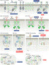
Fig. 2:. Dysregulation of pseudokinase signalling in disease.
a | In normal cells, human epidermal growth factor receptor 3 (HER3) allosterically activates other HER kinases through heterodimerization in a ligand-dependent manner (left panel). Overexpression or mutation of HER3 drives ligand-independent association with its dimerization partners, such as HER2, and activation of signalling pathways that promote tumorigenesis (right panel). b | In normal cells, Janus kinase 2 (JAK2) associates with its cognate receptor at the plasma membrane, and the JAK homology 2 (JH2) pseudokinase domain of JAK2 allosterically inhibits the JH1 kinase domain. This inhibition is relieved upon ligand binding to the receptor, allowing transphosphorylation and activation of the kinase domain (left panel). Mutations in the pseudokinase domain associated with cancers, such as myeloproliferative neoplasms, are believed to disrupt the pseudokinase–kinase domain interaction, causing constitutive activation of the kinase domain (right panel). c | Under normal conditions, kinase suppressor of RAS (KSR) allosterically activates BRAF to promote phosphorylation of MEK and activate mitogen-activated protein kinase (MAPK) signalling. KSR also interacts directly with MEK, and this interaction enhances the allosteric activator function of KSR (left panel). Mutations associated with obesity and insulin resistance impair the ability of KSR to activate the MAPK pathway by disrupting its interactions with MEK and BRAF (right panel). d | Under normal conditions, FAM20A and FAM20C localize to the Golgi apparatus. FAM20A allosterically activates FAM20C, which becomes secreted and phosphorylates extracellular proteins that are important for biomineralization and proper enamel development (left panel). Mutations in FAM20A linked to amelogenesis imperfecta fail to activate FAM20C or promote FAM20C secretion (right panel). e | The transcription factor CCAAT enhancer-binding protein-α (C/EBPα) promotes myeloid cell differentiation. Tribbles homologue 1 (TRIB1) and TRIB2 interact with C/EBPα and the E3 ubiquitin ligase COP1 to mediate ubiquitylation and proteasomal degradation of C/EBPα (left panel). Overexpression of TRIB1 or TRIB2 induces leukaemogenesis by causing excessive degradation of C/EBPα in a COP1-dependent manner (right panel). CRD, cysteine-rich domain; NRG, neuregulin; PI3K, phosphoinositide 3-kinase; STAT, signal transducer and activator of transcription.
Fig. 3:
Fig. 3:. Structural features of pseudokinases.
Left panels show crystal structures of the CDK2, epidermal growth factor receptor (EGFR) and ABL kinase domains in the active, SRC/CDK-like inactive and inactive conformations, respectively. Insets show zoomed views of the active sites. Right panels show zoomed views of the pseudoactive sites in the crystal structures of the indicated pseudokinases. a | In the active conformation (CDK2 is shown), the β3 lysine (K33) forms a salt bridge with E51 in helix αC, and the DFG (Asp-Phe-Gly) aspartate (D145) coordinates Mg2+ in the active site. b | In the SRC/CDK-like inactive conformation (EGFR is shown), helix αC is shifted away from the active site, preventing a salt bridge between the β3 lysine (K721) and E734. c | In the inactive conformation (ABL is shown), the DFG motif is flipped so that phenylalanine (F401) blocks nucleotide binding. d | Helix αC in human epidermal growth factor receptor 3 (HER3) is partially unwound and adopts a SRC/CDK-like inactive conformation. e | Helix αC is unusually short in TRIB1 and adopts a kink, resulting in a shallow nucleotide-binding pocket. f | Pragmin has a disordered helix αC and occluded nucleotide-binding pocket. g | STE20-related adaptor-α (STRADα) coordinates ATP using positively charged residues. h | The ROR2 DLG motif is shifted ~4 Å compared with canonical DFG motifs and interacts with helix αC, pushing it away from the pseudoactive site. i | Mixed lineage kinase domain-like protein (MLKL) has an unusual activation loop helix that interacts with the β3 lysine (K219). The corresponding Protein Data Bank codes for each structure shown are indicated in parentheses. C-lobe, carboxyl-terminal lobe; N-lobe, amino-terminal lobe.
Fig. 4:
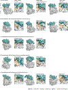
Fig. 4:. Accessibility of the putative nucleotide-binding pocket in different classes of pseudokinases.
Surface representations of crystal structures of active kinases in the active DFG-in (CDK2), SRC/CDK-like inactive (EGFR) and inactive DFG-out conformations (c-ABL) (part a), pseudokinases that do not bind nucleotides or cations (part b), pseudokinases that bind nucleotides in the absence of cations (part c), pseudokinases that bind cations but not nucleotides (part d) and pseudokinases that bind nucleotides and cations (part e). For each structure, bound ATP or ATP analogues are shown as sticks, while Mg2+ ions are shown as spheres. Insets show zoomed views of the putative nucleotide-binding pocket for each kinase or pseudokinase. The corresponding Protein Data Bank codes for each crystal structure shown are indicated in parentheses. αC, helix αC; AMP-PNP, nonhydrolysable ATP analogue; C-lobe, carboxyl-terminal lobe; EGFR, epidermal growth factor receptor; HER3, human EGFR; N-lobe, amino-terminal lobe; JAK2, Janus kinase 2; JH2, JAK homology 2; KSR2, kinase suppressor of RAS 2; ROP2, rhoptry protein 2; STRADα, STE20-related adaptor-α.
Fig. 5:
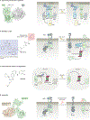
Fig. 5:. Strategies for pharmacological targeting of pseudokinases.
a | Small molecules can be used to allosterically modulate the conformation and interactions of pseudokinases, as illustrated by APS-2–79, an ATP-competitive inhibitor of kinase suppressor of RAS 2 (KSR2), that stabilizes an inactive conformation of KSR2 that is incompatible with heterodimerization with RAF and that blocks RAF-dependent phosphorylation sites on MEK. A crystal structure of KSR2 bound to APS-2–79 and MEK is shown in the left panel. b | Proteolysis-targeting chimaeras (PROTACs) or hydrophobic tagging (HyT) can be used to induce degradation of a protein of interest. The human epidermal growth factor receptor 3 (HER3) inhibitor TX2–121-1 consists of a portion that binds covalently to the HER3 nucleotide-binding pocket (blue) connected to adamantane (red), a hydrophobic moiety that mimics the presence of an unfolded protein. Binding of TX2–121-1 leads to proteasomal degradation of HER3, a process that is aided by chaperones, including HSP70 and HSP90. c | Small molecules other than the bifunctional ligands employed for PROTAC-mediated or HyT-mediated protein degradation can be used to destabilize pseudokinases and induce their degradation. Covalent inhibitors of epidermal growth factor receptor (EGFR) and HER2, such as afatinib, destabilize the pseudokinase TRIB2 and promote its proteasomal degradation. d | Antibodies can be used to target the extracellular domains (ECDs) of receptor tyrosine kinases that possess pseudokinase domains. The monoclonal antibody lumretuzumab (RG7116) locks the HER3 ECD in an inactive conformation that prevents ligand binding. A crystal structure of an antigen-binding fragment, derived from lumretuzumab, bound to the HER3 ECD is shown in the left panel. C/EBPα, CCAAT enhancer-binding protein-α; CRD, cysteine-rich domain; NRG, neuregulin.

References

    1. Futreal PA et al. A census of human cancer genes. Nat Rev Cancer 4, 177–183 (2004). - PMC - PubMed
    1. Wu P, Nielsen TE & Clausen MH FDA-approved small-molecule kinase inhibitors. Trends Pharmacol. Sci. 36, 422–439 (2015). - PubMed
    1. Greenman C et al. Patterns of somatic mutation in human cancer genomes. Nature 446, 153–158 (2007). - PMC - PubMed
    1. Creixell P et al. Kinome-wide decoding of network-attacking mutations rewiring cancer signaling. Cell 163, 202–217 (2015). - PMC - PubMed
    1. Chang MT et al. Identifying recurrent mutations in cancer reveals widespread lineage diversity and mutational specificity. Nature Biotechnology 34, 155–163 (2016). - PMC - PubMed

Publication types