. 2019 May;52(3):e12579.
doi: 10.1111/cpr.12579. Epub 2019 Mar 9.

Induction of neutrophil extracellular traps during tissue injury: Involvement of STING and Toll-like receptor 9 pathways

Affiliations

Induction of neutrophil extracellular traps during tissue injury: Involvement of STING and Toll-like receptor 9 pathways

Li Liu et al. Cell Prolif. 2019 May.

Erratum in

Abstract

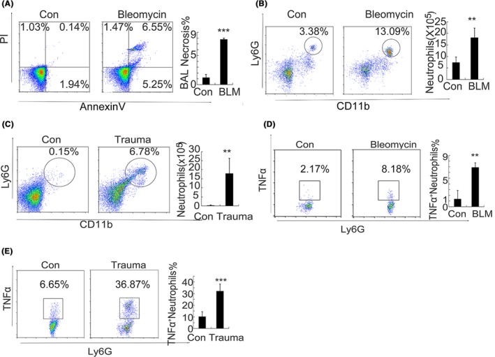
Objectives: Neutrophils are thought to release neutrophil extracellular traps (NETs) to form in response to exogenous bacteria, viruses and other pathogens. However, the mechanisms underlying NET formation during sterile inflammation are still unclear. In this study, we would like to identify neutrophil extracellular traps formation during sterile inflammation and tissue injury and associated pathways and its mechanism.

Materials and methods: We identified different injuries such as chemical-induced and trauma-induced formation of NETs and investigated mechanism of the formation of NETs in vitro and in vivo during the treatment of mtDNA.

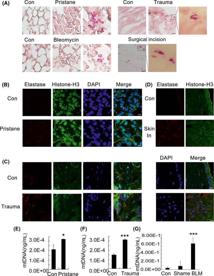
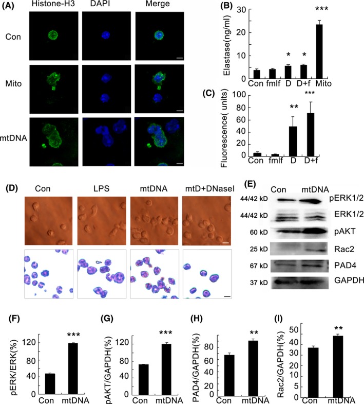
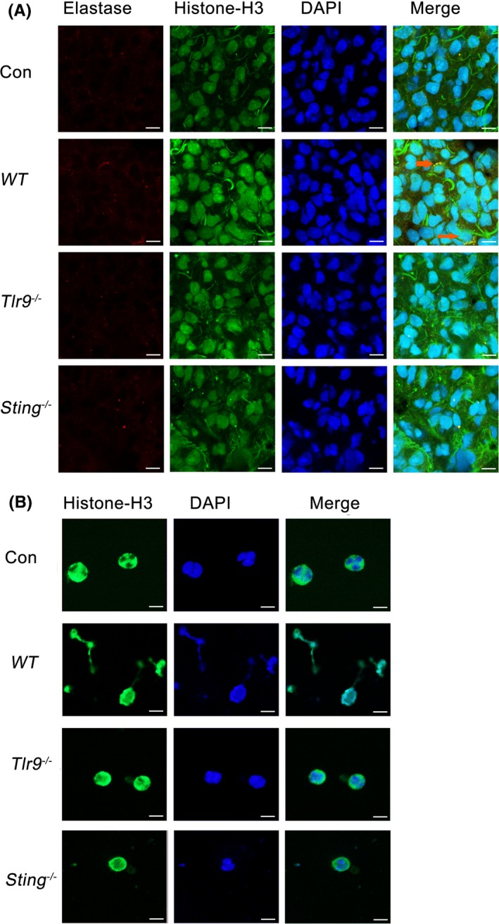
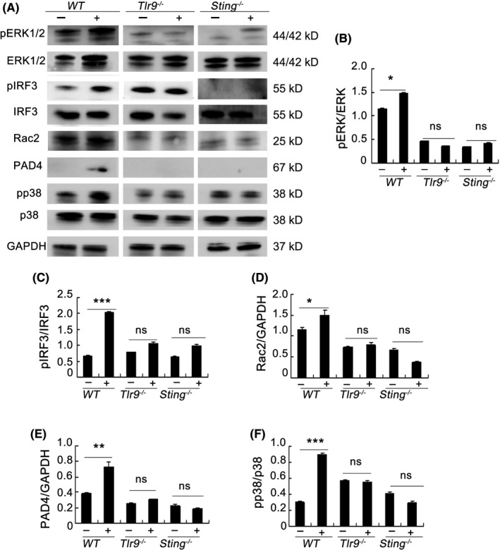
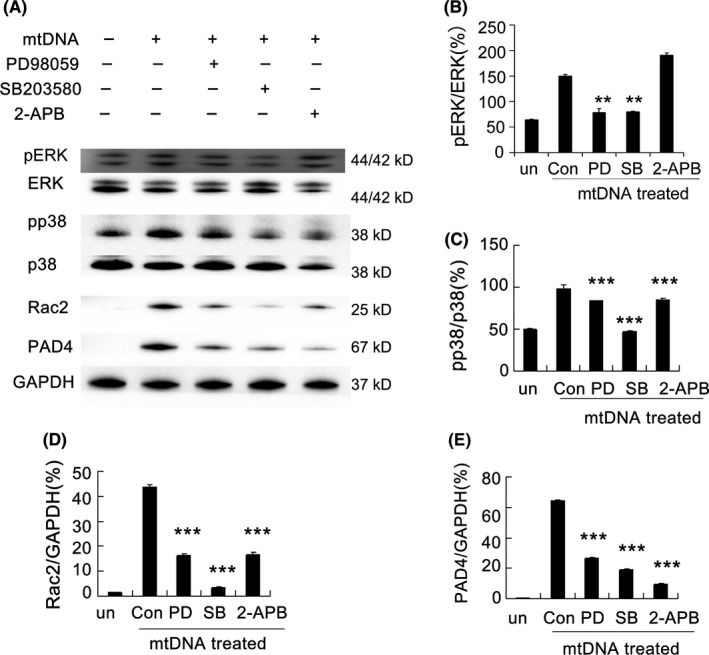
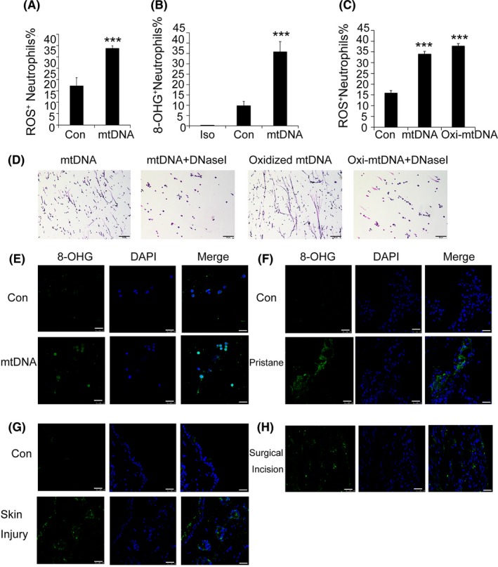
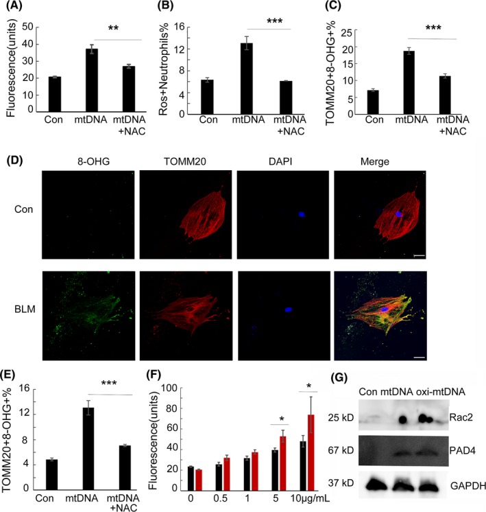
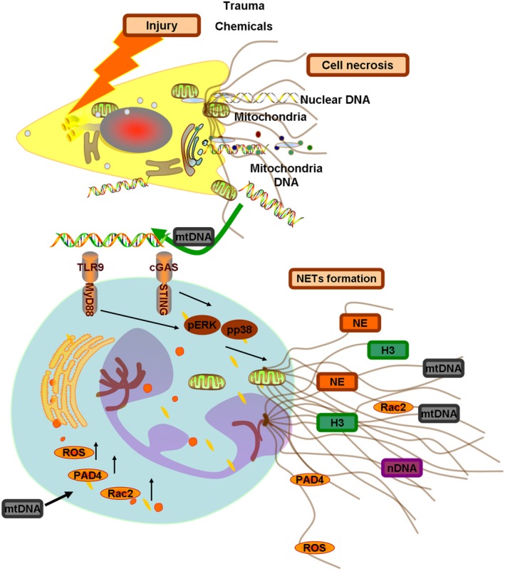
Results: Here, we find the release of mitochondrial DNA (mtDNA) and oxidized mtDNA in acute peripheral tissue trauma models or other chemically induced lung injury, and moreover, endogenous mtDNA and oxidized mtDNA induce the formation of NETs and sterile inflammation. Oxidized mtDNA is a more potent inducer of NETs. Mitochondrial DNA activates neutrophils via cyclic GMP-AMP synthase (cGAS)-STING and the Toll-like receptor 9 (TLR9) pathways and increases the production of neutrophil elastase and extracellular neutrophil-derived DNA in NETs. Mitochondrial DNA also increases the production of reactive oxygen species (ROS) and expression of the NET-associated proteins Rac 2 and peptidylarginine deiminase 4 (PAD4).

Conclusions: Altogether, these findings highlight that endogenous mitochondrial DNA inducted NETs formation and subsequent sterile inflammation and the mechanism associated with NET formation.

Keywords: Toll-like receptor 9; cyclic GMP-AMP synthase; mitochondrial DNA; neutrophil extracellular traps.


Conflict of interest statement

The authors have declared that no conflict of interest exists.

Figures

Figure 1
Figure 1
Inflammatory neutrophil recruitment after trauma‐ and chemical‐induced necrosis. The mice were administered a single intratracheal instillation of bleomycin sulphate dissolved in saline (5 mg/kg body weight). The necrotic cells in the bronchoalveolar lavage fluid (BAL) were examined by flow cytometry, n = 7 (A). The numbers of infiltrated CD45+CD11b+Ly6G+inflammatory neutrophils in lung tissues, n = 7 (B). In the acute peripheral tissue trauma model mice, 24 h after treatment, tissues were removed for a flow cytometric analysis of CD45+CD11b+Ly6G+inflammatory neutrophils infiltrated in the tissues, n = 7 (C). The expression of TNF‐α was detected in the CD45+CD11b+Ly6G+ inflammatory neutrophils in tissues, n = 7 (D, E). Data are representative of three independent experiments, and the results are expressed as the means ± SEM. Statistical comparisons were performed using Student's t test or Dunnett's t test (*P < 0.05; **P < 0.01; ***P < 0.005)
Figure 2
Figure 2
Neutrophil extracellular trap formation during different injury models. C57BL/6 mice were administered a single intratracheal instillation of bleomycin sulphate dissolved in saline (5 mg/kg body weight). C57BL/6 mice received a single 0.5‐mL ip injection of pristane or saline as a control. The acute peripheral tissue trauma model was generated in C57BL/6 mice. Twenty‐four hours after these treatments, tissues were removed and frozen, and human surgical incision tissue was cut for frozen sections. Esterase was detected via stained with a Naphthol AS‐D Chloroacetate Kit (Sigma), and the procedure was performed according to the manufacturer's instruction, n = 8 (A). Sections were stained with anti‐neutrophil elastase (Red) and anti‐histone H3 (Green) antibodies. DAPI (Blue) was used to stain DNA. Images were acquired using a confocal microscope (Zeiss LSM 510 Meta), n = 5 (B‐D). The mtDNA in the serum of mice treated with pristane, or the acute peripheral tissue trauma model or bleomycin were determined at 24 h after treatments by qPCR, n = 6 (E, F, G). Data are representative of three independent experiments, and the results are expressed as the means ± SEM. Statistical comparisons were performed using Student's t test or Dunnett's t test (*P < 0.05; **P < 0.01; ***P < 0.005)
Figure 3
Figure 3
Mitochondrial DNA induces neutrophil extracellular trap formation. Bone marrow neutrophils from C57BL/6 mice were obtained and stimulated with mitochondrial DNA (5 μg/mL) and mitochondria (100 μg/mL) for 2 h. Cells were stained with an anti‐histone H3 primary antibody and the appropriate second antibody. DAPI was used to stain DNA. Images were acquired using a confocal microscope (Zeiss LSM 510 Meta) (A). Neutrophils were cultured (1 × 106 cells/well) and stimulated with fMLF (1 μmol/L), mtDNA (5 μg/mL) or mitochondria (100 μg/mL) for 2 h in 6‐well plate at 37°C. The elastase production in the supernatant was measured by ELISA, n = 6 (B). Sytox Green (5 μmol/L; Life Technologies) was used to detect extracellular DNA. Fluorescence intensity was quantified using the BIOTEK plate reader Synergy HTX, n = 6 (C). Neutrophils were stimulated for 2 h with 5 μg/mL mtDNA, 1 μg/mL LPS and 25 nmol/L DNaseI. The change in neutrophil morphology was observed by Giemsa staining (D). The freshly isolated neutrophils were cultured with mitochondrial DNA (5 μg/mL) at a concentration of 2 × 106cell/ml for 2 hours. Western blot analysis was performed (e). Blot intensities were analysed by Image J (F‐I). Data are representative of three independent experiments, and the results are expressed as the means ± SEM. Statistical comparisons were performed using Student's t test or Dunnett's t test (*P < 0.05; **P < 0.01; ***P < 0.005)
Figure 4
Figure 4
Neutrophil extracellular trap formation induced by mitochondrial DNA depends on the TLR9 and STING pathways. WT mice and Sting−/−, Tlr9−/− mice were intravenously injected with mitochondrial DNA (5 μg/mice) for 24 h. The lung tissue sections were stained with anti‐neutrophil elastase and anti‐histone H3 antibodies, and DAPI was used to stain DNA. Images were acquired using a confocal microscope (Zeiss LSM 510 Meta), n = 6 (A). Bone marrow neutrophils from WT mice and Sting−/−, Tlr9−/− mice were cultured with mitochondrial DNA (5 μg/mL) for 2 h. Cells were stained for NETs, n = 6 (B). Data are representative of three independent experiments
Figure 5
Figure 5
TLR9 and STING pathways are essential for the formation of neutrophil extracellular traps induced by mitochondrial DNA. Bone marrow neutrophils from WT mice and Sting−/−, Tlr9−/− mice were cultured with mitochondrial DNA (5 μg/mL) at a concentration of 2 × 10⁶ cell/mL for 2 h. Western blot analysis was performed (A). Blot intensities were analysed by Image J (B‐F). Data are representative of three independent experiments, and the results are expressed as the means ± SEM. Statistical comparisons were performed using Student's t test or Dunnett's t test (*P < 0.05; **P < 0.01; ***P < 0.005)
Figure 6
Figure 6
MAPK pathways and store‐operated Ca2+entry (SOCE) are necessary for the formation of mtDNA‐induced neutrophil extracellular traps. The neutrophils from C57/BL6 mice were cultured with mitochondrial DNA (5 μg/mL) at a concentration of 2 × 10⁶cell/mL. The inhibitors PD9805 (30 μmol/L), SB203580 (10 μmol/L) and 2‐APB (10 μmol/L) were added to culture medium and incubated for 2 h at 37°C, and Western blot analysis was performed (A). Blot intensities were analysed by Image J (B‐E). Data are representative of three independent experiments, and the results are expressed as the means ± SEM. Statistical comparisons were performed using Student's t test or Dunnett's t test (*P < 0.05; **P < 0.01; ***P < 0.005)
Figure 7
Figure 7
Injured tissues and neutrophil extracellular traps are enriched with oxidized mitochondrial DNA. Bone marrow neutrophils from C57BL/6 mice were obtained and stimulated with mitochondrial DNA (5 μg/mL) for 30 minutes, and ROS production was determined, n = 7 (A). Neutrophils were cultured (1 × 10⁶ cells/well) and stimulated with mtDNA (5 μg/mL) for 2 h in 6‐well plates at 37°C. Oxidized DNA was detected with a biotinylated 8‐OHdG antibody, n = 7 (B). Neutrophils were stimulated with mitochondrial DNA (5 μg/mL) or oxidized mitochondrial DNA (5 μg/mL) for 30 minutes, and ROS production was determined, n = 8 (C). Neutrophils were stimulated for 2 h with 5 μg/mL mtDNA or oxidized mtDNA, in the presence or absence of 25 nM DNaseI. The change in neutrophil morphology was observed by Giemsa staining (D). Freshly isolated neutrophils were cultured with mitochondrial DNA (5 μg/mL) at a concentration of 2 × 106cell/mL for 2 h. 8‐OHdG fluorescence microscopy was performed (E). The 8‐OHdG fluorescence microscopy analysis of pristine‐treated mice, skin‐injured mice and human surgical incision (F‐H). Data are representative of three independent experiments, and the results are expressed as the means ± SEM. Statistical comparisons were performed using Student's t test or Dunnett's t test (*P < 0.05; **P < 0.01; ***P < 0.005)
Figure 8
Figure 8
Reactive oxygen species scavenger inhibits the production of oxidized mitochondrial DNA and NETs. Bone marrow neutrophils were stimulated with mitochondrial DNA (5 μg/mL) for 2 h. Sytox Green (5 μmol/L; Life Technologies) was used to detect extracellular DNA. Fluorescence intensity was quantified using the BIOTEK plate reader Synergy HTX, n = 8 (A). Neutrophils were stimulated with mitochondrial DNA (5 μg/mL) for 30 minutes, and ROS production was determined, n = 7 (B). Neutrophils were stimulated with mtDNA (5 μg/mL) or in the presence of 5 mmol/L NAC for 30 minutes, and oxidized mitochondrial DNA was detected with biotinylated TOMM20 and 8‐OHdG antibodies, n = 7 (C). Primary lung cells were stimulated with bleomycin (20 μg/mL) or in the presence of 5 mmol/L NAC for 24 h. TOMM20 and 8‐OHdG fluorescence microscopy (D) or by flow cytometry was performed, n = 8 (E). Freshly isolated neutrophils were cultured with mitochondrial DNA or oxidized mitochondrial DNA (0.5‐10 μg/mL) for 2 h. Fluorescence intensity of Sytox Green was quantified using the BIOTEK plate reader Synergy HTX, n = 8. Black for mitochondrial DNA and red for oxidized mitochondrial DNA (F). Neutrophils were cultured with mitochondrial DNA or oxidized mitochondrial DNA (5 μg/mL) for 2 h. Western blots for detecting the proteins PAD4 and Rac2 were performed (G). Data are representative of three independent experiments, and the results are expressed as the means ± SEM. Statistical comparisons were performed using Student's t test or Dunnett's t test (*P < 0.05; **P < 0.01; ***P < 0.005)
Figure 9
Figure 9
Outline of the pathways involved in mtDNA‐induced neutrophil extracellular traps. Chemical‐induced and trauma‐induced acute cell necrosis caused the release of the mitochondria and mtDNA from the necrotic cells. The endogenous mitochondrial DNA induced neutrophil extracellular trap formation. Here, we show the underlying mechanisms. Mitochondrial DNA mediated the activation of the p38 MAPK and ERK1/2 signalling responses, which depended on the STING and TLR9 pathways. This activation induced the increased production of NET‐associated proteins Rac2 and PAD4 and the release of ROS, elastase and histone 3 through STING‐ or TLR9‐mediated p38 MAPK and ERK1/2 pathways

References

    1. Brinkmann V, Reichard U, Goosmann C, et al. neutrophil extracellular traps kill bacteria. Science. 2004;303:1532‐1535. - PubMed
    1. Allam R, Kumar SV, Darisipudi MN, et al. Extracellular histones in tissue injury and inflammation. J Mol Med. 2014;92:465‐472. - PubMed
    1. von Kockritz‐Blickwede M, Nizet V. Innate immunity turned inside‐out: antimicrobial defense by phagocyte extracellular traps. J Mol Med. 2009;87:775‐783. - PMC - PubMed
    1. Yousefi S, Mihalache C, Kozlowski E, et al. Viable neutrophils release mitochondrial DNA to form neutrophil extracellular traps. Cell Death Differ. 2009;16:1438‐1444. - PubMed
    1. McIlroy DJ, Jarnicki AG, Au GG, et al. Mitochondrial DNA neutrophil extracellular traps are formed after trauma and subsequent surgery. J Crit Care. 2014;29(1133):e1131‐1135. - PubMed

MeSH terms