

Neuronal PAS Domain 2 (Npas2)-Deficient Fibroblasts Accelerate Skin Wound Healing and Dermal Collagen Reconstruction
- PMID: 30851151
- PMCID: PMC6733676
- DOI: 10.1002/ar.24109
Neuronal PAS Domain 2 (Npas2)-Deficient Fibroblasts Accelerate Skin Wound Healing and Dermal Collagen Reconstruction
Abstract
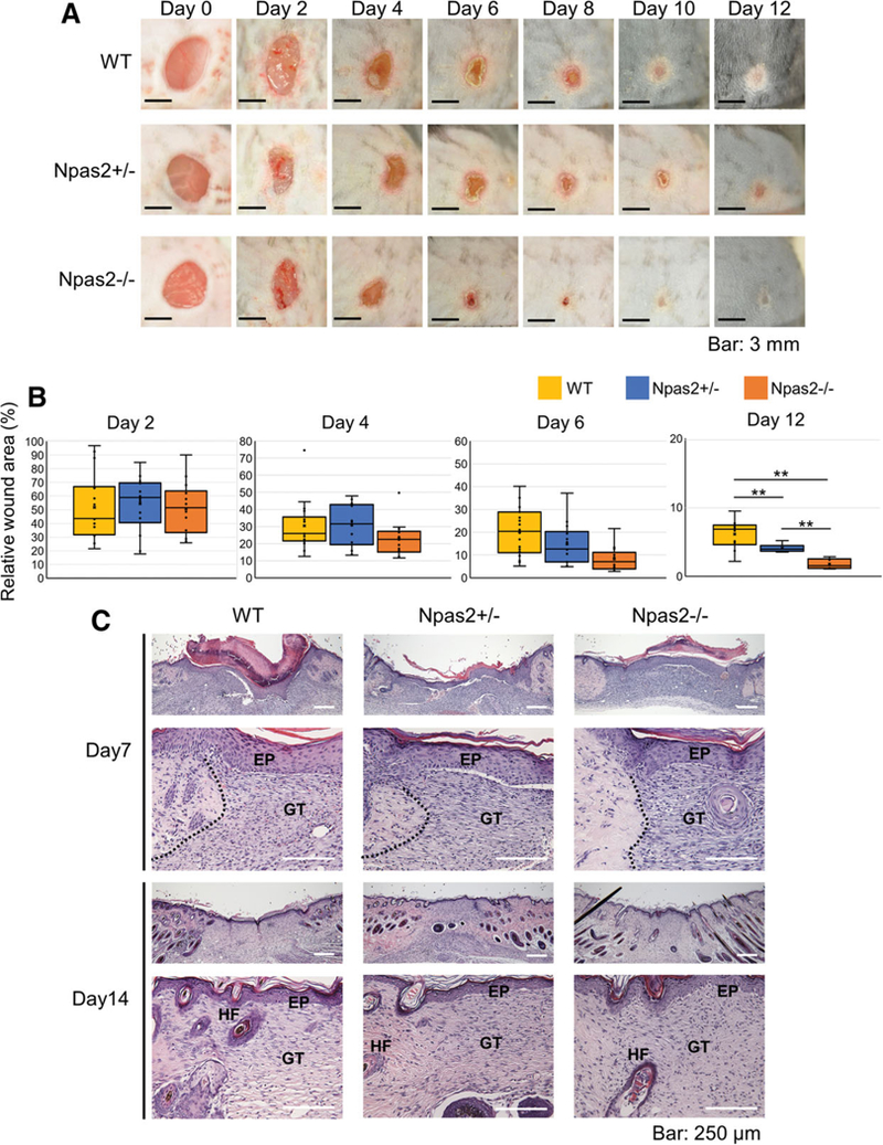
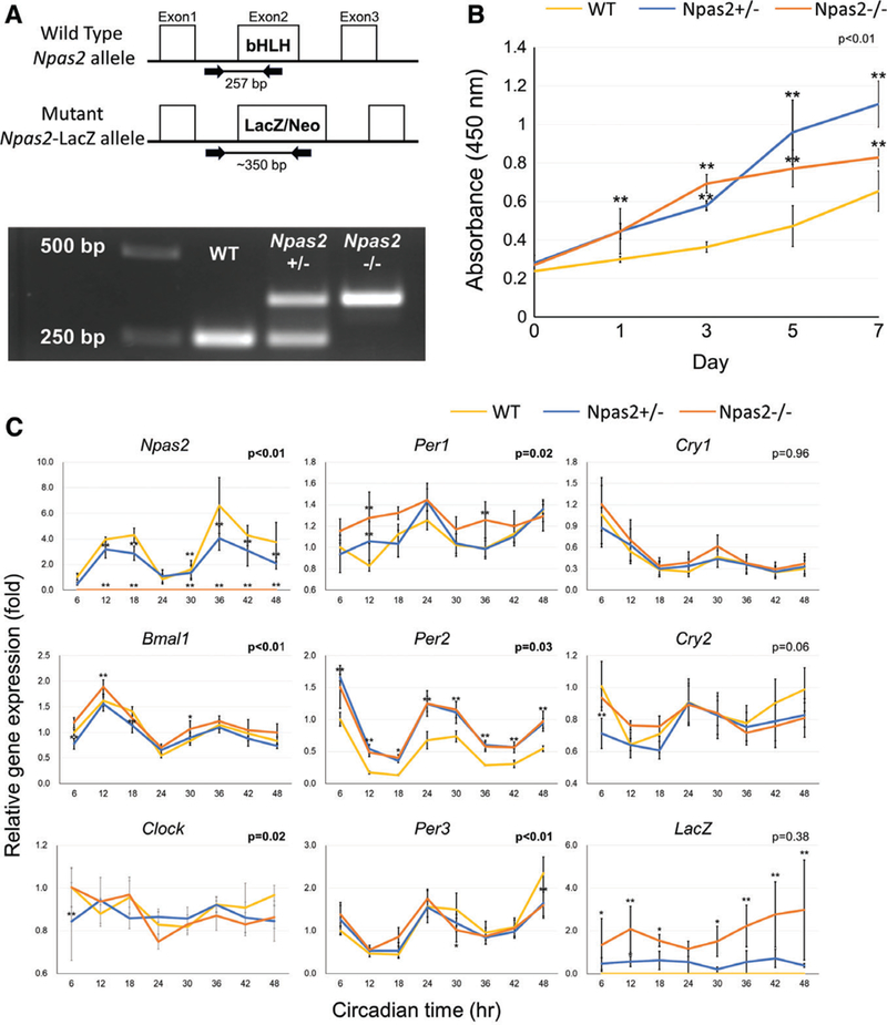
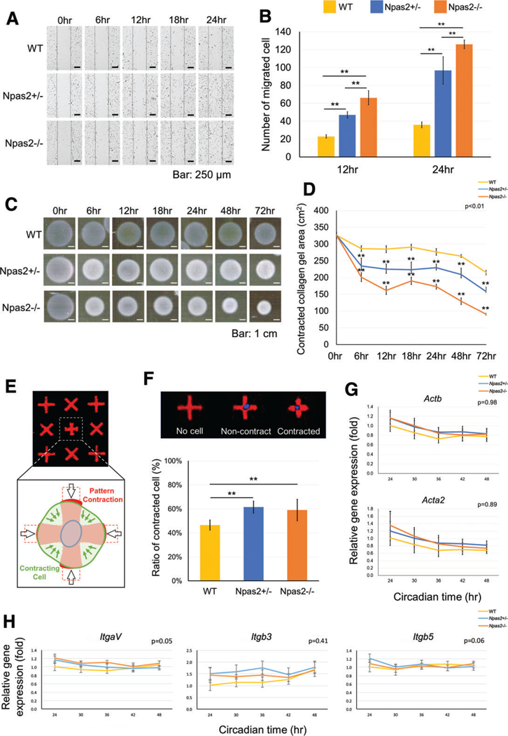
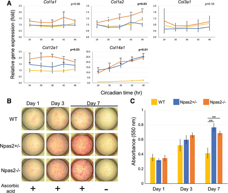
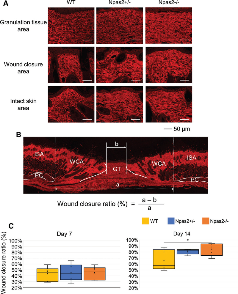
The circadian clock, which consists of endogenous self-sustained and cell-autonomous oscillations in mammalian cells, is known to regulate a wide range of peripheral tissues. The unique upregulation of a clock gene, neuronal PAS domain protein 2 (Npas2), observed along with fibroblast aging prompted us to investigate the role of Npas2 in the homeostasis of dermal structure using in vivo and in vitro wound healing models. Time-course healing of a full-thickness skin punched wound exhibited significantly faster wound closure in Npas2-/- mice than wild-type (WT) C57Bl/6J mice. Dorsal skin fibroblasts isolated from WT, Npas2+/-, and Npas2-/- mice exhibited consistent profiles of core clock gene expression except for Npas2 and Per2. In vitro behavioral characterizations of dermal fibroblasts revealed that Npas2-/- mutation was associated with increased proliferation, migration, and cell contraction measured by floating collagen gel contraction and single-cell force contraction assays. Npas2 knockout fibroblasts carrying sustained the high expression level of type XII and XIV FAICT collagens and synthesized dermis-like thick collagen fibers in vitro. Confocal laser scanning microscopy demonstrated the reconstruction of dermis-like collagen architecture in the wound healing area of Npas2-/- mice. This study indicates that the induced Npas2 expression in fibroblasts may interfere with skin homeostasis, wound healing, and dermal tissue reconstruction, providing a basis for novel therapeutic target and strategy. Anat Rec, 2019. © 2019 Wiley Periodicals, Inc.
Keywords: Npas2; circadian rhythm; collagen; fibroblast; wound healing.
© 2019 Wiley Periodicals, Inc.
Figures
Similar articles
-
Therapeutic downregulation of neuronal PAS domain 2 (Npas2) promotes surgical skin wound healing.Elife. 2022 Jan 18;11:e71074. doi: 10.7554/eLife.71074. Elife. 2022. PMID: 35040776 Free PMC article.
-
NPAS2 Compensates for Loss of CLOCK in Peripheral Circadian Oscillators.PLoS Genet. 2016 Feb 19;12(2):e1005882. doi: 10.1371/journal.pgen.1005882. eCollection 2016 Feb. PLoS Genet. 2016. PMID: 26895328 Free PMC article.
-
Neuronal PAS domain 2 (Npas2) facilitated osseointegration of titanium implant with rough surface through a neuroskeletal mechanism.Biomaterials. 2019 Feb;192:62-74. doi: 10.1016/j.biomaterials.2018.11.003. Epub 2018 Nov 3. Biomaterials. 2019. PMID: 30428407 Free PMC article.
-
Roles of NPAS2 in circadian rhythm and disease.Acta Biochim Biophys Sin (Shanghai). 2021 Oct 12;53(10):1257-1265. doi: 10.1093/abbs/gmab105. Acta Biochim Biophys Sin (Shanghai). 2021. PMID: 34415290 Review.
-
Dermal extracellular matrix molecules in skin development, homeostasis, wound regeneration and diseases.Semin Cell Dev Biol. 2022 Aug;128:137-144. doi: 10.1016/j.semcdb.2022.02.027. Epub 2022 Mar 23. Semin Cell Dev Biol. 2022. PMID: 35339360 Review.
Cited by
-
The molecular clock in the skin, its functionality, and how it is disrupted in cutaneous melanoma: a new pharmacological target?Cell Mol Life Sci. 2019 Oct;76(19):3801-3826. doi: 10.1007/s00018-019-03183-5. Epub 2019 Jun 20. Cell Mol Life Sci. 2019. PMID: 31222374 Free PMC article. Review.
-
Quantifying cellular forces: Practical considerations of traction force microscopy for dermal fibroblasts.Exp Dermatol. 2021 Jan;30(1):74-83. doi: 10.1111/exd.14166. Epub 2020 Sep 21. Exp Dermatol. 2021. PMID: 32767472 Free PMC article. Review.
-
LncRNA Nuclear-Enriched Abundant Transcript 1 Regulates Atrial Fibrosis via the miR-320/NPAS2 Axis in Atrial Fibrillation.Front Pharmacol. 2021 Apr 15;12:647124. doi: 10.3389/fphar.2021.647124. eCollection 2021. Front Pharmacol. 2021. PMID: 34040522 Free PMC article.
-
Therapeutic downregulation of neuronal PAS domain 2 (Npas2) promotes surgical skin wound healing.Elife. 2022 Jan 18;11:e71074. doi: 10.7554/eLife.71074. Elife. 2022. PMID: 35040776 Free PMC article.
-
In vitro assessment of Neuronal PAS domain 2 mitigating compounds for scarless wound healing.Front Med (Lausanne). 2023 Feb 1;9:1014763. doi: 10.3389/fmed.2022.1014763. eCollection 2022. Front Med (Lausanne). 2023. PMID: 36816724 Free PMC article.
References
-
- Akhtar RA, Reddy AB, Maywood ES, Clayton JD, King VM, Smith AG, Gant TW, Hastings MH, Kyriacou CP. 2002. Circadian cycling of the mouse liver transcriptome, as revealed by cDNA microarray, is driven by the suprachiasmatic nucleus. Curr Biol 12: 540–550. - PubMed
-
- Berthod F, Germain L, Guignard R, Lethias C, Garrone R, Damour O, van der Rest M, Auger FA. 1997. Differential expression of collagens XII and XIV in human skin and in reconstructed skin. J Invest Dermatol 108:737–742. - PubMed
-
- Brown RA, Sethi KK, Gwanmesia I, Raemdonck D, Eastwood M, Mudera V. 2002. Enhanced fibroblast contraction of 3D collagen lattices and integrin expression by TGF-beta1 and -beta3: mechanoregulatory growth factors? Exp Cell Res 274:310–322. - PubMed
Publication types
MeSH terms
Substances
Grants and funding
LinkOut - more resources
Full Text Sources
Other Literature Sources
Molecular Biology Databases
